
Pharmacokinetics
Membrane crossing
Passive diffusion (gradient, no energy, most drugs) · filtration (aqueous pores/paracellular; large capillary gaps let even big molecules through) · facilitated diffusion (carrier, no energy, e.g. GLUT4) · active transport (carrier + ATP, against gradient; primary=direct ATP e.g. P-gp, secondary=coupled to another ion, symport/antiport) · vesicular (endo/exocytosis — vit B12-IF complex).
pH/ionization: only unionized fraction diffuses. Weak acid (aspirin) → unionized in acid → absorbed stomach. Weak base (morphine) → unionized in alkaline → absorbed intestine. Ion trapping = drug ionizes on far side of membrane, can’t diffuse back → basis of urine alkalinization in salicylate/barbiturate poisoning.
Absorption
Governed by: dissolution rate (poorly soluble drugs — aspirin, griseofulvin; microfine particles absorb faster) · concentration gradient · surface area (small intestine > stomach) · vascularity/blood flow (heat/exercise ↑, shock ↓) · food (usually delays/reduces — Ca²⁺+tetracycline chelation; exception: fatty food ↑ griseofulvin/lumefantrine) · P-gp efflux in gut wall (limits digoxin, cyclosporine oral absorption; inhibitors-verapamil/erythromycin/quinidine ↑ bioavailability, inducers-rifampin/phenobarbitone ↓) · GI disease (gastroenteritis ↑peristalsis ↓absorption; achlorhydria ↓Fe absorption; CHF mucosal oedema ↓absorption).
Bioavailability (F)
F = fraction of dose reaching systemic circulation unchanged. IV = 100% always.
First-pass metabolism: gut wall + liver metabolize drug before systemic circulation. High-first-pass drugs (GTN, lidocaine, propranolol, morphine, verapamil): oral dose >> parenteral dose, marked inter-individual variation, ↑bioavailability in liver disease.
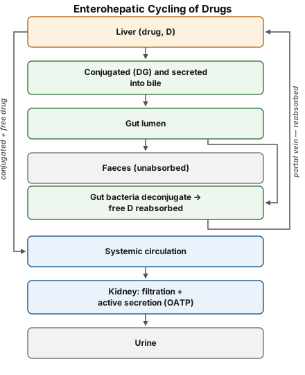
Enterohepatic cycling: liver conjugates → bile → gut bacteria deconjugate → reabsorbed → prolongs action (morphine, doxycycline, OCPs). Antibiotics disrupting gut flora → break cycle → OCP failure.
Bioequivalence: same rate+extent of F under test conditions. Matters most for narrow-margin (digoxin) or precise-dosing drugs (oral anticoagulants/hypoglycaemics).
Distribution
Apparent Vd = total dose ÷ plasma concentration (theoretical, not real volume).
- Low Vd (~0.1-0.2 L/kg): highly protein-bound/large MW, confined to plasma (warfarin, heparin) — poor haemodialysis clearance N/A here (low Vd = actually easier to dialyze; high Vd = poor dialysis clearance)
- ≈ECF (~0.25L/kg): gentamicin, streptomycin
- ≈TBW (~0.6L/kg): ethanol
- Very high Vd (>body mass): digoxin(6L/kg), chloroquine — tissue-sequestered, poorly removed by haemodialysis
Redistribution: lipid-soluble drug → floods high-flow organs (brain) first → rapid redistribution to low-flow high-capacity tissue (muscle/fat) → terminates action despite long elimination t½. Classic: thiopentone (anaesthesia ends in 5-10min via redistribution, NOT metabolism). Repeated dosing → reservoirs fill → longer acting.
BBB: tight junctions + glial investment → only lipid-soluble/unionized drugs cross. Meningitis/encephalitis ↑permeability → penicillin G (normally poor penetration) becomes effective. CTZ + periventricular sites = BBB-deficient (why non-lipid-soluble drugs can still be emetic).
Placenta: lipophilic free passage; NOT absolute barrier — almost any drug can reach fetus at high/prolonged maternal exposure. Exceptions: d-TC (quaternary ammonium), insulin/heparin (large MW).
Plasma protein binding: acidic→albumin, basic→α1-acid glycoprotein. Only free fraction active. Effects: ↑binding→↓Vd, ↑binding→↑duration (unless actively secreted), hypoalbuminemia→↑free drug→toxicity risk (phenytoin, furosemide), displacement interactions (usually transient/minor), highly-bound = poor dialysis removal.
Tissue storage: tetracyclines→bone/teeth · chloroquine→liver/retina/nuclei · digoxin→cardiac+skeletal muscle · thiopentone/DDT→fat · streptomycin→vestibular apparatus.
Biotransformation
Liver = main site (also gut, kidney, lung, plasma, skin, placenta). Lipid-soluble→water-soluble for excretion.
Outcomes: active→inactive (commonest) · active→active metabolite (codeine→morphine, diazepam→oxazepam) · prodrug→active (levodopa→dopamine, enalapril→enalaprilat).
Prodrug uses: ↑bioavailability/BBB penetration (levodopa) · ↑duration (esterified phenothiazines) · ↑taste (clindamycin palmitate) · site-specific activation (methenamine → formaldehyde only in acidic urine = urinary antiseptic).
Phase I (functionalization): oxidation (main, via CYP450+NADPH+O2) · reduction · hydrolysis (esters/amides) · cyclization/decyclization. Metabolite active or inactive.
Phase II (conjugation): glucuronidation (UGT, most important) · acetylation (NAT — isoniazid, sulfonamides) · sulfation · methylation · glycine conjugation · glutathione conjugation (detoxifies reactive quinone/epoxide intermediates — paracetamol’s toxic metabolite; glutathione depletion = mechanism of paracetamol hepatotoxicity). Usually Phase I→II, but isoniazid: Phase II before Phase I (exception).
CYP450: families(1,2,3)→subfamilies(A,B,C)→isoforms. CYP3A4/5 = ~50% of all drug metabolism (liver+gut, contributes to first-pass); CYP2D6, 2C9, 2C19, 1A2 = most of rest. Genetic polymorphism (poor/extensive/ultrarapid metabolizers) = major variability source.
Enzyme induction: rifampin, phenytoin, carbamazepine, phenobarbitone, griseofulvin, chronic alcohol. Onset 4-14 days (new protein synthesis), reversal 1-3 weeks. Consequences: ↓efficacy of inactivated drugs (rifampin→OCP failure) · ↑toxicity of activated/toxic-metabolite drugs (paracetamol+alcohol→↑NAPQI→hepatotoxicity) · autoinduction→tolerance (carbamazepine) · acute intermittent porphyria precipitation (↑ALA synthetase) · therapeutic use: phenobarbitone in neonatal jaundice.
Enzyme inhibition: FAST onset (direct enzyme effect, no synthesis needed) unlike induction. Azoles, macrolides, chloramphenicol, ciprofloxacin, cimetidine. Warfarin+erythromycin/chloramphenicol→bleeding risk. Deliberate use: ritonavir boosts other PIs via CYP3A4 inhibition.
Microsomal (CYP450, UGT — SER of liver/kidney/gut/lung; inducible) vs non-microsomal (esterases, amidases — cytoplasm/mitochondria; NOT inducible, but genetic polymorphism possible). Both deficient at birth (conjugation more so) → grey baby syndrome (chloramphenicol, neonates).
Pharmacogenetics: slow acetylators of INH→↑peripheral neuritis; fast acetylators→need higher dose. Atypical pseudocholinesterase→prolonged succinylcholine apnoea (normal duration 3-6min). G6PD deficiency→haemolysis with primaquine/sulfonamides/dapsone. CYP2D6 poor metabolizers→codeine analgesia lost (can’t convert to morphine).
Hofmann elimination: enzyme-independent spontaneous breakdown at physiological pH/temp. Atracurium — usable in hepatic/renal failure.
Excretion
Kidney = main route. Also lungs, bile/faeces, saliva, sweat, milk.
Renal excretion = GF + tubular secretion − tubular reabsorption
- GF: passive, non-selective (unbound drug only), parallels GFR (↓ after age 50, renal failure)
- Tubular reabsorption: passive, pH-dependent. Weak acids: ↓reabsorption (↑excretion) in alkaline urine. Weak bases: ↓reabsorption (↑excretion) in acidic urine. Only significant for pKa 5-8 drugs. Urine alkalinization = salicylate/phenobarbitone poisoning treatment.
- Active tubular secretion: carrier-mediated (OAT/OCT), unaffected by pH/protein binding (pulls free drug out fast, promotes further dissociation of bound drug). Probenecid blocks penicillin secretion → ↑duration+levels (historically used to extend penicillin action).
Biliary excretion: MW>300, esp. glucuronides → enterohepatic cycling (see above).
Other routes: lungs (volatile anaesthetics/alcohol, by partial pressure) · faeces (purgatives) · skin (As, Hg) · saliva (K-iodide, phenytoin, lithium — used for non-invasive monitoring) · milk (lipid-soluble/less-protein-bound/basic drugs concentrate more; milk pH~7.0 < plasma).
Kinetics of elimination
First-order: constant FRACTION eliminated/time, CL constant, most drugs at therapeutic range (enzymes not saturated). Zero-order (Michaelis-Menten/capacity-limited): constant AMOUNT eliminated/time, CL falls as conc rises. Ethanol always zero-order. Phenytoin, aspirin(OD), theophylline, warfarin: first-order at low dose → zero-order once saturated → disproportionate rise in plasma level with dose ↑ → TDM essential.
t½: time for plasma conc to halve. 1t½=50% gone, 2t½=75%, 3t½=87.5%, 4-5t½=~complete elimination AND steady state. t½ = 0.693×Vd/CL (derived, not fixed — changes if Vd or CL change). Zero-order drugs: t½ not fixed (lengthens as conc falls).
Clearance (CL) = rate of elimination/plasma conc — determines MAINTENANCE dose (not t½ per se, though linked).
Steady state: reached in 4-5 half-lives REGARDLESS of dose size — bigger dose = higher plateau, NOT faster arrival. This is why long-t½ drugs need a loading dose if urgency doesn’t allow waiting.

- Dose rate = (target Cpss × CL) / F
- Loading dose = (target Cp × Vd) / F — depends on Vd only, NOT CL/t½
- Maintenance dose — depends on CL (or t½), NOT Vd
Two-phase (loading+maintenance) dosing: digoxin, lidocaine (acute VT), chloroquine, amiodarone.
Therapeutic Drug Monitoring (TDM)
Useful for: narrow therapeutic index (digoxin, lithium, phenytoin, aminoglycosides) · wide inter-individual variation (TCAs) · renal-impairment-amplified toxicity (aminoglycosides) · poisoning · unexplained treatment failure · compliance check.
NOT useful for: drugs with easy clinical/biochemical endpoint (BP for antihypertensives, glucose, INR) · drugs activated after metabolism (levodopa) · “hit-and-run” drugs (omeprazole, MAOIs, reserpine — effect outlasts plasma presence) · irreversible-action drugs (OP anticholinesterases, phenoxybenzamine).
Prolonging drug action
Slow absorption: SR/CR oral forms · insoluble salt/oily depot (benzathine penicillin, protamine zinc insulin) · vasoconstrictor co-administration (adrenaline+LA — also ↓systemic toxicity, ↓field bleeding). ↑Protein binding: sulfadoxine (weeks) vs sulfadiazine (hours) — same class, different duration by design. ↓Metabolism: chemical modification (ethinyl estradiol) or co-drug inhibiting metabolism (allopurinol+6-MP; cilastatin protects imipenem). ↓Renal excretion: probenecid + penicillin. Targeted devices: liposomes (RES uptake — liposomal amphotericin B) · implants (hormonal IUCD, years) · drug-eluting stents.
Clinical relevance
Phenytoin’s first-order→zero-order shift is the single most bedside-dangerous PK fact in this topic: small dose increase near saturation → disproportionate plasma level rise → toxicity, hence mandatory TDM. Loading dose logic (Vd-driven, urgency-driven) vs maintenance dose logic (CL-driven, balance-driven) is the core reasoning behind virtually every “how much and how often” prescribing decision.
Overview
Pharmacokinetics is what the body does to a drug — its movement in, through, and out of the body, captured by four linked processes: absorption, distribution, metabolism, and excretion. Every one of these processes ultimately comes down to a drug crossing a biological membrane, so the mechanisms of membrane transport underlie the whole subject.
Crossing Membranes
A biological membrane is a phospholipid bilayer studded with proteins that act as carriers, channels, receptors, or enzymes. Drugs cross it by:
- Passive diffusion — movement down a concentration gradient, needing no energy; this accounts for the absorption of most drugs. Rate depends on the drug’s lipid:water partition coefficient and the steepness of the concentration gradient.
- Filtration — passage through aqueous pores or paracellular spaces, relevant mainly for small, lipid-insoluble molecules; capillaries (except in the brain) have large paracellular gaps, so most drugs filter through them regardless of lipid solubility.
- Facilitated diffusion — a carrier moves the drug down its gradient without expending energy (e.g. glucose via GLUT4).
- Active transport — a carrier moves the drug against its gradient, requiring ATP hydrolysis directly (primary active transport, e.g. P-glycoprotein) or indirectly by coupling to another ion’s downhill movement (secondary active transport — symport if both move the same direction, antiport if opposite).
- Vesicular transport (endocytosis/exocytosis) — relevant mainly to very large molecules such as vitamin B12-intrinsic factor complex, not to most conventional drugs.
pH and ionization. Most drugs are weak acids or weak bases, and only the unionized fraction is lipid-soluble enough to diffuse across a membrane. The Henderson-Hasselbalch relationship (pH = pKa + log[ionized]/[unionized]) means a weak acid is more unionized — and better absorbed — in an acidic environment, while a weak base is more unionized in an alkaline one. This is why aspirin (a weak acid) absorbs partly from the acidic stomach, while a base like morphine is essentially all absorbed only once it reaches the more alkaline intestine. It also explains ion trapping: a drug that becomes ionized on one side of a membrane cannot easily diffuse back, so it accumulates there — the basis for urinary alkalinization hastening excretion of weak acids in poisoning.
Absorption
Absorption is movement of drug from its site of administration into the bloodstream. Beyond the membrane-crossing principles above, absorption is influenced by:
- Physical state and dissolution — a drug must be in solution to be absorbed; poorly water-soluble drugs (aspirin, griseofulvin) are absorption-rate-limited by how fast they dissolve, and reducing particle size (microfine formulations) speeds this up.
- Concentration and surface area — a steeper gradient and a larger absorbing surface both accelerate passive diffusion, which is why the small intestine (villi-rich, huge surface area) absorbs faster than the stomach despite gastric emptying being the rate-limiting step for many drugs.
- Vascularity/blood flow — better perfusion maintains the concentration gradient across the absorbing surface; this is why exercise/heat speeds subcutaneous absorption and shock (vasoconstricted) delays it.
- Food — usually delays and reduces absorption (dilution, delayed gastric emptying, chelation — calcium in milk binding tetracyclines), though some exceptions increase it (fatty food and lumefantrine/griseofulvin).
- Efflux transporters — P-glycoprotein in the gut epithelium actively pumps some absorbed drug back into the lumen, limiting oral absorption of digoxin and cyclosporine; P-gp inhibitors (verapamil, erythromycin, quinidine) raise their bioavailability, P-gp inducers (rifampin, phenobarbitone) lower it.
- GI disease — increased peristalsis (gastroenteritis) reduces absorption time; achlorhydria reduces iron absorption; mucosal oedema (as in congestive heart failure) impairs absorption generally.
Bioavailability
Bioavailability (F) is the fraction of an administered dose that reaches the systemic circulation unchanged. An intravenous dose is, by definition, 100% bioavailable; every other route may fall short because of incomplete absorption or because the absorbed drug is destroyed before reaching systemic circulation.
First-pass (presystemic) metabolism is the single biggest determinant of oral bioavailability below 100%. An orally absorbed drug travels: gut lumen → intestinal wall → portal vein → liver → systemic circulation, and both the intestinal wall and the liver can metabolize the drug before it ever reaches the rest of the body. Drugs with high first-pass loss (glyceryl trinitrate, lidocaine, propranolol, morphine, verapamil) need a considerably larger oral dose than their parenteral dose, show marked inter-individual variability in required oral dose, and become more bioavailable in patients with significant liver disease (less hepatic tissue left to extract the drug).
Enterohepatic cycling works the opposite direction on bioavailability: a drug conjugated in the liver and secreted into bile can be deconjugated by gut bacteria, reabsorbed, and sent through the liver again — prolonging both its presence in the body and its apparent bioavailability (morphine, doxycycline, oral contraceptives). Disrupting gut flora with antibiotics can break this cycle — part of the mechanism behind antibiotic-associated oral contraceptive failure.
Bioequivalence. Two formulations of the same drug are bioequivalent if their rate and extent of bioavailability are not significantly different under standard test conditions; formulations that differ meaningfully are bioinequivalent even if they are chemically identical in dose. This matters most for drugs with a narrow safety margin (digoxin) or where dosing precision is critical (oral anticoagulants, oral hypoglycaemics).
Distribution
Once in the bloodstream, a drug distributes into tissues down its concentration gradient until an equilibrium is reached between free drug in plasma and drug in tissue fluid. The extent and pattern of distribution depend on lipid solubility, degree of ionization at physiological pH, plasma and tissue protein binding, presence of tissue-specific transporters, and regional blood flow.
Apparent volume of distribution (Vd) is a theoretical construct: the volume that would be needed to contain the entire administered dose at the same concentration as measured in plasma (Vd = total dose ÷ plasma concentration). It is “apparent” because a drug rarely distributes into a real, definable body-water compartment.
- Low Vd (~0.1–0.2 L/kg) — highly protein-bound or large-molecular-weight drugs confined mostly to plasma (warfarin, heparin).
- Vd ≈ extracellular fluid (~0.25 L/kg) — hydrophilic drugs that cross capillaries but not cell membranes (gentamicin, streptomycin).
- Vd ≈ total body water (~0.6 L/kg) — small, freely diffusible molecules (ethanol).
- Very large Vd, exceeding total body water and even body mass — drugs sequestered heavily in tissue (digoxin ~6 L/kg, chloroquine, propranolol) — the plasma holds only a small fraction of total body drug, which is why these drugs are poorly cleared by haemodialysis in overdose.
Redistribution explains why a highly lipid-soluble drug can have a very short duration of action despite a long elimination half-life: it initially floods high-blood-flow organs (brain, heart, kidney) producing its effect, then rapidly redistributes into low-flow, high-capacity tissue (muscle, fat) as those tissues slowly take up their share — terminating the drug’s action at its original site even though most of the dose is still in the body. Thiopentone’s brief anaesthetic action (5–10 minutes) despite a long half-life is the classic illustration; the same drug given repeatedly becomes progressively longer-acting as the low-flow reservoirs fill up.
Blood-brain barrier (BBB) and blood-CSF barrier. Tight junctions between brain capillary endothelial cells, reinforced by a glial cell investment, restrict entry to lipid-soluble, unionized drugs; efflux transporters (P-gp) reinforce this further. Inflammation (meningitis, encephalitis) increases permeability, letting normally excluded drugs (penicillin G) reach therapeutic brain concentrations — the pharmacological reason penicillin is effective in bacterial meningitis despite being a poor BBB-penetrant under normal conditions. A separate barrier gap exists at the chemoreceptor trigger zone and some periventricular sites, which is why even non-lipid-soluble emetic drugs can act there.
Placental barrier. Lipophilic drugs cross freely; the placenta is not an absolute barrier for anything — even nonlipid-soluble drugs cross to a restricted extent if present at high concentration or for a long duration, so essentially any drug taken by the mother can affect the foetus. Quaternary ammonium compounds (d-tubocurarine) and very large molecules (insulin, heparin) are the practical exceptions that do not cross meaningfully.
Plasma protein binding. Acidic drugs bind mainly to albumin, basic drugs to α1-acid glycoprotein. Only the free (unbound) fraction is pharmacologically active; the bound fraction acts as a temporary reservoir, released as free drug is eliminated. Consequences:
- Highly bound drugs have a smaller Vd (largely confined to plasma).
- Highly bound drugs tend to have a longer duration of action, since bound drug is unavailable for metabolism or filtration (though active tubular secretion can still remove it, because secretion pulls free drug out, prompting further dissociation).
- Hypoalbuminaemia (renal failure, cirrhosis, malnutrition) raises the free fraction and risks toxicity at “normal” total plasma concentrations — relevant for phenytoin and furosemide especially.
- Displacement interactions occur when two drugs compete for the same protein-binding site; the higher-affinity drug displaces the lower-affinity one, transiently raising its free concentration — usually a minor, self-correcting effect unless metabolism/excretion of the displaced drug is also impaired.
- Highly protein-bound drugs are poorly removed by haemodialysis.
Tissue storage/reservoirs. Drugs can accumulate selectively in specific tissues by active transport or binding to tissue constituents, sometimes causing local toxicity: tetracyclines in bone and developing teeth, chloroquine in liver/retina (and nuclei), digoxin in cardiac and skeletal muscle, thiopentone and DDT in adipose tissue (a fat:lean body mass ratio also raises Vd for such drugs), streptomycin on the vestibular apparatus.
Biotransformation (Metabolism)
Biotransformation chemically alters a drug, generally converting a lipid-soluble compound into a more polar, water-soluble one so it can be excreted rather than continuously reabsorbed in the renal tubules. The liver is the principal site; the intestine, kidney, lungs, plasma, skin, and placenta contribute to a lesser degree.
Outcomes of metabolism:
- Active → inactive — the most common outcome (paracetamol, propranolol’s active metabolite eventually inactivated).
- Active → active metabolite — codeine to morphine, diazepam to desmethyldiazepam/oxazepam; the observed effect is the sum of parent drug and metabolite.
- Inactive prodrug → active drug — levodopa to dopamine, enalapril to enalaprilat, prednisone to prednisolone.
Why use a prodrug? Better bioavailability or tissue penetration (levodopa crosses the blood-brain barrier when dopamine itself cannot), prolonged action (esterified phenothiazines), improved taste for paediatric formulations (clindamycin palmitate), or genuinely site-specific activation (methenamine releases formaldehyde only in acidic urine, acting as a urinary antiseptic exactly where it’s needed and nowhere else).
Phase I (non-synthetic/functionalization) reactions expose or introduce a reactive functional group (–OH, –COOH, –NH2, –SH); the resulting metabolite may be active or inactive.
- Oxidation — the dominant phase I reaction, carried out chiefly by the cytochrome P450 (CYP) system in the smooth endoplasmic reticulum, requiring NADPH and molecular oxygen.
- Reduction, hydrolysis (esters, amides — succinylcholine, procaine, lidocaine), cyclization, and decyclization (barbiturates, phenytoin) are the other phase I mechanisms.
Phase II (synthetic/conjugation) reactions attach an endogenous substrate to the drug or its phase I metabolite, almost always producing a polar, inactive, readily excreted product (exceptions: morphine glucuronide and minoxidil sulfate remain active). Glucuronidation (UDP-glucuronosyl transferases) is the most important conjugation pathway quantitatively; others are acetylation (N-acetyltransferase — isoniazid, sulfonamides), sulfation, methylation, glycine conjugation, and glutathione conjugation (which specifically detoxifies reactive quinone/epoxide intermediates, most notably paracetamol’s toxic metabolite — glutathione depletion here is exactly what causes paracetamol hepatotoxicity in overdose).
Most drugs undergo phase I then phase II sequentially, but the order is not fixed — isoniazid is acetylated (phase II) before any phase I oxidation occurs.
The cytochrome P450 system. CYP isoenzymes are grouped into families (1, 2, 3…) and subfamilies (A, B, C…). CYP3A4/5 alone accounts for roughly half of all human drug metabolism and is expressed in both liver and gut wall (contributing directly to intestinal first-pass metabolism); CYP2D6, CYP2C9, CYP2C19, and CYP1A2 handle most of the rest. Genetic polymorphism in these enzymes (extensive vs poor vs ultrarapid metabolizers) is a major source of inter-individual variation in drug response and toxicity.
Enzyme induction. Certain drugs (rifampin, phenytoin, carbamazepine, phenobarbitone, griseofulvin, chronic alcohol) increase the synthesis of microsomal enzymes, accelerating metabolism of themselves (autoinduction) and other co-administered drugs. Onset takes days (4–14 days to peak) since it requires new protein synthesis, and reversal after stopping the inducer takes 1–3 weeks. Consequences worth knowing individually: reduced efficacy of drugs inactivated by metabolism (classic example — rifampin causing oral contraceptive failure), increased toxicity of drugs activated by metabolism or whose toxic metabolite is overproduced (paracetamol hepatotoxicity is worse in chronic alcohol users because CYP2E1 induction generates more of the toxic NAPQI metabolite), tolerance via autoinduction (carbamazepine), precipitation of acute intermittent porphyria (induction upregulates ALA synthetase), and — used therapeutically — acceleration of bilirubin conjugation by phenobarbitone in neonatal jaundice.
Enzyme inhibition is faster in onset than induction, since it acts directly on existing enzyme rather than requiring new synthesis. Azole antifungals, macrolides, chloramphenicol, ciprofloxacin, and cimetidine are common inhibitors; the clinically important consequence is toxicity of the co-administered drug whose metabolism is blocked (warfarin bleeding risk rises sharply with erythromycin or chloramphenicol). Inhibition is occasionally exploited deliberately — low-dose ritonavir is used specifically to inhibit CYP3A4 and boost blood levels of other HIV protease inhibitors.
Microsomal vs non-microsomal enzymes. Microsomal enzymes (CYP450s, UGTs) sit on the smooth endoplasmic reticulum of liver, kidney, intestinal, and lung cells; they carry out most oxidations, reductions, hydrolyses, and glucuronidation, and are the enzymes subject to induction. Non-microsomal enzymes (esterases, amidases, most other conjugases) sit in cytoplasm and mitochondria, are not inducible, but can show genetic polymorphism of their own (acetyltransferase, plasma pseudocholinesterase). Both types are relatively deficient at birth (more so for conjugation than oxidation) — the mechanistic basis of grey baby syndrome with chloramphenicol in neonates.
Pharmacogenetic variation worth naming individually:
- Slow vs fast acetylators of isoniazid — slow acetylators accumulate more unmetabolized drug and are more prone to peripheral neuritis; fast acetylators need a relatively higher dose for the same effect.
- Atypical plasma pseudocholinesterase — metabolizes succinylcholine far more slowly than normal, producing prolonged apnoea from a drug whose normal duration is only 3–6 minutes.
- G6PD deficiency — impairs the RBC’s ability to withstand oxidative stress, precipitating haemolysis on exposure to oxidant drugs (primaquine, sulfonamides, dapsone, high-dose aspirin).
- CYP2D6 poor metabolizers — cannot efficiently convert codeine to morphine, so codeine’s analgesic effect is essentially lost in these individuals.
Factors other than genetics that reduce metabolism: extremes of age (neonatal and elderly hepatic enzyme immaturity/decline), malnutrition, and hepatic disease (cirrhosis prolonging diazepam’s action).
Hofmann elimination is a special, enzyme-independent inactivation mechanism — spontaneous molecular breakdown in body fluids at physiological pH and temperature, not requiring any metabolizing enzyme at all. Atracurium is the standard example, which is precisely why it remains usable in patients with hepatic or renal failure where enzymatic elimination would otherwise be compromised.
Excretion
Excretion removes drug and its metabolites from the body. The kidney is the dominant route for most drugs; the lungs, bile/faeces, saliva, sweat, and milk are minor but occasionally clinically important routes.
Renal excretion is the net result of three processes:
Net renal excretion = Glomerular filtration + Tubular secretion − Tubular reabsorption
- Glomerular filtration — passive, non-selective for any drug small enough and not bound to plasma protein; declines in parallel with GFR (age >50, renal failure).
- Tubular reabsorption — passive diffusion back into blood, governed by lipid solubility and the degree of ionization at the prevailing urinary pH. Weak acids reabsorb less (excrete more) in alkaline urine; weak bases reabsorb less (excrete more) in acidic urine — the basis for alkalinizing urine to hasten elimination of salicylate or phenobarbitone poisoning. This pH-dependence is only clinically significant for drugs with a pKa roughly between 5 and 8; strongly acidic or strongly basic drugs stay ionized (and thus poorly reabsorbed, well excreted) regardless of urinary pH.
- Active tubular secretion — carrier-mediated (organic anion and organic cation transporters) in the proximal tubule; unaffected by urinary pH or protein binding (indeed, active secretion pulls free drug out fast enough to promote further dissociation of bound drug, effectively bypassing protein binding as a barrier to elimination). Competing substrates for the same transporter interact — probenecid blocks penicillin’s tubular secretion, prolonging its action and half-life, which is exactly why probenecid has historically been co-administered with penicillin to extend its effect.
Biliary excretion and enterohepatic cycling. Larger drug molecules (MW > 300), especially glucuronide conjugates, are preferentially excreted into bile. In the gut, bacterial deconjugation frees the drug again for reabsorption — the enterohepatic cycle already described under Bioavailability — before eventual excretion, mostly in urine after re-entering circulation.
Other excretory routes: volatile drugs and general anaesthetic gases via the lungs (rate depending on their partial pressure in blood, independent of lipid solubility); purgatives directly into faeces; heavy metals (arsenic, mercury) via skin; potassium iodide, phenytoin, lithium via saliva (salivary lithium monitoring is used clinically as a non-invasive proxy); and small but clinically relevant amounts of most drugs via breast milk, where lipid-soluble, less-protein-bound, and more basic (milk pH ~7.0, slightly lower than plasma) drugs concentrate preferentially.
Kinetics of Elimination
First-order kinetics — the rate of elimination is directly proportional to the plasma concentration; a constant fraction of the drug present is eliminated per unit time, so clearance stays constant regardless of dose. This applies to the majority of drugs at therapeutic concentrations, because the enzymes/transporters responsible are present in excess and not saturated.
Zero-order (capacity-limited/Michaelis-Menten) kinetics — the eliminating mechanism is saturated, so a constant amount of drug is eliminated per unit time regardless of concentration; clearance falls as concentration rises. Ethanol follows zero-order kinetics throughout its clinical range. Several important drugs — phenytoin, aspirin (in overdose), theophylline, warfarin — follow first-order kinetics at low/therapeutic doses but shift to zero-order once the eliminating enzyme saturates at higher doses; beyond that saturation point, small dose increases cause disproportionately large rises in plasma concentration, which is exactly why phenytoin needs therapeutic drug monitoring rather than simple linear dose titration.
Plasma half-life (t½) is the time for plasma concentration to fall by half. For a drug with rapid distribution and first-order elimination, roughly 50% of the drug is gone after 1 half-life, 75% after 2, 87.5% after 3, and effectively complete elimination (>93%) is reached after 4–5 half-lives — the rule of thumb used throughout pharmacokinetics for “when is a drug essentially cleared” and “when is steady state reached.” Half-life is a derived parameter (t½ = 0.693 × Vd/CL), not a fundamental one — it changes if either volume of distribution or clearance changes, and for a zero-order drug, the concept of a fixed half-life doesn’t strictly apply at all, since t½ itself lengthens as concentration falls further from saturation.
Clearance (CL) is the theoretical volume of plasma completely cleared of drug per unit time (CL = rate of elimination ÷ plasma concentration) — conceptually parallel to creatinine clearance. Clearance, not half-life, is the parameter that actually determines the maintenance dose rate needed for a target steady-state level.
Steady state (plateau principle). With repeated dosing at a fixed interval, plasma concentration rises with each dose (some of the previous dose still present) until the amount eliminated per interval equals the amount administered per interval — a steady-state average concentration (Cpss) with oscillation between trough and peak. Steady state is reached in 4–5 half-lives regardless of dose size, a fact that has two direct clinical consequences: giving a larger dose to “get there faster” does not speed up steady state, it only produces a higher plateau; and for a long-half-life drug, waiting for steady state to build up naturally may be clinically too slow, which is exactly the situation a loading dose is designed to solve.
- Dose rate needed for a target Cpss: Dose rate = (target Cpss × CL) ÷ F.
- Loading dose (a single or briefly-repeated larger dose to reach target concentration immediately): Loading dose = (target Cp × Vd) ÷ F. Loading dose depends only on Vd, not on clearance or half-life.
- Maintenance dose (repeated thereafter to sustain the steady state by balancing ongoing elimination): governed by clearance (or equivalently half-life), not by Vd.
This loading-then-maintenance two-phase strategy is standard for long-half-life or urgently needed drugs — digoxin, lidocaine in acute ventricular arrhythmia, chloroquine, amiodarone.
Without a loading dose, reaching the plateau still takes the same 4–5 half-lives regardless of how large the maintenance dose is — only the height of the eventual plateau changes with dose size, not the time taken to get there. A loading dose sidesteps this delay entirely by placing the patient near the target concentration immediately, after which the regular maintenance dose simply holds it there.
Therapeutic Drug Monitoring (TDM)
Measuring plasma drug concentration is useful specifically when it adds information the clinical picture alone cannot supply:
Indicated for: narrow therapeutic index drugs (digoxin, lithium, phenytoin, aminoglycosides, anticonvulsants generally), drugs with wide inter-individual pharmacokinetic variation (tricyclic antidepressants), drugs whose toxicity is amplified by renal impairment (aminoglycosides), suspected poisoning, unexplained treatment failure, and checking compliance.
Not useful for: drugs whose effect is directly and easily measurable by a clinical or biochemical endpoint instead (blood pressure for antihypertensives, blood glucose for hypoglycaemics, INR for anticoagulants), drugs activated only after metabolism (levodopa — measuring the parent drug tells you little), “hit-and-run” drugs whose effect outlasts their presence in plasma (omeprazole, MAO inhibitors, reserpine), and drugs with irreversible action (organophosphate anticholinesterases, phenoxybenzamine) — since by the time toxicity or efficacy is evident, the plasma level has already done whatever it was going to do.
Prolonging Duration of Drug Action
Extending a drug’s action reduces dosing frequency, improves compliance, and smooths out the peak-trough fluctuation that causes both breakthrough symptoms at the trough and toxicity at the peak. Strategies include:
- Slowing absorption — sustained/controlled-release oral formulations; for injectables, using an insoluble salt or oily depot vehicle (benzathine penicillin extends penicillin’s action from hours to weeks; protamine zinc insulin similarly slows subcutaneous release), or co-administering a vasoconstrictor (adrenaline with local anaesthetic — also usefully reduces systemic toxicity and surgical field bleeding as a side benefit).
- Increasing plasma protein binding — deliberately designing a more highly bound congener that is released slowly in free, active form (sulfadoxine versus sulfadiazine — weeks versus hours of action from the same drug class).
- Slowing metabolism — small chemical modifications can markedly change metabolic rate without changing the pharmacological action itself (ethinyl estradiol vs estradiol), or one drug can be given specifically to inhibit another’s metabolism (allopurinol prolonging 6-mercaptopurine’s action; cilastatin protecting imipenem from renal degradation).
- Slowing renal excretion — blocking active tubular secretion with a competing substrate (probenecid with penicillin).
- Targeted delivery devices — liposomes (selectively taken up by the reticuloendothelial system, useful for liposomal amphotericin B), drug-releasing implants (hormonal IUCDs lasting years), and drug-eluting stents.
Not every drug needs to be made long-acting — a hypnotic meant only to induce sleep, or an analgesic for brief pain relief, is deliberately kept short-acting, and drugs with an inherently long half-life (doxycycline, digoxin, amlodipine) need no special formulation to begin with.
Clinical Relevance
Kinetics is not an abstract exercise — it is the quantitative reasoning behind everyday prescribing decisions. Whether a drug needs a loading dose depends on how urgently a therapeutic level is needed relative to its half-life; whether TDM is worth ordering depends on whether the drug’s effect can otherwise be judged directly; whether a drug is safe in renal or hepatic failure depends on which organ actually clears it and by which kinetic order. The shift from first-order to zero-order kinetics in phenytoin, and the resulting disproportionate rise in plasma level with small dose increases, is one of the most frequently tested single facts in this entire subject precisely because it has a direct, dangerous bedside consequence.
Diagram 1 — Apparent Volume of Distribution (Vd)
What to draw: A single container (“body”) holding a known total amount of drug, with an arrow pointing to a smaller graduated cylinder labelled “plasma sample” showing the measured plasma concentration. Beside it, write the Vd formula (Vd = total dose ÷ plasma concentration) and work through one numeric example (e.g. 1000 mg dose, 50 mg/L plasma concentration → Vd = 20 L).
Labelling requirements: explicitly label Vd as “apparent” and note in one line why — the body is not a real 20 L (or 6 L/kg, or 13,000 L) compartment; label at least one low-Vd drug (warfarin) and one very-high-Vd drug (digoxin or chloroquine) next to the diagram with their actual values, to anchor the concept in real numbers.
Common exam-marking mistakes:
- Treating Vd as a real anatomical volume rather than a calculated ratio — examiners specifically probe this misconception by asking to interpret an unusually large Vd (>total body water).
- Forgetting to state the clinical consequence: large-Vd drugs are poorly removed by haemodialysis in overdose.
Diagram 2 — Blood-Brain Barrier Structure
What to draw: A brain capillary cross-section showing endothelial cells joined by tight junctions, with an investment of glial (astrocyte) foot processes surrounding the capillary from outside.
Labelling requirements: label the tight junctions specifically — they are the actual anatomical basis of the barrier, not the endothelium alone. Show one lipid-soluble drug molecule crossing freely and one lipid-insoluble/ionized drug molecule blocked at the tight junction, to make the selective-permeability point visually rather than only in text.
Common exam-marking mistakes:
- Drawing a “barrier” as a solid wall rather than tight junctions between specific cells — loses the marks tied to the actual structural basis.
- Omitting the glial process investment, which is the second component of the barrier alongside the endothelial tight junctions.
Diagram 3 — Enterohepatic Cycling
What to draw: A cycle from liver (drug conjugated to its glucuronide) → bile → gut lumen → bacterial deconjugation → free drug reabsorbed via the portal vein → back to liver, with a side branch showing the fraction that escapes into faeces unabsorbed, and a separate path showing conjugated/free drug entering systemic circulation for renal excretion.
Labelling requirements: the cycle itself must be drawn as a closed loop, not a linear sequence — the entire teaching point is that the drug returns to the liver rather than being eliminated on first pass through bile. Name at least one drug example on the diagram (morphine, doxycycline, or oral contraceptives).
Common exam-marking mistakes:
- Drawing biliary excretion as a dead end (straight to faeces) without showing the reabsorption loop — this misses the entire concept being tested.
- Not connecting the cycle to its clinical consequence: prolonged drug action, and the specific mechanism of antibiotic-induced oral contraceptive failure (gut flora disruption breaks the cycle).
Diagram 4 — Steady State: With vs Without a Loading Dose
What to draw: Two plasma concentration-vs-time curves on the same axes, x-axis marked in multiples of half-life (0 to 5–6). One curve rises gradually from zero to plateau over 4–5 half-lives (no loading dose); the other starts near the target line immediately and oscillates there from the first dose (with loading dose). Mark the target steady-state concentration as a dashed horizontal reference line.
Labelling requirements: the x-axis must be marked in half-lives, not in absolute time units — the “4–5 half-lives to steady state” rule is independent of what the actual half-life value is, and that generality is the point of the diagram.
Common exam-marking mistakes:
- Drawing the no-loading-dose curve as reaching steady state faster with a bigger maintenance dose — a bigger maintenance dose changes the height of the plateau, not the time (in half-lives) taken to reach it. This is the single most common misconception this diagram exists to correct.
- Omitting the oscillation (peak-trough fluctuation) at steady state and drawing a flat line instead, which loses the connection to dosing interval and Cmax/Cmin reasoning.
Short Essays (5)
Short Answers (30)
Short Notes (15)
Multiple Choice Questions (10)
My Sticky Notes
Personal revision notes, mnemonics and reminders.
