
Bacterial Morphology and Physiology
Shapes and Arrangement
Cocci: round. Bacilli: rods. Vibrios: short comma-shaped. Spirilla: rigid spirals. Spirochetes: flexible spirals. Actinomycetes: branching filaments.
Cluster = Staphylococcus. Chain = Streptococcus. Lancet-shaped pairs = pneumococcus. Kidney-shaped pairs = gonococcus. Tetrads = Micrococcus. Bamboo-stick chain = B. anthracis. Chinese-letter pattern = C. diphtheriae.
Spirochetes, Mycoplasma, Rickettsia, Chlamydia — do not stain well with Gram stain.
Cell Wall
Peptidoglycan = NAM + NAG chains, cross-linked.
Gram-positive: thick peptidoglycan (16–80 nm). Cross-link via pentaglycine bridge, L-lysine involved. Has teichoic acid (wall + lipoteichoic). No outer membrane, no LPS.
Gram-negative: thin peptidoglycan (2 nm). Cross-link direct, meso-diaminopimelic acid instead of L-lysine. Has outer membrane (porins, Braun’s lipoprotein) + LPS. No teichoic acid.
LPS = Lipid A (endotoxin) + core polysaccharide + O antigen (serotyping).
Periplasmic space: between outer membrane and plasma membrane, Gram-negative only (or vestigial in Gram-positive).
Cell Membrane
Fluid mosaic bilayer, 5–10 nm. No sterols except Mycoplasma. Site of respiration, transport, lipid/wall synthesis.
Cytoplasmic Contents
Ribosomes: 70S (30S+50S). Site of translation.
Mesosomes: membrane infoldings, more in Gram-positive, respiratory enzymes, mitochondria-like role.
Inclusions: appear in starvation, disappear when fed. Volutin (metachromatic) granules = polyphosphate, classic in C. diphtheriae, stain with Albert’s/Neisser’s stain.
Nucleoid: single circular dsDNA, no membrane, no histones. V. cholerae has two chromosomes. Plasmids = extrachromosomal DNA, carry resistance/toxin genes.
Capsule and Slime Layer
Capsule = organized, adherent. Slime layer = loose, easily removed. S. salivarius has both.
Mostly polysaccharide. Exception: B. anthracis capsule is polypeptide (poly-D-glutamate).
Functions: blocks phagocytosis, blocks complement lysis, resists desiccation, forms biofilms (catheters, prosthetic valves), basis of pneumococcal/meningococcal/Hib vaccines.
Detection: India ink (negative stain, clear halo), Quellung reaction (pneumococcus swells with antiserum), latex agglutination (direct antigen detection, e.g. CSF).
Flagella
Motility organ. Monotrichous = one polar (V. cholerae, Pseudomonas). Lophotrichous = tuft polar (Spirillum). Amphitrichous = one at each pole. Peritrichous = all over (Salmonella Typhi, E. coli).
Parts: filament (flagellin), hook, basal body (motor).
H antigen = flagellar antigen, used for serotyping, not protective.
Detected indirectly: motility medium, hanging drop, Craigie tube. Characteristic movement: darting (V. cholerae), tumbling (Listeria), swarming (Proteus).
Fimbriae (Pili)
Shorter, thinner, more numerous than flagella. Function: adhesion, not motility. Made of pilin.
Sex pilus: fewer, longer, only on donor cells, forms conjugation tube for DNA transfer.
Bacterial Spores
Survival form, not reproduction — one cell makes one spore. Bacillus, Clostridium mainly.
Layers inside-out: core → cortex → coat → exosporium.
Resistance comes from calcium + dipicolinic acid in the cortex.
Position: central/subterminal/terminal. Bulging or non-bulging — fixed per species. Bacillus: usually non-bulging. Clostridium: usually bulging.
Sterilization indicators: Geobacillus stearothermophilus spores for autoclave, Bacillus atrophaeus spores for hot air oven/ethylene oxide.
B. anthracis spores — used in 2001 bioterrorism attack.
Staining: unstained/refractile on Gram stain. Weakly acid-fast — modified ZN stain or Schaeffer-Fulton stain.
Atypical Forms
Pleomorphism: variable shape/size, same species (Haemophilus, Proteus).
Involution forms: swollen, aberrant, ageing culture + high salt (gonococcus, plague bacillus).
L forms: cell wall lost entirely, become spherical. Named after Lister Institute. Unstable = reverts when inducing agent removed. Stable = permanent. Mycoplasma resembles stable L form but is not one. Linked to relapsing pyelonephritis.
Growth Requirements
Water ~80% of cell mass. Need C, N, H, O, trace ions (S, P, Na, K, Mg, Fe, Mn).
Fastidious organisms need vitamins (bacterial growth factors) — e.g. Brucella, H. influenzae need niacin.
Classification:
- Carbon source: autotroph (CO2) vs heterotroph (organic).
- Electron source: lithotroph (inorganic) vs organotroph (organic).
- Energy source: chemotroph (chemical) vs phototroph (light).
Most pathogens = chemoorganoheterotroph (like E. coli).
Cell Division
Binary fission. Generation time: E. coli/most pathogens ~20 min. M. tuberculosis 10–15 hr. M. leprae 12–13 days.
Growth Curve
Lag: no division, no death, cells enlarge. Log: division fastest, smallest cells, best time for Gram stain/biochemical tests. Stationary: division = death (balanced), total count still rising, viable count flat. Sporulation, exotoxin, antibiotic, bacteriocin production happen here. Decline: no division, death continues, viable count falls, involution forms appear.
Factors Affecting Growth
Oxygen: obligate aerobe (needs O2: TB, Pseudomonas), obligate anaerobe (killed by O2: C. tetani), facultative anaerobe (either: most pathogens), microaerophilic (low O2: Campylobacter, Helicobacter), aerotolerant anaerobe (tolerates but doesn’t use O2).
CO2: capnophilic organisms need 5–10% CO2 (Brucella, pneumococcus).
Temperature: psychrophile <20°C, mesophile 25–40°C (most pathogens), thermophile 55–80°C.
pH: most pathogens 7.2–7.6. Lactobacilli tolerate pH <4. V. cholerae tolerates pH up to 8.9.
Osmotic effect: hypertonic saline → plasmolysis (shrink). Distilled water → plasmoptysis (swell/rupture).
Desiccation: T. pallidum, gonococcus die fast on drying. M. tuberculosis, S. aureus survive weeks. Lyophilization used to preserve stock cultures.
Shape and Arrangement
Bacteria fall into a small set of basic shapes, and the shape itself is often the first clue to identity on a Gram-stained smear.
- Cocci — spherical or oval cells.
- Bacilli — rod-shaped cells.
- Vibrios — short, comma-shaped curved rods.
- Spirilla — rigid spiral rods.
- Spirochetes — long, flexible spiral organisms.
- Actinomycetes — branching, filamentous forms.
Cell arrangement after division is equally characteristic, because daughter cells separate incompletely in a pattern fixed for each species.
| Arrangement | Example organism |
|---|---|
| Clusters (grape-like) | Staphylococcus |
| Chains | Streptococcus |
| Pairs, lancet-shaped | Pneumococcus |
| Pairs/short chains, kidney- or lens-shaped | Gonococcus, meningococcus |
| Tetrads | Micrococcus |
| Packets of eight | Sarcina |
| Chain, bamboo-stick pattern | Bacillus anthracis |
| Cuneiform / Chinese-letter pattern | Corynebacterium diphtheriae |
| Palisades | Diphtheroids |
| Branching filaments | Actinomyces, Nocardia |
A few groups resist ordinary Gram staining altogether and need special methods to be seen at all: spirochetes (too thin for the light microscope’s resolving power), Mycoplasma (no cell wall to take up the stain), and Rickettsia/Chlamydia (obligate intracellular organisms).
The Bacterial Cell Envelope
Cell Wall
The cell wall sits just outside the plasma membrane and is the structure most responsible for a bacterium’s shape and mechanical strength. It resists osmotic lysis, carries surface antigens, and is the target of several major antibiotic classes. Its architecture divides bacteria into two broad groups that behave very differently in the Gram stain, in pathogenesis, and in drug susceptibility.
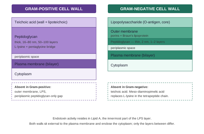
Peptidoglycan is the load-bearing scaffold in both groups — alternating N-acetylmuramic acid (NAM) and N-acetylglucosamine (NAG) sugars, cross-linked by short peptide chains — but its thickness and cross-linking chemistry differ sharply:
- In Gram-positive organisms it is thick (16–80 nm, tens of layers) and cross-links via a pentaglycine bridge between the L-lysine of one tetrapeptide chain and the D-alanine of the next.
- In Gram-negative organisms it is a single thin sheet (about 2 nm), and the cross-link is a direct bond between D-alanine and meso-diaminopimelic acid, which sits at the position L-lysine occupies in Gram-positive cell walls.
Teichoic acid is unique to the Gram-positive wall — glycerol or ribitol polymers that thread through the peptidoglycan (wall teichoic acid) or anchor into the membrane (lipoteichoic acid) — and functions as a major surface antigen.
Gram-negative bacteria compensate for their thin peptidoglycan with an outer membrane, a second phospholipid bilayer anchored to the peptidoglycan by Braun’s lipoprotein. Porins embedded in it act as size-selective channels for small solutes and double as phage receptors. The outer leaflet of this membrane carries lipopolysaccharide (LPS), built from three parts:
- Lipid A — buried in the membrane, and the actual endotoxin: responsible for fever, complement activation, and the lethal effects of Gram-negative sepsis.
- Core polysaccharide — a short connecting chain.
- O antigen — the outermost, highly variable chain that defines serotype and is a target for antibody.
Between the outer membrane and the plasma membrane lies the periplasmic space, a compartment absent (or vestigial) in Gram-positive cells, which holds the thin peptidoglycan layer along with periplasmic enzymes.
Clinically, this structural split explains a great deal at once: Gram-positive organisms lack an LPS-driven endotoxin response but can shed toxic capsular/wall polypeptide (as in anthrax); beta-lactams that block peptidoglycan cross-linking cripple both groups, but drugs that must cross an outer membrane porin to reach their target work far better against Gram-positives, which have none in the way.
Cell Membrane
The plasma membrane lies beneath the cell wall and is essential to viability regardless of Gram reaction. It follows the fluid mosaic model — a phospholipid bilayer, roughly 5–10 nm thick, studded with integral and peripheral proteins. Unlike eukaryotic membranes it lacks sterols (the one exception being Mycoplasma, which incorporates cholesterol scavenged from the host), though many bacteria instead stabilize the membrane with sterol-like hopanoids.
Functionally, the membrane is where a bacterium does most of its metabolic work: it acts as a selective osmotic barrier, houses the permease systems that move nutrients in and wastes out, and is the site of respiration, lipid synthesis, and cell-wall precursor assembly — the roles a mitochondrion and endoplasmic reticulum would split between them in a eukaryotic cell.
Cytoplasmic Contents
Bacterial cytoplasm has no membrane-bound organelles, which is the central structural fact separating prokaryotes from eukaryotes. What it does contain:
- Ribosomes — 70S particles (30S + 50S subunits), the site of translation, strung along mRNA as polysomes. Their difference from the eukaryotic 80S ribosome is exploited pharmacologically by antibiotics that target bacterial translation selectively.
- Mesosomes — infoldings of the plasma membrane, more prominent in Gram-positive organisms, that carry respiratory enzymes (a rough functional parallel to mitochondria) and appear to coordinate cell-wall septum formation with chromosome segregation during division.
- Cytoplasmic inclusions — storage depots that appear under nutrient stress and vanish once the deficiency is corrected. Organic examples include glycogen and polyhydroxybutyrate granules; the classic inorganic example is the volutin (metachromatic) granule of Corynebacterium diphtheriae — polymetaphosphate deposits that stain reddish-purple with Albert’s or Neisser’s stain against a background of a different color, the basis of the polar-body appearance used to identify the organism.
- Nucleoid — a single circular, supercoiled, double-stranded DNA chromosome with no surrounding membrane and no histone-type packaging protein. A small number of species carry a linear chromosome or more than one chromosome (Vibrio cholerae has two). Extrachromosomal DNA — plasmids — often carries drug resistance or toxin genes and can move between cells independent of chromosomal replication.
Cell Wall Appendages
Capsule and Slime Layer
Many bacteria secrete a viscid layer external to the wall (glycocalyx). When it forms a sharply defined, adherent structure it is called a capsule; when it is loose and easily washed off, a slime layer; some organisms — Streptococcus salivarius is the standard example — produce both.
Most capsules are polysaccharide. The classic exception is Bacillus anthracis, whose capsule is a polypeptide (poly-D-glutamate).
Capsules matter clinically for several linked reasons:
- They block phagocytosis and complement-mediated lysis, which is the single biggest reason encapsulated organisms are more virulent than their unencapsulated mutants.
- They protect against desiccation and against lysozyme/phage attack.
- They seed biofilm formation — adherent bacterial communities embedded in a self-made polysaccharide matrix that colonize catheters, prosthetic valves, and other implanted devices, and that resist both host defenses and antibiotics far better than free-living bacteria do.
- Because the polysaccharide is antigenic, purified capsular material is the basis of several vaccines (pneumococcal, meningococcal, Haemophilus influenzae type b).
Capsules can be shown by negative staining (India ink — the capsule appears as a clear halo against a dark background), by the Quellung reaction (anti-capsular antiserum makes the pneumococcal capsule swell and appear more refractile), or by latex agglutination for capsular antigen directly in a specimen such as CSF.
Flagella
Flagella are the organs of bacterial motility — long, thin protein filaments extending from the cell surface. Their arrangement is species-characteristic and diagnostically useful:
| Pattern | Example |
|---|---|
| Monotrichous — single polar flagellum | Vibrio cholerae, Pseudomonas |
| Lophotrichous — tuft of polar flagella | Spirillum |
| Amphitrichous — one flagellum at each pole | Alcaligenes faecalis |
| Peritrichous — flagella over the whole surface | Salmonella Typhi, E. coli |
Structurally, a flagellum has three parts: a hollow filament built from a single protein (flagellin), a basal body embedded in the cell envelope that acts as the rotary motor, and a flexible hook connecting the two. Flagellar (H) antigens are strongly immunogenic and are used in serotyping, though the resulting antibodies are not protective.
Because flagella lie below the light microscope’s resolving power, their presence is usually inferred indirectly — from spreading growth on semisolid motility medium, from the hanging-drop or Craigie tube methods, or from the organism’s own characteristic movement pattern (the darting motility of Vibrio cholerae, the tumbling motility of Listeria, swarming on solid agar by Proteus).
Fimbriae (Pili)
Fimbriae are shorter, thinner, more numerous surface filaments than flagella (up to a thousand per cell) and serve a completely different purpose — adhesion, not locomotion. Built from a protein called pilin, common fimbriae let bacteria stick to epithelial surfaces and initiate colonization; many fimbriated Gram-negative organisms also agglutinate red cells, a property (hemagglutination) used as an indirect assay for their presence.
The sex pilus is a distinct, longer, and much less numerous structure (as few as one to ten per cell) found only on donor (“male”) Gram-negative bacteria. It forms the conjugation tube through which chromosomal or plasmid DNA passes to a recipient cell during bacterial conjugation.
Bacterial Spores
Spore formation is a survival strategy, not reproduction — one vegetative cell produces exactly one spore, and no cell division occurs during the process. Only a limited group of genera can do it, chiefly Bacillus and Clostridium.
Sporulation begins when nutrients run low and takes roughly ten hours to complete. From the inside out, the mature spore is layered as core → cortex → coat → exosporium; deposition of calcium and dipicolinic acid into the cortex is what gives the finished spore its extraordinary resistance to heat, desiccation, and most disinfectants. Spores can be central, subterminal, or terminal in position, and either contained within the width of the parent cell or bulging out of it — a fixed, species-specific pattern useful for identification (Bacillus spores typically do not bulge; Clostridium spores typically do).
Because ordinary disinfection cannot be relied on to kill spores, they double as biological indicators of sterilization: failure of Geobacillus stearothermophilus spores to grow after autoclaving, or of Bacillus atrophaeus spores after hot-air/ethylene-oxide sterilization, confirms the cycle actually worked. The same resistance is what made anthrax spores usable as a bioterrorism agent.
Spores appear as unstained, refractile areas on a Gram stain (they resist the dye) but are weakly acid-fast and can be picked up with a modified Ziehl–Neelsen stain, or more specifically with the Schaeffer–Fulton technique.
Atypical and Cell-Wall-Deficient Forms
- Pleomorphism — wide variation in the size and shape of individual cells of one species (Haemophilus, Proteus), usually reflecting defective cell wall synthesis.
- Involution forms — swollen, aberrant cells that appear in ageing cultures under high salt concentration (gonococcus, plague bacillus); many are non-viable.
- L forms — bacteria that have lost their cell wall entirely, named for the Lister Institute where they were first described in Streptobacillus moniliformis. Loss of the wall makes every L form spherical regardless of the parent’s original shape. Unstable L forms revert to normal morphology once the inducing agent (typically penicillin) is withdrawn; stable L forms retain the aberrant form indefinitely through subculture. Mycoplasma, which lacks a cell wall permanently by nature rather than by induced loss, resembles a stable L form but is not considered one on genetic and biochemical grounds. L forms are implicated in the persistence of some chronic infections, such as relapsing pyelonephritis.
Physiology: Growth and Nutrition
Growth Requirements
Water makes up roughly 80% of bacterial mass, and normal growth further requires sources of carbon, nitrogen, hydrogen, oxygen, and trace inorganic ions (sulfur, phosphorus, sodium, potassium, magnesium, iron, manganese). Fastidious species additionally need specific organic growth factors — often B-group vitamins — supplied in the medium; Brucella and Haemophilus influenzae need niacin, for instance, and several lactobacilli need pyridoxine or vitamin B12.
Metabolically, bacteria are classified along three independent axes:
- Carbon source — autotrophs fix atmospheric CO₂; heterotrophs use pre-formed organic molecules.
- Source of reducing power — lithotrophs draw electrons from inorganic compounds; organotrophs draw them from organic ones.
- Energy source — chemotrophs use chemical energy; phototrophs use light.
Nearly every pathogen of medical importance is a chemoorganoheterotroph — it derives energy, carbon, and reducing power all from organic compounds, exactly as E. coli does.
Cell Division and Generation Time
Bacteria reproduce by binary fission: the chromosome replicates and segregates, then a transverse septum closes across the cell and the two daughter cells separate (often incompletely, which is what produces the chains and clusters described earlier). The generation time — time for one cell to become two — varies enormously by species: about 20 minutes for E. coli and most pathogens, 10–15 hours for Mycobacterium tuberculosis, and 12–13 days for Mycobacterium leprae. This single number explains why TB and leprosy cultures take weeks, while a stool or urine culture is read the next day.
Bacterial Growth Curve
Plotting viable count against time after inoculating a fresh liquid medium produces a curve with four distinct phases.
| Phase | Cell division | Cell death | Total count | Viable count | What is happening |
|---|---|---|---|---|---|
| Lag | No | No | Flat | Flat | Enzymes and metabolites accumulate; cells reach maximum size |
| Log (exponential) | Yes | No | Rising | Rising | Fastest division; smallest cell size; best time for Gram stain and biochemical tests |
| Stationary | Yes | Yes (balanced) | Still rising | Flat | Nutrient exhaustion begins; sporulation, exotoxin, antibiotic and bacteriocin production occur here |
| Decline | No | Yes | Flat | Falling | Nutrients exhausted, toxic products accumulate; involution forms appear |
Factors Affecting Growth
Oxygen requirement splits bacteria into several physiological classes: obligate aerobes (Mycobacterium tuberculosis, Pseudomonas) grow only with oxygen; obligate anaerobes (Clostridium tetani) are killed by it; facultative anaerobes (most pathogens, including E. coli and S. aureus) grow either way; microaerophiles (Campylobacter, Helicobacter) need a low-oxygen atmosphere; and aerotolerant anaerobes survive oxygen exposure without actually using it.
Carbon dioxide — a few species (Brucella, Streptococcus pneumoniae) grow best with 5–10% CO₂ and are called capnophilic.
Temperature sorts organisms into psychrophiles (below 20°C), mesophiles (25–40°C, the range that covers essentially every human pathogen, since it spans body temperature), and thermophiles (55–80°C).
pH — most pathogens prefer 7.2–7.6; lactobacilli tolerate acidic pH below 4, while Vibrio cholerae tolerates alkaline pH up to 8.9 (a fact exploited in selective media for stool culture).
Osmotic pressure — the cell wall lets bacteria tolerate a wide range of external osmolarity, but sudden exposure to strong hypertonic saline can shrink the cell (plasmolysis), while distilled water can swell and rupture it (plasmoptysis).
Desiccation — survival varies widely by species: Treponema pallidum and Neisseria gonorrhoeae die within minutes of drying, while Mycobacterium tuberculosis and Staphylococcus aureus can survive for weeks. Controlled drying under vacuum (lyophilization) exploits the same principle deliberately, to preserve laboratory stock cultures.
Diagram 1: Gram-Positive vs Gram-Negative Cell Wall
What to draw: Two side-by-side cross-sections, outermost layer at the top of each stack, cytoplasm at the bottom.
- Gram-positive stack (outside → in): teichoic acid layer sitting on top of a thick peptidoglycan band → thin periplasmic gap → plasma membrane → cytoplasm.
- Gram-negative stack (outside → in): LPS (O-antigen + core) → outer membrane (mark the porins) → thin peptidoglycan sheet → periplasmic space → plasma membrane → cytoplasm.
- Label the peptidoglycan cross-link chemistry difference explicitly on each side: pentaglycine bridge / L-lysine (Gram-positive) vs direct D-alanine–meso-DAP bond (Gram-negative) — this is the single most commonly tested distinguishing fact and the most common labelling mistake (students frequently reverse which side carries the pentaglycine bridge).
- Mark Lipid A as the endotoxin-active component within the LPS band, not the O-antigen — a frequent exam confusion.
Common exam-marking mistakes to avoid when hand-drawing this:
- Do not draw teichoic acid on the Gram-negative side or LPS/outer membrane on the Gram-positive side — these are mutually exclusive and examiners specifically check for this swap.
- Do not label the O-antigen as the toxic component; only Lipid A is the endotoxin.
- Do not omit the periplasmic space on the Gram-negative side — it is a real, named compartment, not empty space.
- Keep peptidoglycan thickness visually proportionate: Gram-positive should look conspicuously thicker than Gram-negative, since the numeric contrast (16–80 nm vs 2 nm) is itself an examined fact.
Other structures — hand-drawing notes only (no rendered diagram)
These structures are better drawn as simple labelled sketches than as multi-step process diagrams — each is a single static structure, not a mechanism or pathway, so a rendered flowchart would add nothing a labelled hand sketch doesn’t already give.
Flagellar arrangement patterns — draw a single bacillus four times, varying only flagellar placement: one polar flagellum (monotrichous), a tuft at one pole (lophotrichous), one flagellum at each pole (amphitrichous), flagella covering the whole surface (peritrichous). Label each pattern with its example organism. Common mistake: confusing lophotrichous (a tuft, several flagella at one pole) with amphitrichous (single flagella, but at both poles).
Bacterial spore structure — a single oval spore in cross-section, labelled core (centre, contains DNA) → cortex → coat → exosporium (outermost), plus its position within the parent cell (central, subterminal, or terminal) and whether it bulges the cell outline. Common mistake: labelling the layers in the wrong order, or forgetting that not every genus produces a bulging spore.
Bacterial growth curve — this is deliberately not rendered as a plotted graph (per the no-fabricated-quantitative-graph rule): the four phases have no real numeric data to plot accurately, and the notes.md table already conveys the same information without pretending to axis-level precision. If hand-drawing it for revision, sketch only the qualitative shape (flat → rising steeply → plateau → falling) and label each segment with its name — do not put numbers on the axes.
Short Essays (4)
Short Answers (1)
Short Notes (29)
Multiple Choice Questions (10)
My Sticky Notes
Personal revision notes, mnemonics and reminders.
