
Type I Hypersensitivity
Definition
Rapid (15-30min) IgE-mediated reaction to allergen in a sensitised person. = anaphylactic/atopic hypersensitivity.
Aetiology (atopy)
Genetic (↑IgE, ↑IL-4 Th2, HLA-B8, +ve family history in 50%) + environmental (pollutants, viral URI, hygiene hypothesis) + genes (HLA, cytokines, FcεRI component, ADAM33). 20-30% = non-atopic (temperature/exercise-triggered, no Th2/IgE).
Pathogenesis
- Sensitisation (1st exposure) — Th2 (IL-4,5,13) → B cells → IgE. IgE binds FcεRI on mast cells/basophils (high affinity — always occupied despite low serum IgE)
- Re-exposure — allergen cross-links surface IgE → degranulation
- Mediators released (3 groups):
- Preformed amines: histamine (vasodilatation, ↑permeability, smooth muscle contraction, mucus) + tryptase (→ kinins, C5a) + heparin (storage matrix)
- Newly synthesised lipids: PGD2 (bronchospasm, mucus — most abundant mast cell COX product), LTC4/LTD4 (1000s-fold more potent than histamine for permeability/bronchoconstriction), LTB4 (neutrophil/eosinophil/monocyte chemotaxis)
- Cytokines: TNF, chemokines → late phase
Two phases
- Immediate (5-30min, gone by 1h) — amines+lipids → vasodilatation, leakage, spasm
- Late-phase (2-8h, lasts days) — cytokines → eosinophil/neutrophil/Th2 infiltrate, real tissue injury, self-sustaining even without more allergen
Manifestations
Systemic anaphylaxis — antisera (ATS), drugs (penicillin), stings → itching, urticaria, bronchoconstriction, laryngeal oedema, GI symptoms, shock, death.
Local: hay fever · bronchial asthma · food allergy · cutaneous (urticaria/wheal-flare) · angioedema (AD inherited, C1-INH deficiency).
Atopic triad/march: eczema → allergic rhinitis → asthma (sequential in same patient).
Treatment (mechanism-mapped)
Corticosteroids (late phase) · antihistamines (immediate) · leukotriene antagonists · bronchodilators · epinephrine (anaphylaxis) · anti-IgE/anti-Th2-cytokine biologics.
Clinical relevance
Biphasic reaction (immediate + delayed relapse) → extended observation post-anaphylaxis. Angioedema (hereditary, C1-INH) ≠ ordinary local anaphylaxis — antihistamines less effective. Atopic march predicts trajectory from eczema.
Definition
Type I (immediate, anaphylactic/atopic) hypersensitivity is a rapidly developing immune response to an antigen — termed an allergen — in a person previously sensitised to it. The reaction is mediated by IgE antibodies bound to mast cells and basophils, and classically appears within 15–30 minutes of re-exposure, though a distinct second wave of inflammation follows hours later. It is the immunologic basis of allergy, ranging from mild seasonal rhinitis to fatal anaphylaxis.
Aetiology
Susceptibility to Type I reactions — termed atopy — is genetically determined: atopic individuals have higher serum IgE and more IL-4-producing Th2 cells than the general population, a positive family history is present in about half of atopic individuals, and HLA type (notably HLA-B8) and low suppressor T-cell activity have been linked to the tendency. Genes implicated include those encoding HLA molecules, cytokines controlling Th2 responses, a component of the IgE receptor (FcεRI), and ADAM33 (airway remodelling). Environmental factors are at least as important: pollutant-driven increases in mucosal permeability raise allergen entry, concurrent viral upper respiratory infection can precipitate allergic response in a susceptible individual, and the hygiene hypothesis proposes that reduced early-life microbial exposure predisposes to allergy in later life by under-educating the immune system. Around 20–30% of immediate hypersensitivity reactions are triggered by non-antigenic stimuli (temperature extremes, exercise) via abnormally sensitive mast cells rather than a true Th2/IgE mechanism — non-atopic allergy.
Pathogenesis
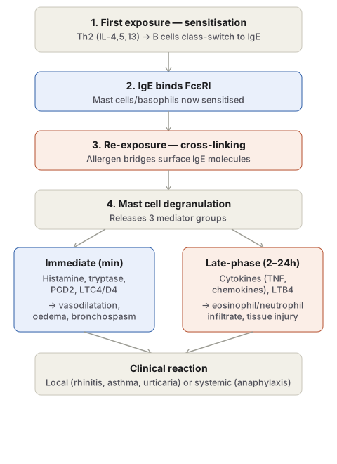
Sensitisation (first contact). On first exposure to allergen, allergen-specific Th2 cells are activated and secrete IL-4, IL-5 and IL-13: IL-4 and IL-13 drive B cells to class-switch to IgE production, IL-5 activates recruited eosinophils, and IL-13 stimulates epithelial mucus secretion. The IgE produced binds with very high affinity to FcεRI receptors on the surface of mast cells (and circulating basophils), which — because of this affinity — remain IgE-coated even though serum IgE concentration itself is low. The mast cell is now fully sensitised.
Re-exposure and degranulation. On subsequent exposure, allergen binds and cross-links the mast-cell-surface IgE, triggering a signalling cascade through FcεRI that culminates in mast cell degranulation, releasing three groups of mediators:
- Preformed vasoactive amines, released within seconds — chiefly histamine (vasodilatation, increased vascular permeability, smooth muscle contraction, increased mucus secretion), along with neutral proteases (e.g. tryptase, which generates kinins and cleaves complement to produce further chemotactic factors such as C5a) and heparin/proteoglycans that serve as the amine storage matrix.
- Newly synthesised lipid mediators, generated via the same arachidonic acid pathways as other leukocytes — prostaglandin D2 (the most abundant COX product of mast cells, causing intense bronchospasm and mucus secretion), and the leukotrienes LTC4/LTD4 (several thousand-fold more potent than histamine, molar for molar, at increasing vascular permeability and bronchial smooth muscle contraction) and LTB4 (highly chemotactic for neutrophils, eosinophils and monocytes).
- Cytokines, including TNF and various chemokines, responsible for driving the delayed, late-phase component of the reaction.
Two phases of the reaction:
- Immediate reaction — evident within 5–30 minutes, subsiding by about an hour, driven by the preformed amines and lipid mediators: vasodilatation, vascular leakage, smooth muscle spasm.
- Late-phase reaction — beginning 2–8 hours later and lasting days, driven mainly by cytokines: an inflammatory infiltrate dominated by eosinophils, neutrophils and Th2 lymphocytes, with genuine tissue injury (e.g. mucosal epithelial damage) that can sustain the reaction even without further allergen exposure — the reason antihistamines alone are often insufficient and anti-inflammatory therapy (corticosteroids) is also needed in chronic allergic disease.
The overall effects of the released mediators are: increased vascular permeability, smooth muscle contraction, an early transient vasoconstriction followed by vasodilatation, shock (in severe systemic reactions), increased gastric/nasal/lacrimal secretion, and recruitment of eosinophils and neutrophils both locally and systemically (producing peripheral eosinophilia/neutrophilia).
Clinical Manifestations
Systemic anaphylaxis follows antigen entering the circulation — administration of antisera (e.g. anti-tetanus serum) or drugs (classically penicillin), or insect stings (wasp, bee). Clinical features include itching, urticaria, erythema, bronchoconstriction with respiratory distress, laryngeal oedema (risking airway obstruction), vomiting, abdominal cramps, diarrhoea, and — without prompt treatment — systemic vasodilatation, hypotension (anaphylactic shock), circulatory collapse and death within minutes.
Local (atopic) reactions occur when the antigen is confined to the site of entry:
- Hay fever (allergic rhinitis) — pollen sensitisation of conjunctiva and nasal mucosa.
- Bronchial asthma — allergy to inhaled allergens such as house dust.
- Food allergy — ingested allergens (fish, cow’s milk, eggs).
- Cutaneous anaphylaxis — antigen contact with skin, producing urticaria, wheal and flare.
- Angioedema — an autosomal dominant disorder with laryngeal, eyelid, lip, tongue and truncal oedema.
Atopic dermatitis, allergic rhinitis and asthma frequently develop sequentially in the same individual — the atopic triad, and this sequential progression is termed the atopic march. Ingested or inhaled allergens can still trigger a systemic reaction if absorbed into the circulation (as in peanut allergy), so the site of exposure does not strictly predict whether a reaction stays local or becomes systemic.
Treatment Correlate
Because the pathogenesis maps directly onto distinct pharmacological targets, therapy is organised around the mechanism: corticosteroids reduce the late-phase inflammatory component; antihistamines counter histamine’s immediate effects; leukotriene antagonists block the LTC4/LTD4/LTB4 axis; bronchodilators treat asthmatic bronchospasm; epinephrine reverses the vasodilatation and hypotension of anaphylaxis; and newer biologic agents block Th2 cytokines, their receptors, or IgE itself.
Clinical Relevance
- The two-phase kinetics (immediate mediator release versus a later cytokine-driven inflammatory wave) explain why a patient can appear to recover from an acute allergic reaction only to relapse hours later (biphasic anaphylaxis), and why observation periods after anaphylaxis treatment are recommended well beyond apparent resolution of symptoms.
- Recognising angioedema specifically as autosomal dominant (hereditary C1 inhibitor deficiency being the underlying complement-regulatory mechanism, see Chemical Mediators of Inflammation) distinguishes it from ordinary IgE-mediated local anaphylaxis, with direct implications for management, since antihistamines are far less effective in the hereditary form.
- The atopic march is clinically useful for anticipating disease trajectory in a child presenting with eczema, since it predicts a raised likelihood of subsequent allergic rhinitis and asthma rather than being three unrelated diagnoses.
Diagram 1: Mechanism of Type I Hypersensitivity
Draw a single downward column of four stages, then split into two side-by-side boxes for the immediate versus late-phase reaction, converging into a final “clinical reaction” box.
- First exposure — sensitisation: Th2 cytokines drive B-cell class switching to IgE.
- IgE binds FcεRI on mast cells/basophils.
- Re-exposure — cross-linking: allergen bridges adjacent surface IgE molecules.
- Degranulation, releasing three mediator groups.
- Branch: Immediate reaction (minutes, amines/lipid mediators) versus late-phase reaction (hours, cytokine-driven, eosinophil/neutrophil infiltrate).
- Converge to clinical reaction — local or systemic.
Labels required
- The sensitisation step must be shown as requiring a first exposure separate from the reaction-triggering second exposure — a very common error is describing Type I hypersensitivity as occurring on first contact with an allergen.
- The two-phase split (immediate vs late-phase) must be shown as parallel outputs of the same degranulation event, each with its own named mediators and timeframe, since conflating them into a single undifferentiated “reaction” loses the exam-relevant point about biphasic anaphylaxis.
Errors commonly made
- Describing IgE as the mediator of tissue injury directly, rather than as the sensitising antibody that must first bind mast cells before any injury-producing mediator is released.
- Omitting the late-phase reaction and its distinct cytokine-driven mediators, leaving the impression that Type I hypersensitivity resolves entirely within the first hour.
Short Answers (2)
Multiple Choice Questions (5)
My Sticky Notes
Personal revision notes, mnemonics and reminders.
