
Chronic and Granulomatous Inflammation
Definition and causes
Prolonged inflammation — active inflammation, destruction and healing simultaneous. Arises: (1) after acute inflammation (extensive destruction/organism persists), (2) after recurrent acute attacks, (3) de novo (low-pathogenicity organism, e.g. TB). Also: hypersensitivity disease (autoimmune, allergic), prolonged toxic exposure (silica, endogenous lipid in atherosclerosis).
General features
Mononuclear infiltrate (macrophages, lymphocytes, plasma cells) + tissue destruction + angiogenesis/fibrosis (healing attempt), all simultaneous.
Acute vs chronic — cells: neutrophils vs lymphocytes/plasma cells/macrophages/giant cells. Systemic: high fever/neutrophilic leucocytosis vs mild fever/lymphocytic leucocytosis/↑ESR/anaemia/amyloidosis (long-standing).
2 types: chronic non-specific (granulation tissue, fibrous healing, no distinct pattern — e.g. chronic osteomyelitis; suppurative variant retains abscess/polymorphs, e.g. actinomycosis) vs chronic granulomatous (TB, leprosy, syphilis, sarcoidosis).
Macrophages
Dominant cell — from monocytes via same adhesion/chemokine machinery as neutrophils, but survive longer → dominate by 48h.
- M1 (classical) — TLR ligands + IFN-γ → NO, ROS, ↑lysosomal enzymes → microbicidal, pro-inflammatory
- M2 (alternative) — IL-4/IL-13 → not microbicidal, drives repair: angiogenesis, fibroblast activation, collagen synthesis, anti-inflammatory
Lymphocytes
CD4+ T subsets:
- Th1 → IFN-γ → drives M1
- Th2 → IL-4/5/13 → eosinophils, drives M2 (parasites, allergy)
- Th17 → IL-17 → neutrophil recruitment
Macrophage↔T cell bidirectional loop (Ag presentation + IL-12 ↔ cytokines) sustains chronic inflammation. Eosinophils: IgE/parasitic — major basic protein (toxic to parasites, injures epithelium too).
Granulomatous inflammation
Granuloma = ~1mm circumscribed collection of epithelioid cells (modified macrophages) rimmed by lymphocytes.
Two types:
- Immune granuloma — poorly degradable antigen, T-cell-mediated (type IV hypersensitivity) — TB, leprosy
- Foreign body granuloma — indigestible material, too large to phagocytose, NO T-cell involvement — talc, sutures
Pathogenesis (immune granuloma)
- Macrophages engulf antigen, fail to digest → transform to epithelioid cells
- Present antigen to CD4+ T cells → activation
- Cytokines: IL-1/IL-2 (more T cells), IFN-γ (macrophage activation), TNF-α (fibroblasts + endothelial PGs), TGF-β/PDGF (fibroblast growth)
- Granuloma: epithelioid cells ± giant cells centrally (± caseation, e.g. TB) → lymphocytes peripherally → fibrosis
Composition
- Epithelioid cells — slipper-shaped nucleus, weakly phagocytic
- Giant cells — Langhans’ (peripheral/ring nuclei, TB) vs foreign body (scattered nuclei, foreign material)
- Lymphocytes, ± plasma cells
- Necrosis (caseation in TB) — not universal
- Fibrosis at periphery
Tubercle = classic example: central caseation → epithelioid cells + Langhans’ giant cells → lymphocyte cuff → fibrous margin.
Clinical relevance
M1/Th1 → tissue-destructive chronic disease (TB, RA); M2/Th2 → fibrotic/allergic disease (asthma, parasites). Immune vs foreign-body granuloma distinction changes diagnostic workup. ↑ESR + mild fever + anaemia ± amyloidosis = systemic signature of chronic inflammation.
Definition
Chronic inflammation is prolonged inflammation in which active inflammation, tissue destruction and attempts at healing all proceed simultaneously, rather than in the sequential phases seen in an acute episode. It arises in one of three settings:
- Following acute inflammation, when tissue destruction has been extensive or when the causative organism survives in small numbers at the site — as when pneumonia terminates in a lung abscess.
- Following recurrent attacks of acute inflammation, as in recurrent urinary tract infection progressing to chronic pyelonephritis, or repeated cholecystitis becoming chronic.
- Starting de novo, when infection with a low-pathogenicity organism is chronic from the outset — the paradigm example being Mycobacterium tuberculosis.
Persistent infection by organisms difficult to eradicate (mycobacteria, certain fungi, viruses, parasites) is the classical cause, but chronic inflammation is equally central to hypersensitivity disease — autoimmune conditions such as rheumatoid arthritis, in which self-antigens drive a self-perpetuating reaction, and allergic disease such as asthma, in which the trigger is an otherwise harmless environmental substance — and to prolonged exposure to toxic agents, whether exogenous (inhaled silica in silicosis) or endogenous (cholesterol and lipid deposition driving the chronic inflammatory process of atherosclerosis).
General Features
Unlike acute inflammation’s vascular changes, oedema and neutrophilic infiltrate, chronic inflammation is recognised by three features occurring together:
- Mononuclear cell infiltration — macrophages, lymphocytes and plasma cells, sustained by continued recruitment, local proliferation of macrophages, and their longer survival at the site compared with the short-lived neutrophil.
- Tissue destruction, driven by substances released from activated macrophages — proteases, elastase, collagenase, lipase, reactive oxygen species, cytokines (IL-1, IL-8, TNF-α) and nitric oxide.
- Attempted healing, through proliferation of small blood vessels (angiogenesis) and fibroblasts, forming granulation tissue that matures into fibrosis and scarring.
The two forms of chronic inflammation are directly contrasted with acute inflammation across every axis that matters for examination purposes:
| Feature | Acute inflammation | Chronic inflammation |
|---|---|---|
| Onset/duration | Rapid, short | Delayed, prolonged |
| Cardinal signs | Present | Usually imperceptible |
| Main cells | Neutrophils, eosinophils, late lymphomononuclear cells | Lymphocytes, plasma cells, macrophages (epithelioid cells in granulomas), giant cells |
| Plasma exudation | Present | Variable |
| Systemic effects | High fever, neutrophilic/eosinophilic leucocytosis, lymphangitis | Mild fever, lymphocytic/monocytic leucocytosis, raised ESR, anaemia, amyloidosis (long-standing) |
| Morphology | Abscess, ulcer, blood-borne spread | Chronic non-specific or granulomatous inflammation |
| Fate | Resolution, healing, chronicity | Resolution, healing, dystrophic calcification |
Systemic effects specific to chronic inflammation include mild persistent fever with weight loss, anaemia of chronic disease, a leucocytosis with relative lymphocytosis rather than neutrophilia, a consistently raised ESR, and — in long-standing chronic suppurative disease — the risk of secondary systemic (AA) amyloidosis.
Two subtypes are recognised: chronic non-specific inflammation, in which the response is granulation tissue and fibrous healing without a distinctive histologic pattern (chronic osteomyelitis, chronic ulcer, lung abscess — chronic suppurative inflammation is a variant retaining polymorph infiltration and abscess formation, as in actinomycosis); and chronic granulomatous inflammation, in which the injurious agent provokes a specific, recognisable tissue reaction — granuloma formation — as in tuberculosis, leprosy, syphilis, actinomycosis and sarcoidosis.
Role of Macrophages
Macrophages are the dominant cell of chronic inflammation. Circulating monocytes migrate into tissue and differentiate into macrophages using the same adhesion-molecule and chemokine machinery that governs neutrophil emigration; because macrophages survive far longer in tissue than neutrophils, they typically become the dominant population within 48 hours of onset. Beyond phagocytosis, activated macrophages secrete cytokines and eicosanoids that sustain the inflammatory reaction, initiate repair and fibrosis, and present antigen to T lymphocytes — the last of these setting up the feedback loop with lymphocytes that is central to sustaining chronic inflammation.
Macrophages follow one of two activation pathways, and distinguishing them is a frequently tested point:
- Classical (M1) activation — driven by microbial products acting through TLRs and by IFN-γ from T cells — produces macrophages that generate NO and reactive oxygen species and upregulate lysosomal enzymes, maximising their microbicidal capacity and their ability to potentiate inflammation.
- Alternative (M2) activation — driven by IL-4 and IL-13 rather than IFN-γ — produces macrophages that are not primarily microbicidal but instead drive tissue repair: they secrete growth factors promoting angiogenesis, activate fibroblasts, stimulate collagen synthesis, and tend to inhibit ongoing inflammation. A plausible (though not strictly proven) sequence is classical activation first to destroy the offending agent, followed by alternative activation to terminate inflammation and begin repair.
Role of Lymphocytes
T and B lymphocytes are activated by microbial and other antigens and amplify chronic inflammation once engaged; some of the most severe chronic inflammatory reactions, including granulomatous inflammation, depend specifically on lymphocyte–macrophage interaction. CD4+ T cells divide functionally into three subsets with distinct downstream effects: Th1 cells secrete IFN-γ, driving classical (M1) macrophage activation; Th2 cells secrete IL-4, IL-5 and IL-13, recruiting eosinophils and driving alternative (M2) activation — important in defence against helminths and in allergic inflammation; and Th17 cells secrete IL-17, recruiting neutrophils into what is nominally a chronic inflammatory reaction. Macrophages and T cells interact bidirectionally — macrophages present antigen and secrete IL-12 to stimulate T cells, while activated T cells produce cytokines that recruit and further activate macrophages — creating a self-sustaining cycle that is the mechanistic reason chronic inflammation, once fully established, tends to persist. Activated B lymphocytes and plasma cells are also frequently present, and in some settings (long-standing rheumatoid synovium, Hashimoto thyroiditis) the accumulated lymphoid cells organise into tertiary lymphoid structures resembling lymph-node follicles.
Eosinophils are a further characteristic cell of chronic inflammation driven by IgE-mediated immunity or parasitic infection, recruited via eotaxin and related chemokines; their granule major basic protein is toxic to parasites but also injures host epithelium, explaining their dual role in parasite defence and in allergic tissue damage.
Granulomatous Inflammation
A granuloma is defined as a small (roughly 1 mm), circumscribed collection of modified macrophages called epithelioid cells, rimmed peripherally by lymphoid cells. Granulomatous inflammation is the host’s characteristic response to an agent that is poorly digestible — either because it provokes a strong T-cell-mediated (delayed-type, type IV hypersensitivity) immune response, as with persistent microbes such as M. tuberculosis and M. leprae, or because it is an indigestible foreign body too large to be phagocytosed, such as suture material or talc, in which case no T-cell-mediated immune response is required at all. This distinction — immune (hypersensitivity) granuloma versus foreign body granuloma — is worth keeping explicit, since the two are frequently conflated despite differing in whether antigen-specific T-cell activation is actually involved.
Pathogenesis of the Immune Granuloma
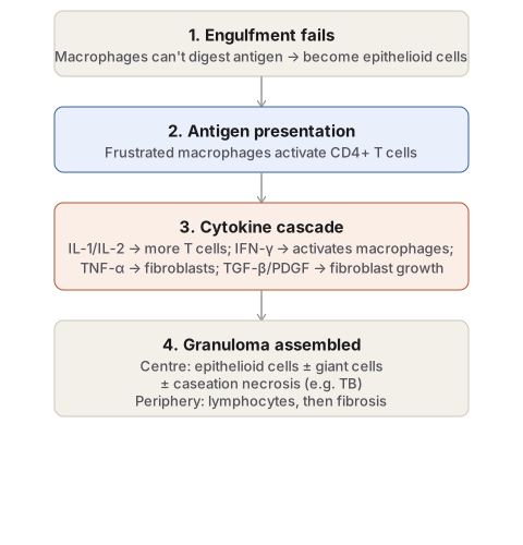
- Engulfment. Macrophages and monocytes phagocytose the antigen but, because it is poorly degradable, fail to destroy it; instead of clearing the antigen, the macrophages themselves undergo morphologic transformation into epithelioid cells.
- Antigen presentation to CD4+ T cells. The frustrated macrophages, acting as antigen-presenting cells, present the antigen to CD4+ T lymphocytes, activating them.
- Cytokine cascade. Activated T cells release IL-1 and IL-2, which stimulate further T-cell proliferation, and interferon-γ, which activates macrophages (the classical/M1 pathway described above); activated macrophages in turn release TNF-α, which promotes fibroblast proliferation and stimulates endothelial prostaglandin secretion, and growth factors (TGF-β, PDGF) that further stimulate fibroblast growth.
- Granuloma assembly. The result is a granuloma with transformed macrophages (epithelioid cells) and occasional multinucleate giant cells at the centre, a peripheral rim of lymphocytes (mainly T cells), and — depending on the granuloma’s age — a healing response of fibroblasts and collagen at the margin.
Composition of a Granuloma
- Epithelioid cells — elongated, weakly phagocytic modified macrophages with a characteristic slipper-shaped vesicular nucleus and abundant pale cytoplasm with hazy, closely apposed cell margins, giving an epithelium-like appearance under the microscope (hence the name).
- Multinucleate giant cells, formed by fusion of adjacent epithelioid cells and containing 20 or more nuclei: Langhans’ giant cells, with nuclei arranged peripherally in a horseshoe or ring or clustered at the two poles, typical of tuberculosis; and foreign body giant cells, with nuclei scattered centrally/throughout the cytoplasm, typical of foreign body reactions. Both types are weakly phagocytic but secrete products that assist in removing the invading agent.
- Lymphoid cells, reflecting the cell-mediated immune basis of the reaction; plasma cells may also be present where a humoral component contributes.
- Necrosis, present in some but not all granulomatous conditions — classically caseation necrosis (a dry, cheese-like appearance) at the centre of the tubercle in tuberculosis.
- Fibrosis, at the periphery, representing the healing response of proliferating fibroblasts.
The tubercle of tuberculosis is the classical fully developed example: a roughly 1 mm lesion with central caseation necrosis, surrounded by epithelioid cells and one or more Langhans’ giant cells, in turn surrounded by a peripheral lymphocytic cuff and bounded by fibrous tissue.
Clinical Relevance
- The distinction between M1 and M2 macrophage activation, and between Th1/Th2/Th17-driven inflammation, is now the standard framework for explaining why some chronic diseases are dominated by tissue destruction (Th1/M1-driven, as in tuberculosis or rheumatoid arthritis) while others are dominated by fibrosis or allergic tissue damage (Th2/M2-driven, as in asthma or parasitic disease) — the same macrophage lineage produces opposite tissue effects depending on which cytokine activated it.
- Recognising whether a granuloma is immune-type (T-cell-mediated, antigen persists because it resists digestion) or foreign-body type (no T-cell involvement, antigen persists only because it is too large to engulf) determines the correct diagnostic reasoning — a granuloma around inert, polarisable foreign material should not be worked up as an infectious or autoimmune process.
- A consistently raised ESR with mild fever, anaemia and, in long-standing cases, systemic AA amyloidosis is the systemic signature that should raise suspicion of an underlying chronic inflammatory or chronic suppurative process even before a causative organism or lesion has been identified.
Diagram 1: Pathogenesis of the Immune Granuloma
Draw a single downward column of four stages.
- Engulfment fails — macrophages ingest the antigen but cannot digest it, and transform morphologically into epithelioid cells.
- Antigen presentation — the frustrated macrophages present the antigen to CD4+ T cells, activating them.
- Cytokine cascade — name each cytokine against its specific action: IL-1/IL-2 drive further T-cell proliferation, IFN-γ activates macrophages, TNF-α drives fibroblast proliferation, TGF-β/PDGF stimulate fibroblast growth.
- Granuloma assembled — draw a small cross-section within this final box: a central core of epithelioid cells (with giant cells and, where relevant, caseation necrosis), a surrounding ring of lymphocytes, and an outer margin of fibrosis.
Labels required
- Each cytokine in step 3 must be paired with its specific downstream effect — a generic “cytokines are released” box loses the exam-relevant detail of which cytokine does what.
- The final granuloma cross-section must show the three-zone architecture (epithelioid centre, lymphocytic rim, fibrous margin) explicitly, since this layout is itself a common spot-diagnosis image.
Errors commonly made
- Presenting granuloma formation as a direct macrophage response to antigen without the intervening CD4+ T-cell activation step, which is the actual basis for calling this a type IV hypersensitivity reaction.
- Failing to note that this specific pathway describes the immune granuloma only — a foreign body granuloma reaches a similar end architecture without requiring T-cell involvement at all, because the stimulus is persistent activation by material too large to phagocytose rather than an undegradable antigen.
Short Essays (6)
Short Answers (1)
Multiple Choice Questions (4)
My Sticky Notes
Personal revision notes, mnemonics and reminders.
