
Acute Leukaemia
Overview
Neoplastic WBC precursors — MATURATION DEFECT (not fast proliferation) defines acute leukaemia. Blasts stuck at myeloblast/promyelocyte stage (AML) or lymphoblast stage (ALL). Blast generation time is actually PROLONGED, not shortened — cells just fail to differentiate/clear normally.
General pathogenesis (both AML+ALL)
- Genetic damage to single clone — translocations/mutations. Causes: heredity, radiation, chemical carcinogens, drugs (NOT viruses for AML). Multi-step; may follow pre-existing MDS/MPD.
- Maturation defect — arrest at specific stage
- Myelosuppression — accumulating blasts suppress normal stem cells (crowding + cell-mediated/humoral inhibition) → marrow failure. Normal stem cells can recover post-treatment.
- Organ infiltration — nodes, liver, spleen, skin, gonads, CNS
Risk factors: genetic syndromes (Down, Bloom, Klinefelter, Wiskott-Aldrich, Fanconi, ataxia telangiectasia) · radiation (↑CML/AML/ALL, NOT CLL/hairy cell) · chemicals (benzene, tobacco) · chemo drugs · immunodeficiency.
AML
Adults, median age 50. Mutation blocks myeloid maturation. Key translocations: t(8;21)[M2], t(15;17)[M3], inv(16)[M4].
FAB M0-M7:
- M0 minimally differentiated — MPO negative
- M1 without maturation — myeloblasts, MPO+
- M2 with maturation — myeloblasts+promyelocytes, Auer rods, MPO+++
- M3 acute promyelocytic — hypergranular, multiple Auer rods, MPO+++
- M4 myelomonocytic (Naegeli) — mixed, NSE+
- M5 monocytic (Schilling) — M5a blasts/M5b differentiated, NSE++
- M6 erythroleukaemia (Di Guglielmo’s) — erythroblasts>50%, PAS+
- M7 megakaryocytic — undifferentiated, platelet peroxidase+
FAB criterion: ≥30% marrow blasts. WHO: ≥20% blasts, relies more on cytogenetics/molecular/flow cytometry.
Clinical features:
- Marrow failure: anaemia, bleeding (thrombocytopenia), infections, fever
- Organ infiltration: bone pain (sternal tenderness), lymphadenopathy, splenomegaly (moderate), hepatomegaly, gum hypertrophy (M4/M5), chloroma (greenish MPO+ tumour mass), meningeal involvement, testicular/mediastinal involvement
~25% have preleukaemic phase (months-years before overt disease).
Labs: severe normochromic anaemia, thrombocytopenia (<50k, serious bleeding risk <20k), M3 = DIC risk, WBC subnormal(25%) to >100k. Marrow: hypercellular (dry tap possible→trephine), ≥30%(FAB)/≥20%(WHO) blasts, dyserythropoiesis, ↓megakaryocytes.
ALL
Most common cancer in children <4yr. Pre-B (90%) vs Pre-T (10%). Differentiation stage ≠ aggressiveness, but Pre-T MORE aggressive.
Pre-B: usually leukaemic in children, early extranodal (nodes, hepatosplenomegaly, CNS, testis, skin). Pre-T: thymic origin → mediastinal mass + pleural effusion, rapid marrow/blood spread, marrow failure features, more aggressive.
B/T-ALL morphologically identical, need immunophenotyping. Blood: anaemia, thrombocytopenia, lymphoblasts >20% (round-convoluted nuclei, high N:C, no granules). Marrow: 20-95% blasts (WHO >20%). Cytochem: PAS+, focal acid phosphatase+.
AML vs ALL comparison
| AML | ALL | |
|---|---|---|
| Age | Adults 15-40yr; 20% childhood leukaemias | Children<15yr; 80% childhood leukaemias |
| Splenomegaly/nodes | + | ++ |
| Special finding | Gum hypertrophy | CNS involvement |
| Predominant cell | Myeloblasts | Lymphoblasts |
| Dx criteria | FAB M0-M7, WHO≥20%blasts | FAB L1-L3, Pre-B90%/Pre-T10%, WHO≥20%blasts |
| Cytochem | MPO+, Sudan black+, NSE+(M4/M5) | PAS+, focal acid phosphatase+ |
| Immunophenotype | CD13,33,41,42 | TdT+ both; PreB:CD19,20; PreT:CD1,2,3,5,7 |
| Cytogenetics | M3:t(15;17), M4:inv(16) | variable |
| Therapy | Cytarabine, anthracyclines, 6-thioguanine | Vincristine, prednisolone, anthracyclines, L-asparaginase |
Clinical relevance
FAB(30%)/WHO(20%) blast threshold = frequently tested number, shift reflects move to molecular Dx. Gum hypertrophy→M4/M5; mediastinal mass+pleural effusion→Pre-T ALL = exam-findable clues before labs return. M3 = specific DIC emergency risk, needs coag screen. MPO/Sudan black(AML) vs PAS(ALL) = fastest practical test to separate the two before immunophenotyping/cytogenetics available.
Overview
Acute leukaemias are neoplastic proliferations of white cell precursors characterised by a maturation defect — leukaemic blasts fail to mature beyond the myeloblast/promyelocyte stage (acute myeloid leukaemia, AML) or the lymphoblast stage (acute lymphoblastic leukaemia, ALL) — with a rapidly downhill natural history if untreated. Critically, it is the maturation block, not accelerated proliferation, that defines acute leukaemia: leukaemic blast generation time is actually somewhat prolonged relative to normal precursors, but blocked cells simply accumulate rather than being cleared by normal differentiation and turnover.
AML and ALL share overlapping clinical features and can be difficult to distinguish by clinical presentation alone; laboratory and cytochemical distinction is essential.
General Pathogenesis (Shared by AML and ALL)
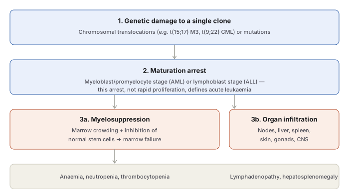
- Genetic damage to a single clone — malignant transformation of one myeloid or lymphoid clone, from chromosomal translocation, mutation, or insertional mutagenesis (implicated agents: heredity, ionising radiation, chemical carcinogens, certain drugs; viruses are not implicated in AML but are in some lymphoid malignancies). Leukaemogenesis is multi-step, and acute leukaemia sometimes follows a pre-existing myelodysplastic or myeloproliferative disorder.
- Maturation defect — the leukaemic clone is arrested at a specific developmental stage.
- Myelosuppression — accumulating leukaemic cells suppress normal haematopoietic stem cells, partly by physical marrow crowding and partly by cell-mediated/humoral inhibition (some AML patients have a hypocellular marrow despite leukaemia, showing crowding alone doesn’t explain the failure); surviving normal stem cells can restore normal hematopoiesis after effective treatment.
- Organ infiltration — leukaemic cells circulate and infiltrate lymph nodes, liver, spleen, skin, gonads, and CNS.
Risk associations (both types): genetic syndromes (Down, Bloom, Klinefelter, Wiskott-Aldrich, Fanconi anaemia, ataxia telangiectasia), ionising radiation (atomic bomb survivors, therapeutic/occupational exposure — particularly increases CML, AML, ALL, but not CLL or hairy cell leukaemia), chemical carcinogens (benzene, tobacco), long-term chemotherapy/alkylating agent exposure, and immunodeficiency states.
Acute Myeloid Leukaemia (AML)
A heterogeneous disease, mainly of adults (median age 50), from mutation-driven blockade of myeloid stem cell maturation. Key chromosomal abnormalities: t(8;21) [M2], t(15;17) [M3, acute promyelocytic leukaemia], inv(16) [M4].
FAB Classification (M0–M7)
| Subtype | Old name | Key feature | Cytochemistry |
|---|---|---|---|
| M0 | Minimally differentiated | Blasts lack definite features but show myeloid antigens | Myeloperoxidase negative |
| M1 | AML without maturation | Myeloblasts predominate, few granules | Myeloperoxidase + |
| M2 | AML with maturation | Myeloblasts + promyelocytes, Auer rods may be present | Myeloperoxidase +++ |
| M3 | Acute promyelocytic leukaemia | Hypergranular promyelocytes, multiple Auer rods per cell | Myeloperoxidase +++ |
| M4 | Acute myelomonocytic (Naegeli) | Mixed myeloid + monocytic maturation | MPO ++, non-specific esterase + |
| M5 | Acute monocytic (Schilling) | M5a: poorly-differentiated monoblasts; M5b: differentiated promonocytes/monocytes | Non-specific esterase ++ |
| M6 | Acute erythroleukaemia (Di Guglielmo’s) | Erythroblasts >50% | Erythroblasts PAS+, myeloblasts MPO+ |
| M7 | Acute megakaryocytic leukaemia | Pleomorphic undifferentiated blasts | Platelet peroxidase + |
FAB criterion for AML: ≥30% marrow blasts. WHO classification lowers this threshold to ≥20% blasts and relies more on cytogenetic/molecular/immunophenotypic features (multiparametric flow cytometry) than morphology alone.
Clinical Features
Divided by mechanism:
I. Bone marrow failure: anaemia (pallor, lethargy, dyspnoea); bleeding (thrombocytopenia — bruising, petechiae, gum bleeding); infection (mouth, throat, skin, respiratory, perianal sites); fever (often infective, sometimes without an identifiable source).
II. Organ infiltration: bone pain/tenderness (sternal tenderness, bone infarcts, subperiosteal infiltrates); lymphadenopathy, tonsillar enlargement; moderate splenomegaly (occasionally infarction, subcapsular haemorrhage, rare rupture); hepatomegaly (usually without functional impairment); renal infiltration (usually asymptomatic); gum hypertrophy — classic for M4/M5; chloroma (granulocytic sarcoma) — a localised, myeloperoxidase-driven greenish tumour mass in skin or orbit; meningeal involvement (more typical of ALL, but can occur); testicular/mediastinal involvement.
Roughly 25% of AML patients have a preleukaemic phase (anaemia/cytopenias) preceding overt leukaemia by months to years.
Laboratory Findings
- Blood: severe, progressive, normochromic anaemia; thrombocytopenia (usually <50,000/µL, with serious spontaneous haemorrhage risk below 20,000/µL); M3 carries a specific risk of DIC; total WBC ranges from subnormal (1,000–4,000/µL in ~25% of patients) to markedly elevated (>100,000/µL in advanced disease), with blasts predominating and accompanying neutropenia from marrow infiltration.
- Marrow: typically hypercellular (occasionally a “dry tap” from cellular adhesiveness/reticulin enmeshment, requiring trephine biopsy); tightly packed with leukaemic blasts (≥30% by FAB, ≥20% by WHO); dyserythropoiesis, megaloblastic change, ring sideroblasts common; megakaryocytes reduced/absent; cytogenetic analysis for the characteristic translocations.
Acute Lymphoblastic Leukaemia (ALL)
The most common cancer of children under 4 years — lymphoid malignancy of precursor B or T cells. Pre-B ALL accounts for ~90% of cases; pre-T for the remaining 10%. Stage of differentiation does not correlate with aggressiveness, but pre-T disease is clinically more aggressive than pre-B.
Precursor B-cell disease: usually presents as leukaemia in children; extranodal involvement early (lymphadenopathy, hepatosplenomegaly, CNS infiltration, testicular enlargement, cutaneous infiltration); cytopenia-related infections.
Precursor T-cell disease: since T-cell precursors differentiate in the thymus, often presents as a mediastinal mass with pleural effusion, progressing rapidly to marrow/blood involvement; marrow failure features (anaemia, neutropenia, thrombocytopenia); lymphadenopathy, hepatosplenomegaly, CNS involvement common; more aggressive than pre-B disease.
Morphologic and Laboratory Features
B and T-precursor ALL are morphologically indistinguishable and require immunophenotyping for subtype. Blood shows anaemia, thrombocytopenia, and variable TLC with circulating lymphoblasts (typically >20%) — round-to-convoluted nuclei, high nucleocytoplasmic ratio, no cytoplasmic granularity. Marrow shows 20–95% malignant precursor cells (WHO threshold: >20% blasts); megakaryocytes reduced/absent. Cytochemistry: PAS positive, focal acid phosphatase positive (contrast AML’s myeloperoxidase/Sudan black positivity and diffuse acid phosphatase in M4/M5).
AML versus ALL — Comparative Summary
| Feature | AML | ALL |
|---|---|---|
| Common age | Adults 15–40 yrs; ~20% of childhood leukaemias | Children <15 yrs; ~80% of childhood leukaemias |
| Splenomegaly/hepatomegaly/lymphadenopathy | + | ++ |
| Bony tenderness | + | + |
| Other | Gum hypertrophy | CNS involvement |
| Predominant cell | Myeloblasts/promyelocytes | Lymphoblasts |
| Diagnostic criteria | FAB M0–M7; WHO >20% blasts | FAB L1–L3; WHO Pre-B (90%)/Pre-T (10%); WHO >20% blasts |
| Cytochemistry | Myeloperoxidase+, Sudan black+, NSE+ (M4/M5), diffuse acid phosphatase+ (M4/M5) | PAS+, focal acid phosphatase+ |
| Immunophenotyping | CD13, 33, 41, 42 | TdT+ (both); Pre-B: CD19, 20; Pre-T: CD1, 2, 3, 5, 7 |
| Cytogenetics | M3: t(15;17); M4: inv(16) | Variable |
| Specific therapy | Cytarabine, anthracyclines (daunorubicin, doxorubicin), 6-thioguanine | Vincristine, prednisolone, anthracyclines, L-asparaginase |
Clinical Relevance
- The FAB/WHO blast-percentage threshold (30% vs 20%) is a concrete, frequently tested numeric criterion, and the shift to WHO’s lower threshold reflects a broader move toward cytogenetic/molecular diagnosis over morphology alone.
- Gum hypertrophy specifically pointing to M4/M5 subtype, and mediastinal mass with pleural effusion specifically pointing to precursor T-cell ALL, are both examination-level physical findings that narrow the leukaemia subtype before any laboratory result returns.
- M3 (acute promyelocytic leukaemia) carrying a specific DIC risk is a genuine clinical emergency consideration distinct from the general bleeding risk of thrombocytopenia in other AML subtypes — coagulation screening is specifically indicated when M3 is suspected.
- The AML/ALL cytochemistry pairing (myeloperoxidase/Sudan black for AML vs PAS for ALL) is the fastest practical bedside-adjacent test to separate the two when blast morphology alone is ambiguous, well before immunophenotyping or cytogenetic results are available.
Diagram 1: General Pathogenesis of Acute Leukaemia
Draw a single downward sequence: genetic damage → maturation arrest → forking into two parallel consequences (myelosuppression, organ infiltration) → converging into a shared results line.
- Genetic damage to a single clone — translocations/mutations.
- Maturation arrest — myeloblast/promyelocyte stage (AML) or lymphoblast stage (ALL). 3a. Myelosuppression — marrow crowding + inhibition of normal stem cells. 3b. Organ infiltration — nodes, liver, spleen, skin, gonads, CNS.
- Results — anaemia/neutropenia/thrombocytopenia (from 3a) and lymphadenopathy/hepatosplenomegaly (from 3b).
Labels required
- Step 2 must state explicitly that it is the arrest, not accelerated proliferation, that defines acute leukaemia — and that blast generation time is actually prolonged, not shortened. This is the single most commonly misunderstood point in the whole topic.
- Both consequences (3a and 3b) must be shown as parallel, independent outcomes of the same accumulated blast population, not sequential steps of each other.
Errors commonly made
- Describing acute leukaemia as caused by “rapid proliferation” of blasts — the actual defect is failure to mature/differentiate and clear normally, not an accelerated cell cycle.
- Treating marrow failure as caused only by physical overcrowding — some AML marrows are hypocellular despite leukaemia, showing that cell-mediated/humoral suppression of normal stem cells is a separate, additional mechanism.
- Omitting that surviving normal stem cells (not destroyed by the leukaemic process) are what allow marrow recovery after effective treatment — the marrow failure is suppressive, not a permanent destruction of all normal precursors.
My Sticky Notes
Personal revision notes, mnemonics and reminders.
