
Myocardial Infarction
Definition
Coagulative necrosis of cardiac muscle from prolonged severe ischaemia, arising from an imbalance between myocardial oxygen demand and coronary supply.
Risk factors
Age (rises steadily, peak 40–65), male sex (gap narrows but persists after menopause), hypertension, diabetes, smoking, dyslipidaemia. About 5% occur under 40, usually in the presence of major risk factors.
Pathogenesis
~90% of infarcts arise on coronary atherosclerosis narrowing a major trunk by more than 75%. Plaque stability matters as much as size:
- Stable plaque: thick collagenous cap, minimal inflammation, small core — rarely ruptures.
- Vulnerable plaque: thin cap, large lipid/foam-cell core, active inflammation — prone to rupture.
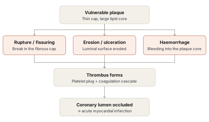
Acute plaque change (the usual trigger) — three patterns: rupture/fissuring, erosion/ulceration, or haemorrhage into the plaque core. Any of these exposes thrombogenic material → platelet adhesion/aggregation → tissue factor release → coagulation cascade → thrombus → occlusion within minutes; coronary vasospasm compounds the narrowing.
Non-atherosclerotic causes (~10%): vasospasm alone, embolism (mural thrombus, valve vegetations, paradoxical embolism), arteritis, hypercoagulable states, dissection, severe hypotension/hypoxaemia.
Reversible vs irreversible injury
| Reversible | Irreversible | |
|---|---|---|
| Window | First 20–30 min | Beyond ~20–40 min |
| Biochemical | ↓ATP, lactate accumulation | Sarcolemmal breakdown → leak of myoglobin, LDH, CK, troponins |
| Functional | Loss of contractility within 60 sec | Arrhythmias |
| Structural | Mitochondrial swelling, glycogen loss | Coagulative necrosis complete by ~6 hours |
Necrosis begins in the subendocardium (least well perfused zone) and advances outward toward the epicardium with continued occlusion — the wavefront phenomenon. This is why early reperfusion salvages muscle even after some myocardium has already died.
Classification
By wall thickness: transmural (full thickness, majority, STEMI) vs subendocardial (inner third–half, NSTEMI). By territory: LAD 40–50% (anterior wall/apex/anterior septum) · RCA 30–40% (inferior/posterior wall, posterior septum) · LCX 15–20% (lateral wall).
Gross timeline
0–12h: no visible change (TTC stain reveals it within 2–3h) → 12–24h: pale reddish-blue, blotchy → 1–3d: yellow-tan, soft, hyperaemic border → 3–7d: mottled, softest point (rupture risk peaks here) → 10d–2wk: vascularised granulation rim → beyond 2wk: firm grey-white scar, mature by ~6 weeks.
Microscopic timeline
<30 min: EM only (glycogen loss, mitochondrial swelling → later amorphous densities, sarcolemmal disruption) → 6–12h: coagulative necrosis begins, wavy fibres, early neutrophils → 12–24h: preserved cell outlines, eosinophilic cytoplasm, pyknosis → 1–3d: neutrophils peak, nuclei disappear, contraction bands at margin → 3–7d: macrophages remove debris, granulation begins → 1–2wk: vascularised granulation tissue, pigmented macrophages (lipofuscin, haemosiderin) → 2–8wk: collagen deposition increases → beyond 2 months: dense acellular scar, no longer datable.
Healing proceeds margin → centre. Extension of infarct reverses this (more advanced healing centrally than peripherally) — a histologic clue.
Diagnosis
Clinical: crushing retrosternal pain radiating to arm/neck/jaw/back, sweating, apprehension; ~10–15% silent (elderly, diabetics). ECG: ST elevation + T inversion + Q waves (transmural); non-ST elevation (subendocardial).
| Marker | Onset | Peak | Normalises | Note |
|---|---|---|---|---|
| Myoglobin | 1–3h | 8–12h | 24–36h | Earliest, not cardiac-specific |
| CK-MB | 2–4h | 24h | 48–72h | Sensitive, not fully specific |
| Troponin I/T | 2–4h | 24–48h | 7–10d / 10–14d | Most sensitive and specific |
| LDH | 24–48h | 3–6d | 7–14d | Obsolete marker |
Reperfusion
Salvages muscle if achieved within 20–30 min. Reperfused infarct: grossly haemorrhagic (leaky microvasculature); microscopically shows contraction band necrosis. Reperfusion injury: reactive oxygen species, calcium overload, inflammatory burst.
Complications
Arrhythmia (most common; VF = leading cause of sudden death) · CHF (~50%) · cardiogenic shock (>40% LV lost) · rupture (peak day 3–7: free wall → tamponade; septum → VSD; papillary muscle → acute MR) · mural thrombus/thromboembolism · ventricular aneurysm (late, large transmural infarcts) · pericarditis (early fibrinous, day 2–3; late Dressler syndrome, immune-mediated) · extension of infarct · chronic ischaemic heart disease.
Definition
Myocardial infarction is coagulative necrosis of cardiac muscle caused by prolonged, severe ischaemia, most often the end result of a sudden reduction in coronary blood flow. It is the most feared consequence of ischaemic heart disease — the group of cardiac conditions arising from an imbalance between the myocardial demand for oxygenated blood and the supply delivered through the coronary arteries.
Epidemiology and Risk
Myocardial infarction accounts for a large share of deaths in developed countries and correlates closely with the prevalence of coronary atherosclerosis in a population. Incidence rises steadily with age, though a meaningful minority of attacks occur in people under forty, almost always those carrying major atherosclerotic risk factors — hypertension, diabetes, smoking, and dyslipidaemia. Men carry a substantially higher risk than women throughout life; the gap narrows after menopause but never fully closes, a pattern attributed to the protective effect of oestrogen during the reproductive years.
Etiopathogenesis
Coronary Atherosclerosis as the Underlying Substrate
Roughly ninety percent of infarcts occur on a background of coronary atherosclerosis severe enough to compromise more than seventy-five percent of the luminal cross-sectional area of one or more of the three major coronary trunks. The extent of fixed narrowing alone, however, does not fully determine risk — the structural stability of the plaque matters as much as its size.
- Stable plaques carry a dense, collagen-rich fibrous cap, minimal inflammation, and a negligible necrotic lipid core, and rarely rupture.
- Vulnerable plaques carry a thin fibrous cap over a large necrotic core rich in foam cells and extracellular lipid, with active inflammation eroding the cap’s collagen faster than smooth muscle cells can replace it. These are the plaques that precipitate acute events.
Acute Plaque Change
The trigger for most infarcts is a sudden structural change in a previously partially occlusive plaque, converting it into the substrate for total occlusion. Three patterns of acute plaque change are recognised:
- Rupture or fissuring, exposing highly thrombogenic plaque material to the bloodstream.
- Erosion or ulceration, exposing the thrombogenic subendothelial basement membrane.
- Haemorrhage into the plaque core, from rupture of the fibrous cap or of fragile new vessels formed within the plaque, which enlarges the plaque abruptly and narrows the lumen without necessarily breaching the surface.
Any of these changes exposes thrombogenic material to circulating platelets, which adhere, activate, and aggregate to form a mural thrombus; tissue factor released at the site activates the coagulation cascade, and the thrombus can progress to complete luminal occlusion within minutes. Coronary vasospasm, triggered by mediators released from activated platelets and endothelium, narrows the lumen further.
Non-Atherosclerotic Causes
In roughly ten percent of cases, infarction occurs without significant fixed coronary atherosclerosis, due to coronary vasospasm alone, embolism (from a left atrial or ventricular mural thrombus, from valve vegetations, or paradoxically from the venous circulation through a patent foramen ovale), arteritis, hypercoagulable states, vascular dissection, or profound hypotension and hypoxaemia reducing myocardial perfusion despite a patent coronary tree.
Consequences of Myocardial Ischaemia
Ischaemia produces a spectrum of change that depends critically on how long perfusion remains inadequate.
Reversible injury, within the first twenty to thirty minutes: aerobic glycolysis ceases within seconds, ATP production falls and toxic metabolites such as lactate accumulate, myocardial contractility is lost within about sixty seconds — enough on its own to precipitate acute heart failure — and at the ultrastructural level, glycogen is depleted, mitochondria swell, and myofibrils relax. If perfusion is restored within this window, the myocardium can be fully preserved.
Irreversible injury, once ischaemia persists beyond roughly twenty to forty minutes: the sarcolemmal membrane breaks down, releasing intracellular proteins — myoglobin, lactate dehydrogenase, creatine kinase, and troponins — into the blood, where their appearance becomes the basis for laboratory diagnosis. Coagulative necrosis of the myocardium is essentially complete within about six hours of onset.
Necrosis does not begin uniformly across the ventricular wall. Because the subendocardium is the zone least well perfused under normal conditions, it is the first to become irreversibly injured; with continuing occlusion, the wave of necrosis then advances outward through the full thickness of the wall toward the epicardium. This time-dependent, subendocardium-to-epicardium progression is the reason early reperfusion can still salvage a substantial portion of an evolving infarct even after the earliest myocardium has already died.
Classification
| Basis | Types |
|---|---|
| Thickness of wall involved | Transmural (full thickness, the great majority of infarcts, corresponding to ST-elevation on ECG) vs subendocardial (inner third to half, corresponding to non-ST-elevation) |
| Anatomic region | Anterior, posterior/inferior, lateral, septal, and combinations |
| Age of the lesion | Recent (fresh) vs old (healed) |
| Coronary territory occluded | Left anterior descending (40–50% of infarcts — anterior wall, apex, anterior septum), right coronary (30–40% — inferior/posterior wall, posterior septum), left circumflex (15–20% — lateral wall) |
Transmural infarcts are unifocal and solid, distributed along the territory of a specific coronary artery, and are usually associated with occlusive thrombus, pericarditis, and — later — cardiac aneurysm. Subendocardial infarcts tend to be multifocal, patchy, and circumferential rather than confined to one territory, more often the result of a coronary thrombus that has undergone partial lysis, or of profound and prolonged hypotension superimposed on chronic stenosis; pericarditis and true aneurysm formation are not features of this pattern.
Morphology
Gross Evolution
| Time | Appearance |
|---|---|
| 0–12 hours | No change visible to the naked eye; triphenyltetrazolium chloride staining of a fresh slice can reveal the infarct within two to three hours, since normal myocardium turns brick-red from dehydrogenase activity while the infarcted zone remains unstained |
| 12–24 hours | Pale, reddish-blue, blotchy discolouration from stagnated blood |
| 1–3 days | Sharply defined, yellow-tan, and soft, with a hyperaemic border |
| 3–7 days | Mottled, with a pale yellow necrotic centre bordered by a hyperaemic zone of granulation tissue; the myocardium is at its softest and weakest during this window, which is when the risk of rupture is greatest |
| 10 days–2 weeks | Soft, rimmed by vascularised, reddish-purple granulation tissue |
| Beyond 2 weeks | Progressively firmer, grey-white, and contracted, maturing into a dense fibrous scar by about six weeks |
Microscopic Evolution
Changes are similar in transmural and subendocardial infarcts and follow a well-defined timetable useful for estimating the age of an infarct at autopsy or biopsy.
- Under 30 minutes: no light-microscopic change; electron microscopy alone shows glycogen loss and mitochondrial swelling (reversible), followed after roughly thirty to sixty minutes by mitochondrial amorphous densities and sarcolemmal disruption (irreversible).
- 6–12 hours: coagulative necrosis begins; wavy fibres appear at the margin (stretched, non-contractile dead myofibrils), with interstitial oedema and haemorrhage, and early neutrophil margination.
- 12–24 hours: classic coagulative necrosis is established — preserved cell outlines, intensely eosinophilic cytoplasm, and pyknotic nuclei.
- 1–3 days: neutrophilic infiltrate is well developed at the margins; nuclei disappear and cross-striations fade; contraction band necrosis may be seen at the margins.
- 3–7 days: neutrophils die off and macrophages begin removing necrotic debris; granulation tissue starts forming at the periphery.
- 1–2 weeks: vascularised granulation tissue with proliferating capillaries and fibroblasts is well established at the margins, advancing toward the centre; pigmented macrophages containing lipofuscin and haemosiderin, along with eosinophils, lymphocytes, and plasma cells, are prominent.
- 2–8 weeks: progressive collagen deposition with declining cellularity.
- Beyond 2 months: a dense, largely acellular collagenous scar — a fully healed infarct can no longer be reliably dated on morphology alone.
Healing always advances from the margins toward the centre; if a fresh episode of necrosis develops adjacent to an existing infarct (extension of infarct), the resulting lesion paradoxically shows more advanced healing centrally than peripherally, the reverse of the usual pattern, which is itself a clue to recognising extension on histology.
Diagnosis
Diagnosis rests on the combination of clinical features, electrocardiographic change, and serum biomarkers.
Clinical features: sudden, severe, crushing retrosternal or precordial pain, often radiating to the arm, neck, jaw, or back; associated sweating, nausea, and a sense of apprehension; and, in more severe cases, hypotension, oliguria, and low-grade fever. A meaningful minority of infarcts — more common in elderly and diabetic patients — are silent, without typical pain.
Electrocardiographic changes: ST-segment elevation (in transmural infarction), T-wave inversion, and the development of pathological Q waves; subendocardial infarction typically produces non-ST-elevation changes instead.
Serum biomarkers, released as the damaged sarcolemma allows intracellular proteins to leak into the circulation:
| Marker | Onset | Peak | Normalises | Notes |
|---|---|---|---|---|
| Myoglobin | 1–3 hours | 8–12 hours | 24–36 hours | Earliest to rise, but lacks cardiac specificity |
| CK-MB | 2–4 hours | 24 hours | 48–72 hours | Sensitive, but also raised by skeletal muscle injury |
| Troponin I / T | 2–4 hours | 24–48 hours | 7–10 days (I) / 10–14 days (T) | Most sensitive and specific marker in current use |
| Lactate dehydrogenase | 24–48 hours | 3–6 days | 7–14 days | Non-specific; now largely of historical interest |
Reperfusion
Restoring coronary flow — by thrombolytic therapy, angioplasty and stenting, or bypass surgery — can salvage myocardium if achieved within the first twenty to thirty minutes of onset, and reduces final infarct size even when achieved later. Reperfused myocardium has a distinctive appearance: grossly haemorrhagic rather than pale, because the microvasculature injured during ischaemia leaks once flow is restored, and microscopically marked by contraction band necrosis — thick, irregular, transverse, intensely eosinophilic bands within dead myofibres, reflecting massive uncontrolled contraction on reintroduction of oxygen and calcium. Reperfusion also carries its own risk of injury, mediated by a burst of reactive oxygen species, further calcium overload, and an intensified inflammatory response.
Complications
| Complication | Notes |
|---|---|
| Arrhythmia | The most common complication overall; ranges from sinus bradycardia to ventricular fibrillation, which is the leading mechanism of sudden death after infarction |
| Congestive heart failure | Develops in about half of patients; responsible for a large share of deaths |
| Cardiogenic shock | Follows loss of a large mass of functioning myocardium, roughly forty percent of the left ventricle; carries a high mortality |
| Myocardial rupture | Most frequent three to seven days after infarction, when the necrotic wall is softest; may involve the free wall (haemopericardium and tamponade), the interventricular septum (acute left-to-right shunt), or a papillary muscle (acute severe mitral regurgitation) |
| Mural thrombosis and thromboembolism | From endocardial damage and regional wall akinesis promoting stasis; may embolise to the brain, kidney, spleen, or elsewhere |
| Ventricular aneurysm | A late complication of large transmural infarcts, from outward bulging of thin, non-contractile scar tissue; predisposes to mural thrombus |
| Pericarditis | Early, fibrinous pericarditis around the second to third day over a transmural infarct; a later, immunologically mediated form — postmyocardial infarction (Dressler) syndrome — may appear weeks later |
| Extension of infarct | Fresh necrosis adjacent to an existing infarct; healing is more advanced centrally than peripherally, the reverse of a simple infarct |
| Chronic ischaemic heart disease | Progressive heart failure from cumulative myocardial fibrosis after one or more infarcts |
Clinical Relevance
- The subendocardium-first pattern of necrosis is the biological rationale for the phrase “time is muscle” — the sooner flow is restored, the more of the still-viable outer myocardium can be saved before the wave of necrosis reaches it.
- The choice and timing of serum markers guides both diagnosis and the detection of reinfarction: troponin’s long window makes it the preferred diagnostic marker, while CK-MB’s faster return to baseline makes it more useful for identifying a second infarct occurring within days of the first.
- Recognising that myocardial rupture peaks between the third and seventh day, precisely when the infarct is at its softest, explains why patients are monitored closely through this period even after apparent early stabilisation.
Diagram 1: Coronary Territories and Corresponding Infarct Regions
Draw two views of the heart side by side: an anterior external view and a transverse (short-axis) section through the ventricles at mid-level, both showing the three major coronary territories in different shading.
On the anterior view: trace the left anterior descending artery running down the interventricular groove toward the apex, the right coronary artery along the right atrioventricular groove, and the left circumflex artery curving around the left side toward the posterior surface.
On the transverse section: divide the ventricular wall into three shaded zones matching the three territories — anterior wall and anterior two-thirds of the septum (LAD), lateral wall (LCX), and inferior/posterior wall with the posterior third of the septum (RCA).
Labels required
- Each artery must be labelled by name and abbreviation.
- Each shaded territory must be labelled with its approximate frequency as the culprit vessel: LAD 40–50%, RCA 30–40%, LCX 15–20%.
- A small note beside the septum indicating it is supplied by both the LAD (anterior two-thirds) and RCA (posterior third), since septal infarcts are frequently misassigned to a single vessel.
Errors commonly made
- Assigning the entire septum to one artery instead of splitting it between the LAD and RCA territories.
- Reversing the relative frequencies of the three vessels.
- Omitting the thin preserved rim of subendocardial myocardium in the transverse section — even a fully transmural infarct spares a very thin strip nourished directly by ventricular blood, and its omission is a commonly missed labelling detail.
Diagram 2: Timeline of Gross and Microscopic Change
Draw a single horizontal timeline divided into labelled time-zones (0–12h, 12–24h, 1–3d, 3–7d, 10d–2wk, >2wk), with two rows of small illustrations beneath it — a gross cut-surface view of the ventricular wall in the top row, and a corresponding microscopic field in the bottom row, for each time-zone.
Gross row: unchanged pale myocardium → reddish-blue blotchy area → sharply defined yellow-tan soft area with hyperaemic border → mottled area with pale centre and hyperaemic rim (mark this zone with a small warning symbol for rupture risk) → soft area with a vascularised reddish-purple rim → firm grey-white contracted scar.
Microscopic row: normal fibres → wavy fibres with interstitial oedema/haemorrhage and early neutrophils → coagulative necrosis with preserved outlines, eosinophilic cytoplasm, pyknotic nuclei → dense neutrophilic infiltrate with fading striations → macrophages clearing debris, early granulation tissue → mature granulation tissue with pigmented macrophages → dense collagenous scar.
Labels required
- Every time-zone individually labelled on both rows.
- The 3–7 day zone explicitly flagged as the period of maximal softness and rupture risk.
- The transition from neutrophil-dominant to macrophage-dominant infiltrate must be labelled by name at the correct time-zone, since the order of inflammatory cell types is frequently tested.
Errors commonly made
- Placing coagulative necrosis features at the wrong time-zone — students frequently draw pyknotic, eosinophilic fibres too early (before 12 hours) when nothing is yet visible even by light microscopy.
- Failing to mark the rupture-risk window at 3–7 days, which is the clinical payoff of this entire diagram.
- Drawing granulation tissue starting at the centre of the infarct rather than at its margin — healing always advances margin-to-centre, and reversing this direction is a common error worth specifically avoiding.
Diagram 3: Sequence of Acute Plaque Change to Occlusion
Draw a short flowchart of four cross-sectional views of a coronary artery, each showing the vessel lumen and the atheromatous plaque within its wall.
- Intact vulnerable plaque: thin fibrous cap over a large lipid core, narrowing the lumen partially.
- Branch into three small panels side by side showing the three types of acute plaque change: (a) a break in the cap surface labelled “rupture/fissuring,” (b) a frayed, eroded luminal surface labelled “erosion/ulceration,” (c) an enlarged plaque with a dark red pocket inside it labelled “haemorrhage into the plaque.”
- All three panels converge into one drawing showing a platelet-fibrin thrombus forming over the disrupted surface.
- Final panel: the lumen completely occluded by thrombus.
Labels required
- All three types of acute plaque change must be individually named, not merged into a single “plaque rupture” label.
- The thrombus formation step must note both platelet aggregation and tissue factor-driven activation of the coagulation cascade.
Errors commonly made
- Naming only “plaque rupture” and omitting erosion/ulceration and intraplaque haemorrhage as separate mechanisms — all three are independently examinable.
- Skipping the vulnerable-plaque structure (thin cap, large core) at the start, which is the substrate that explains why this particular plaque was prone to acute change in the first place.
Long Essays (1)
Short Essays (2)
Multiple Choice Questions (3)
My Sticky Notes
Personal revision notes, mnemonics and reminders.
