
Tuberculosis
Organism
M. tuberculosis (Koch’s bacillus) — Gram-neutral, strict aerobe, favours high-O2 tissue (lung apex). No exotoxin/endotoxin — all damage is host response. Virulence factors: cord factor, Wax-D (adjuvant).
Detection: ZN staining (acid-fast — resists 20% H2SO4, vs 5% for M. leprae) · fluorescent (auramine/rhodamine) · culture (LJ medium 8-12wk; HPLC 2-3wk) · PCR (species confirmation, distinguishes NTM).
NTM (atypical mycobacteria) — environmental, less virulent, classified by growth speed/pigment. 5 patterns: pulmonary, lymphadenitis, skin ulcers, abscess, bacteraemia (AIDS).
Transmission and spread
Inhalation (main) · ingestion (tonsillar/intestinal) · inoculation (rare) · transplacental (rare).
Spread: local (macrophages) · lymphatic (primary route — regional lymphadenitis) · haematogenous (miliary TB) · natural passages (pleura, bronchus, tube→peritoneum, larynx, gut, ureter).
Pathogenesis
No toxin → damage = type IV (delayed) hypersensitivity + CD4+ T-cell immunity, both together.
Guinea pig model: Primary = no reaction 10-14d → nodule → ulcerates, heals poorly, nodes involved. Secondary (Koch phenomenon) = indurates in 1-2d → ulcerates, heals fast, nodes spared → shows hypersensitivity + immunity.
BCG = attenuated bovine strain → CMI + delayed hypersensitivity, tuberculin+.
Mantoux (PPD) — induration >15mm@72h = positive (can’t distinguish infection vs disease). False +: prior BCG, atypical mycobacteria. False -: anergy, sarcoidosis, viral infections, Hodgkin, very recent exposure (8-10wk), disseminated/fulminant TB.
Evolution of tubercle (sequence)
- Neutrophils (overwhelmed)
- Macrophage infiltration (~12h, complement-opsonised)
- Phagocytosis → NO + TNF-α/IL-1 → more macrophages
- CD4+ T cells → IFN-γ, IL-2
- Epithelioid transformation (2-3d)
- Granuloma + giant cells (Langhans’=peripheral/ring nuclei; foreign body=central)
- Hard tubercle (lymphocyte cuff, fibroblasts, NO necrosis)
- Soft tubercle (10-14d, central caseation — hallmark; AFB sparse centrally)
Fate: cold abscess (no pus cells!) · sinuses (bone/joint/node/epididymis) · coalescence+fibrosis · dystrophic calcification ± ossification.
Primary TB (Ghon complex)
Unsensitised host, mostly children, almost exclusively lung. 3 components: Ghon focus (subpleural, upper lobe-lower part) + lymphatics + hilar nodes (caseating). Ingested→tabes mesenterica.
Fate: heal (fibrosis→calcification) · progressive primary (bronchial spread) · primary miliary (haematogenous) · reactivation → progressive secondary.
Secondary TB
Sensitised host (endogenous reactivation or exogenous reinfection), children+adults, widespread organs.
| Primary | Secondary | |
|---|---|---|
| Site | Lung only | Lung + everywhere |
| Lesion | Ghon complex | Cavitation, miliary, fibrocaseous |
Apical consolidation (high O2) → caseation + peripheral fibrosis. HIV reactivation → looks primary-like (hilar, not apical/cavitary).
4 patterns:
- Fibrocaseous — cavitary (open, infectious, complications: aneurysm→haemoptysis, bronchopleural fistula, empyema, pleural thickening) vs non-cavitary
- Caseous pneumonia — exudative, hypersensitive host, numerous AFB
- Miliary — millet-seed lesions, pulmonary artery(lung-confined) vs pulmonary vein(systemic)
- Empyema/pleurisy — serous/fibrinous → fibrosis or caseous empyema
Diagnosis
AFB smear · culture (LJ/rapid) · PCR · CBC (lymphocytosis, ↑ESR) · CXR · Mantoux · FNAC of node (replaced biopsy).
Death: pulmonary insufficiency, haemorrhage, miliary sepsis, cor pulmonale, amyloidosis.
Clinical relevance
ZN decolourisation strength discriminates TB vs leprosy on unlabelled AFB. Koch phenomenon explains why primary/secondary TB look different despite same organism (host sensitisation, not bacillary difference). Mantoux false-negative settings matter clinically (don’t over-trust in sick/anergic/recently-exposed patients). HIV reactivation mimics primary pattern radiologically.
Definition and Significance
Tuberculosis is the classical example of chronic granulomatous inflammation in humans, caused by infection with Mycobacterium tuberculosis, and remains a major public health problem worldwide despite advances in chemotherapy — the burden falls disproportionately on developing countries of Asia, Africa and Latin America, with India and China together accounting for roughly half of all global cases. Malnutrition, poverty, overcrowding, chronic debilitating disease (uncontrolled diabetes, alcoholism) and immunocompromise all raise incidence; in the West, HIV/AIDS has driven a resurgence. HIV-infected individuals both acquire tuberculosis at markedly higher rates and, once infected, progress to active disease within weeks rather than months to years; their disease is more often extra-pulmonary (lymph nodes, pleura, pericardium, meninges) and more often sputum-smear negative despite being culture positive, and they are also more susceptible to M. avium-intracellulare infection.
Causative Organism
Mycobacterium tuberculosis (Koch’s bacillus, after Robert Koch’s 1882 discovery) is a slender, Gram-neutral, strictly aerobic rod that thrives best in tissue with high oxygen tension — which is why the lung apex, the site of highest regional oxygen tension, is its preferred site of secondary disease. Of the M. tuberculosis complex, M. tuberculosis hominis (human strain) is now overwhelmingly the common pathogen; M. tuberculosis bovis (bovine strain), once significant via unpasteurised milk, is now rare.
The organism’s acid-fastness — its defining diagnostic property — comes from mycolic acids and other lipids cross-linked in its cell wall, which resist decolourisation once stained. Diagnostic demonstration methods include:
- Ziehl–Neelsen (acid-fast) staining, resisting decolourisation by acid and alcohol; the organism is decolourised only by 20% sulphuric acid (compared with the weaker 5% needed for the less acid-fast M. leprae, a useful point of contrast between the two mycobacteria).
- Fluorescent staining (auramine, rhodamine), a reliable and economical alternative exploiting mycobacterial autofluorescence.
- Culture on Löwenstein–Jensen medium (highly specific but slow, 8–12 weeks by the conventional method; newer methods such as Bactec culture or HPLC of mycolic acids shorten this to 2–3 weeks).
- Molecular methods (PCR/nucleic acid amplification), now the most useful approach for species confirmation and for distinguishing M. tuberculosis from non-tuberculous mycobacteria, which require different treatment.
Two virulence-associated lipid constituents are worth naming specifically because they explain the organism’s pathogenesis: cord factor (a mycoside essential for growth and virulence) and Wax-D (a glycolipid in the cell wall that acts as an adjuvant alongside tuberculoprotein). Notably, the bacillus itself produces no exotoxin or endotoxin — every tissue change seen in tuberculosis is the host’s response to the organism, not direct bacterial toxicity.
Non-tuberculous (atypical) mycobacteria are environmental organisms distinct from M. tuberculosis complex and M. leprae, classified by growth speed and pigment production (rapid growers such as M. fortuitum; slow growers such as M. avium-intracellulare and M. kansasii, further split into photochromogens, scotochromogens and non-chromogens). They cause disease similar to but far less virulent than tuberculosis, acquired directly from the environment rather than person-to-person, in five recognised patterns: pulmonary disease, lymphadenitis, ulcerated skin lesions, abscesses, and — in AIDS — bacteraemia.
Transmission and Spread
Infection is acquired by inhalation of organisms in cough droplets or dried sputum from an open pulmonary case (the dominant route); by ingestion, producing tonsillar or intestinal tuberculosis, from self-swallowed sputum or bovine bacilli in milk; rarely by inoculation into skin from infected tissue; and rarely transplacentally, causing congenital tuberculosis.
Once established, the organism spreads within the body by: local spread, macrophages carrying bacilli into surrounding tissue; lymphatic spread, the primary route, since tuberculosis is fundamentally a disease of lymphoid tissue — bacilli reach lymphoid follicles and regional lymph nodes, producing the regional lymphadenitis typical of childhood infection; hematogenous spread, from bacillaemia or caseous material eroding a vein wall, producing millet-seed-sized lesions across multiple organs (miliary tuberculosis); and spread via natural passages — lung to pleura (tuberculous pleurisy), airway to airway (transbronchial spread), fallopian tube to peritoneum (tuberculous peritonitis), sputum to larynx (laryngitis), swallowed sputum to ileocaecal region, and kidney to bladder via the ureter.
Pathogenesis: Hypersensitivity and Immunity
Because the organism produces no toxin, tuberculous tissue damage is entirely a consequence of the host’s own delayed-type (type IV) hypersensitivity response, developing alongside — and closely linked to — protective cell-mediated immunity, both triggered by CD4+ T cells sensitised against mycobacterial antigens (tuberculoprotein), which then release lymphokines that increase macrophage microbicidal activity.
The classical demonstration of this dual response is the guinea pig experiment (a species with no natural resistance to M. tuberculosis, making the host response easy to isolate):
- Primary infection: intradermal bacilli produce no visible reaction for 10–14 days, after which a nodule forms, ulcerates and heals poorly, with regional lymph node tubercle formation — comparable to primary childhood tuberculosis (which, unlike the guinea pig, usually heals in humans).
- Secondary infection (bacilli injected 4–6 weeks after prior sensitisation): the site indurates and darkens within 1–2 days, ulcerates but heals quickly, and regional nodes are spared — the Koch phenomenon, evidence of both hypersensitivity and acquired immunity. Old tuberculin (OT) reproduces the same pattern and underlies the human skin test.
BCG vaccination (attenuated bovine bacilli) induces protective cell-mediated immunity with a delayed hypersensitivity reaction that heals, leaving the host tuberculin-positive and immune; it is routine at birth where prevalence is high but not used in the low-prevalence US, partly because it confounds skin-test interpretation.
Tuberculin (Mantoux) skin test: intradermal PPD produces induration >15 mm at 72 hours in a sensitised individual — a positive test indicates hypersensitivity to mycobacterial antigen but cannot distinguish past infection from active disease. False positives occur with prior BCG or atypical mycobacterial infection; false negatives occur in cutaneous anergy, sarcoidosis, some viral infections, Hodgkin disease, very recent infection (8–10 weeks), and fulminant/disseminated disease (where circulating tuberculoprotein masks the local hypersensitivity reaction).
Evolution of the Tubercle
The formation of a fully developed tubercle follows a defined sequence, classically demonstrated by intravenous inoculation in the guinea pig:
- An initial, ineffective neutrophil response, rapidly overwhelmed by the organism.
- By ~12 hours, progressive macrophage infiltration — bacilli opsonised by complement fragments C3b (and related factors) attract macrophages, which dominate the lesion from this point on (inhaled bacilli reaching alveoli directly recruit macrophages from the outset).
- Macrophages phagocytose the bacilli; those that die release nitric oxide (antimycobacterial) and cytokines (TNF-α, IL-1), driving local macrophage proliferation and further monocyte recruitment from blood.
- T- and B-cell activation: CD4+ T cells release IFN-γ and IL-2, shaping the macrophage response and establishing the delayed-type hypersensitivity reaction (CD4+ deficiency in HIV explains the disproportionate susceptibility to disseminated disease); B cells contribute little protective humoral immunity, though anti-LAM antibodies may limit dissemination in children.
- Within 2–3 days, activated macrophages transform into epithelioid cells (pale eosinophilic cytoplasm, elongated vesicular nuclei).
- Epithelioid cells aggregate into granulomas, driven by cytokines from sensitised CD4+ T cells and mycobacterial cell wall constituents.
- Macrophages unable to destroy the bacilli fuse into giant cells — Langhans’ type (peripheral horseshoe/ring or bipolar nuclei) or foreign body type (central nuclei).
- A peripheral zone of lymphocytes and plasma cells forms around the epithelioid/giant-cell core, itself bounded by fibroblasts — this stage, lacking central necrosis, is the hard tubercle.
- Within 10–14 days, the centre undergoes caseation necrosis — cheesy, lipid-rich, structureless, eosinophilic and granular with nuclear debris — producing the soft tubercle, the hallmark tuberculous lesion, whose formation reflects both CD4+/CD8+ T-cell–mediated mechanisms and direct mycobacterial toxicity to macrophages. Acid-fast bacilli are sparse within the mature soft tubercle (found chiefly at the margins of recent necrosis and cavity walls), since the caseous environment does not favour rapid bacillary proliferation.
A granuloma’s subsequent fate is variable: liquefied caseous material may discharge on a surface as a cold abscess (no pus cells despite the name); in bone, joint, node or epididymal disease it may form draining sinuses; adjacent granulomas may coalesce with progressive fibrosis; or the caseous material within a fibrous capsule may undergo dystrophic calcification and, over years, even ossify.
Primary Tuberculosis
Infection of a previously unexposed, unsensitised individual — also called Ghon’s complex or childhood tuberculosis. The primary complex is the lesion at the portal of entry plus the draining lymphatic and nodal foci, most often in the lung and hilar nodes, occasionally tonsil/cervical node or, with ingested bacilli, small intestine/mesenteric node. Progressive disseminated primary tuberculosis is particularly common in immunocompromised hosts (e.g. AIDS).
The pulmonary Ghon complex has three components: the Ghon focus — a 1–2 cm solitary subpleural focus of tuberculous pneumonia, typically in the upper part of the lower lobe, under a patch of pleurisy; the lymphatic vessel component — draining lymphatics carrying bacillus-laden phagocytes, occasionally forming beaded miliary tubercles; and the lymph node component — matted, caseating hilar/tracheobronchial nodes, which remain a potential source of later reinfection. Microscopically: tuberculous granulomas with peripheral fibrosis and extensive central caseation; older lesions show fibrosis and calcification. Ingested bacilli produce a small intestinal focus with mesenteric node enlargement (tabes mesenterica), which may rupture into the peritoneum causing tuberculous peritonitis.
Fate of the primary complex: (1) most commonly, healing by fibrosis and later calcification/ossification; (2) progressive primary tuberculosis — continued local growth with bronchial dissemination of caseous material to other lung regions; (3) primary miliary tuberculosis — haematogenous spread via an eroded vessel to liver, spleen, kidney, brain, bone marrow; (4) reactivation of an apparently healed lesion under lowered resistance or heightened hypersensitivity, producing progressive secondary tuberculosis.
Secondary (Post-Primary) Tuberculosis
Infection or reactivation in a previously sensitised individual — endogenous (reactivation of dormant primary disease) or exogenous (fresh reinfection). Distinguishing primary from secondary disease is a frequent examination point:
| Feature | Primary tuberculosis | Secondary tuberculosis |
|---|---|---|
| Age | Mostly children | Children and adults |
| Organs | Almost exclusively lung | Lung, lymph nodes, and widely elsewhere (genitourinary tract, bone, meninges, brain, eye, liver, spleen, intestine, skin) |
| Lesions | Ghon complex (lung focus + lymphatics + hilar nodes) | Tubercles, extensive caseation, miliary lesions, cavitation, fibrocaseous disease, caseous pneumonia, pleurisy/effusion |
| Fate | Heals by fibrosis/calcification; may reactivate later | Consolidation, nodules, thickened pleura, amyloidosis; may itself reactivate under impaired immunity |
Secondary pulmonary disease characteristically begins as a 1–2 cm apical area of consolidation (lymphohaematogenous spread from the primary complex favours the apex, where oxygen tension is highest), progressing to central caseation with peripheral fibrosis — histologically the same caseating granuloma seen elsewhere. In HIV-related reactivation, the pattern instead resembles primary disease, with hilar node rather than apical cavitary involvement.
Progressive secondary pulmonary tuberculosis produces four recognised patterns:
- Fibrocaseous tuberculosis — peripheral healing with massive central caseation, either breaking into a bronchus to form a cavity (cavitary/open disease, favouring further bacillary proliferation because of high oxygen tension, and the source of “open” infectiousness), or remaining undrained as non-cavitary chronic fibrocaseous disease. A cavity has a thick fibrous wall lined by caseous material, thrombosed vessels traversing the lumen, and surrounding consolidation; microscopically, coalesced granulomas with caseation, epithelioid and Langhans’ giant cells, and an outer fibrous wall, sometimes with dystrophic calcification. Complications include aneurysm of vessels crossing the cavity wall (a cause of haemoptysis), bronchopleural fistula, tuberculous empyema, and pleural thickening.
- Tuberculous caseous pneumonia — caseous material spreading through the lung in a highly hypersensitive host, with an exudative reaction (oedema, fibrin, polymorphs, monocytes) containing numerous demonstrable bacilli.
- Miliary tuberculosis — lymphohaematogenous dissemination producing millet-seed-sized (1 mm), yellowish lesions without grossly visible caseation, but showing minute tubercle structure with caseation microscopically; may be confined to the lung (pulmonary artery route) or systemic (pulmonary vein route, reaching liver, spleen, kidney, brain, bone marrow, genitourinary tract).
- Tuberculous empyema/pleurisy — reactive serous or fibrinous pleural exudate, which may heal by fibrosis (thickened pleura) or, occasionally, accumulate caseous material as frank empyema.
Clinical Features and Diagnosis
Pulmonary features include productive cough (with possible haemoptysis), pleural effusion, dyspnoea and orthopnoea, with apical changes, nodularity or miliary/diffuse infiltrate on chest x-ray; systemic features include fever, night sweats, fatigue, and weight/appetite loss, with secondary systemic amyloidosis a risk in long-standing untreated disease. Diagnosis rests on AFB microscopy of sputum or aspirate, mycobacterial culture (traditional LJ medium, 4–8 weeks, or rapid HPLC-based methods in 2–3 weeks), PCR, complete haemogram (lymphocytosis, raised ESR), chest x-ray, the Mantoux test, and fine-needle aspiration cytology of an enlarged node, which has largely replaced biopsy for diagnostic confirmation. Death results chiefly from pulmonary insufficiency, pulmonary haemorrhage, sepsis from disseminated miliary disease, cor pulmonale, or secondary amyloidosis.
Clinical Relevance
- The Ziehl–Neelsen decolourisation strength (20% sulphuric acid for M. tuberculosis versus 5% for the less acid-fast M. leprae) is a small but genuinely useful discriminator when a histology question presents an ambiguous acid-fast organism without naming it.
- The Koch phenomenon is the conceptual key to understanding why primary and secondary tuberculosis look so different pathologically despite sharing the same organism — the host’s prior sensitisation, not a difference in the bacillus, is what changes the tissue response from slow-developing, node-involving primary disease to a fast, locally-contained, node-sparing secondary reaction.
- A negative Mantoux test does not exclude active disease — recognising the specific settings where it is falsely negative (cutaneous anergy, sarcoidosis, disseminated/fulminant tuberculosis, very recent exposure) is what prevents this test from being over-relied upon in a sick, immunosuppressed, or recently exposed patient.
- Distinguishing primary from secondary tuberculosis by site (apex versus hilar/lower zone) and node involvement has direct diagnostic value, since HIV-related reactivation reverts to a primary-like pattern and can be misread radiologically as fresh primary infection rather than reactivation.
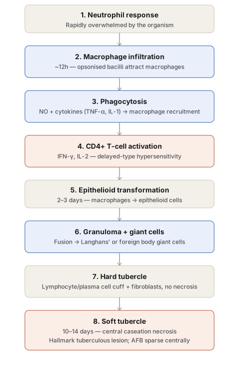
Diagram 1: Evolution of the Tubercle
Draw a single downward column of eight numbered stages.
- Neutrophil response — brief, ineffective, rapidly overwhelmed.
- Macrophage infiltration — around 12 hours, driven by complement opsonisation of the bacilli.
- Phagocytosis — dying macrophages release NO and cytokines (TNF-α, IL-1), recruiting more macrophages.
- CD4+ T-cell activation — IFN-γ and IL-2 shape the developing delayed-type hypersensitivity reaction.
- Epithelioid transformation — by 2–3 days, activated macrophages take on the pale, elongated, vesicular-nucleus morphology of epithelioid cells.
- Granuloma and giant cell formation — epithelioid cells aggregate; some fuse into Langhans’ (peripheral/ring nuclei) or foreign body (central nuclei) giant cells.
- Hard tubercle — a lymphocyte/plasma cell cuff and bounding fibroblasts form, with no necrosis yet.
- Soft tubercle — by 10–14 days, the centre undergoes caseation necrosis: this is the diagnostic, hallmark lesion of tuberculosis.
Labels required
- The timeline (12 hours; 2–3 days; 10–14 days) must be marked at the relevant steps, since exam questions frequently ask “by what stage does X occur.”
- The hard-tubercle-to-soft-tubercle transition must be explicit, since “hard” (no necrosis) versus “soft” (caseation present) is a specific, examinable distinction, not just two names for the same lesion.
- Langhans’ giant cells must be distinguished from foreign body giant cells by nuclear arrangement, since both can appear in this sequence for different reasons.
Errors commonly made
- Skipping straight from “macrophages present” to “granuloma forms,” omitting the intervening CD4+ T-cell activation step that is the actual basis for classifying this as a type IV hypersensitivity reaction.
- Describing caseation necrosis as present from the beginning of granuloma formation rather than as a later event that defines the transition from hard to soft tubercle.
Short Essays (2)
Short Answers (1)
Multiple Choice Questions (4)
My Sticky Notes
Personal revision notes, mnemonics and reminders.
