
Rheumatic Heart Disease
Definition
Rheumatic fever is an immune-mediated multisystem disease following group A streptococcal pharyngitis; chronic pancarditis with lasting valvular damage is rheumatic heart disease. The heart, not the joints, sustains the permanent damage.
Epidemiology
Children aged five to fifteen; both sexes affected equally; incidence tracks the prevalence of untreated streptococcal pharyngitis, hence common in overcrowded, low-resource settings including the Indian subcontinent.
Etiopathogenesis
Preceding group A β-haemolytic streptococcal pharyngitis, two to three weeks before onset. Organism is never found in the lesions — the mechanism is autoimmune, not infective. Three lines of evidence support this:
- Environmental: temporal link to pharyngitis, outbreaks precede clusters of cases, antibiotics reduce incidence and recurrence.
- Host susceptibility: familial clustering, HLA class II association.
- Immunologic: elevated antistreptolysin O, antistreptokinase, antistreptohyaluronidase, anti-DNAase B titres; molecular mimicry between streptococcal M protein/cell wall antigens and human myosin, keratin, actin, laminin, vimentin, N-acetylglucosamine.
Net mechanism: antibodies raised against streptococcal antigens cross-react with cardiac, joint, skin, and neural tissue because of shared epitopes.
Aschoff body — the pathognomonic lesion
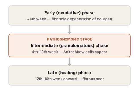
1–2 mm perivascular nodule in the interstitium of the heart, evolving through three phases (a single heart may show all three at once):
- Early/exudative (~4th week): oedema, collagen fragmentation, fibrinoid degeneration.
- Intermediate/granulomatous (4th–13th week): pathognomonic stage. Fibrinoid focus surrounded by lymphocytes, plasma cells, and Anitschkow cells (owl’s-eye nucleus in cross-section, caterpillar chromatin in longitudinal section); fused multinucleate forms are Aschoff giant cells.
- Late/healing (12th–16th week onward): spindled, palisaded cells, eventual perivascular fibrous scar.
Active and healed Aschoff bodies coexisting in one heart = ongoing rheumatic activity.
Rheumatic pancarditis
Endocarditis: small (1–3 mm), warty, grey-brown vegetations along the line of valve closure — firmly attached, rarely embolise (unlike infective endocarditis). Mitral alone or mitral + aortic account for most cases; tricuspid and pulmonary rarely and mildly involved. Chronic healing → fibrosis, calcification, chordal fusion → fish-mouth/buttonhole mitral stenosis. Mural lesion: MacCallum’s patch, posterior left atrial wall above the posterior mitral leaflet.
Myocarditis: soft, flabby left ventricle; Aschoff bodies concentrated in interventricular septum, left ventricle, left atrium — proximity to conduction system explains arrhythmias; most immediately life-threatening component of acute carditis.
Pericarditis: fibrinous exudate — “bread-and-butter” appearance; may organise into adhesive pericarditis.
Extracardiac lesions
Migratory polyarthritis (large joints, most common presenting feature) · subcutaneous nodules (painless, over extensor surfaces, histologically a miniature Aschoff body) · erythema marginatum (migratory rash, central clearing) · rheumatic arteritis · Sydenham chorea (delayed CNS involvement, more common in girls) · rheumatic pneumonitis/pleuritis (uncommon).
Diagnostic criteria
| Major | Minor |
|---|---|
| Carditis | Fever |
| Polyarthritis | Arthralgia |
| Sydenham chorea | Previous rheumatic fever |
| Erythema marginatum | Raised ESR/CRP, leucocytosis |
| Subcutaneous nodules | Prolonged PR interval |
Diagnosis: evidence of preceding streptococcal infection + (2 major) or (1 major + 2 minor).
Chronic sequelae and causes of death
Cardiac failure (most common; valvular deformity in the young, superimposed coronary disease in the old) · infective endocarditis on the damaged valve · embolism (mural thrombus in a fibrillating/stenotic left atrium — brain, kidney, spleen, lungs) · sudden death (ball-valve thrombus, or acute coronary insufficiency with coexisting aortic stenosis).
Definition
Rheumatic fever is a multisystem inflammatory disease that follows an episode of group A streptococcal pharyngitis, affecting the heart, joints, central nervous system, skin, and subcutaneous tissue through an immune-mediated rather than an infective mechanism. When the acute illness involves all three layers of the heart, the resulting pancarditis and its long-term valvular consequences are termed rheumatic heart disease. Though the name emphasises joint involvement, and though migratory joint pain is the presenting complaint in most patients, it is the heart that sustains the lasting damage — an old teaching aphorism captures this precisely: rheumatic fever licks the joints but bites the heart.
Epidemiology
Rheumatic fever occurs mainly in children between five and fifteen years of age, at the age when streptococcal throat infection is most frequent, and affects both sexes roughly equally. Its incidence tracks the frequency and severity of streptococcal pharyngitis in a population, which is why it has declined sharply in developed countries with improved living conditions and prompt antibiotic treatment, while remaining a significant public health problem across the Indian subcontinent and other developing regions where overcrowding and poor access to care allow streptococcal throat infections to go untreated.
Etiopathogenesis
A preceding throat infection with group A β-haemolytic streptococci is now firmly established as the trigger, typically two to three weeks before the onset of rheumatic fever — the interval needed for an immune response to develop. Yet the organism itself is never found within the inflamed tissues, and only a small fraction of infected individuals — under three percent — go on to develop the disease. This gap between exposure and effect is explained by three converging lines of evidence.
Environmental evidence. A clear temporal link exists between streptococcal pharyngitis and the onset of rheumatic fever; outbreaks of streptococcal infection in schools and training camps are followed by clusters of rheumatic fever; antibiotic treatment of the preceding throat infection lowers both the incidence and the recurrence rate of the disease; and experimental repeated streptococcal infection reproduces similar cardiac lesions in animals.
Host susceptibility. The disease clusters in families and in identical twins, and is more common in individuals carrying particular HLA class II alleles, pointing to an inherited predisposition rather than a matter of exposure alone.
Immunologic evidence. Patients show elevated antibody titres against streptococcal antigens — antistreptolysin O and S, antistreptokinase, antistreptohyaluronidase, and anti-DNAase B — and these antibodies cross-react with human tissue components. Group A streptococcal cell wall polysaccharide and the streptococcal M protein in particular share epitopes with human myosin, keratin, actin, laminin, vimentin, and N-acetylglucosamine.
Taken together, the pathogenesis is one of molecular mimicry: a genetically susceptible individual mounts an antibody response against streptococcal antigens, and because those antigens closely resemble self-molecules in the heart, joints, skin, and brain, the resulting autoimmune attack damages the patient’s own tissues rather than the organism that triggered it.
Cardiac Lesions
The Aschoff Body
The hallmark lesion of rheumatic pancarditis is the Aschoff nodule or Aschoff body — a small, spheroidal or fusiform focus, one to two millimetres across, found in the interstitium near small blood vessels of the myocardium and endocardium, and occasionally in the pericardium and the adventitia of the proximal aorta. A single heart may show Aschoff bodies at every stage of development simultaneously, since the lesion evolves through three phases over the course of an illness.
- Early (exudative) phase, apparent by about the fourth week of illness: oedema and accumulation of ground substance separate the collagen fibres of the connective tissue, which then fragment and take on the staining appearance of fibrin — a change called fibrinoid degeneration.
- Intermediate (proliferative or granulomatous) phase, the stage that is pathognomonic of rheumatic disease, seen from the fourth to the thirteenth week: the fibrinoid focus becomes surrounded by lymphocytes, plasma cells, and distinctive large mononuclear cells called Anitschkow cells (cardiac histiocytes), whose nuclear chromatin appears as a caterpillar-like serrated ridge in longitudinal section and as an owl’s-eye central body in cross-section. Some Anitschkow cells fuse into multinucleate Aschoff giant cells, which are themselves pathognomonic of the disease.
- Late (healing or fibrous) phase, occurring twelve to sixteen weeks into the illness: the nodule becomes spindle-shaped, the Anitschkow cells lose their vesicular character and arrange in a palisade, and over months to years the lesion is replaced by a small perivascular fibrocollagenous scar.
The presence of active Aschoff bodies alongside old, healed lesions in the same heart is taken as evidence of ongoing rheumatic activity.
Rheumatic Pancarditis
All three layers of the heart may be affected, though the intensity varies between them.
Endocarditis. Rheumatic valvulitis is the lesion responsible for the lasting cardiac damage of chronic disease. In the acute phase, the valve leaflets thicken and lose their translucency, and small, warty vegetations — one to three millimetres, grey-brown and translucent — form along the line of closure of the cusps, so closely spaced that the free margin appears as a continuous rough ridge. Unlike the vegetations of infective endocarditis, these are firmly attached and rarely embolise. All four valves may be involved, but with markedly unequal frequency: the mitral valve alone, or mitral together with aortic, account for the great majority of cases, while the tricuspid and pulmonary valves are only infrequently and mildly affected — a distribution attributed to the greater mechanical stress borne by the valves of the left heart. With chronic healing, the valve undergoes diffuse fibrous thickening, calcification, and fusion and shortening of the chordae tendineae, producing the characteristic “fish-mouth” or “buttonhole” deformity of the mitral orifice. Mural endocarditis may also occur, most typically as MacCallum’s patch, a map-like area of thickened, wrinkled endocardium on the posterior wall of the left atrium just above the posterior mitral leaflet.
Myocarditis. The myocardium of the left ventricle is soft and flabby in the acute stage. Aschoff bodies are scattered through the interstitial tissue, most densely in the interventricular septum, left ventricle, and left atrium, and their location near the conduction system accounts for the arrhythmias and conduction disturbances that make myocarditis the most immediately life-threatening component of acute carditis.
Pericarditis. A fibrinous exudate coats the pericardial surfaces, giving the classic “bread-and-butter” appearance when the visceral and parietal layers are peeled apart. If the exudate organises rather than resolving, fibrous adhesions form between the two layers, producing chronic adhesive pericarditis.
Extracardiac Lesions
- Migratory polyarthritis, affecting large joints in a fleeting pattern where inflammation in one joint subsides as another becomes involved, is the most common presenting feature, occurring in the great majority of adult cases.
- Subcutaneous nodules, small, painless, and attached to tendons, ligaments, or periosteon over extensor surfaces of the wrists, elbows, knees, and ankles, are histologically miniature Aschoff bodies with a central fibrinoid zone surrounded by a palisade of histiocytes and fibroblasts.
- Erythema marginatum, a non-itchy, migratory rash with central clearing and raised red margins, on the trunk and proximal limbs.
- Rheumatic arteritis, involving coronary and other visceral arteries, occasionally showing fibrinoid necrosis or poorly formed Aschoff-like foci near the vessel wall.
- Sydenham chorea (chorea minor, Saint Vitus’ dance), a delayed manifestation from central nervous system involvement, producing involuntary jerky movements and emotional lability, more common in girls, with underlying small haemorrhages, oedema, and perivascular lymphocytic infiltration in the basal ganglia and brainstem.
- Rheumatic pneumonitis and pleuritis, uncommon, with oedema, capillary haemorrhage, and fibrinous exudate in the alveoli, though Aschoff bodies are not typically found here.
Clinical Diagnosis
No single laboratory test confirms rheumatic fever; diagnosis rests on a combination of major and minor clinical criteria alongside evidence of a preceding streptococcal infection, first formalised by Jones and periodically revised.
| Category | Criteria |
|---|---|
| Major | Carditis, polyarthritis, Sydenham chorea, erythema marginatum, subcutaneous nodules |
| Minor | Fever, arthralgia, previous history of rheumatic fever, elevated ESR/CRP and leucocytosis, prolonged PR interval on ECG |
| Supportive evidence of streptococcal infection | Positive throat culture, elevated streptococcal antibody titres |
A diagnosis is supported by evidence of preceding streptococcal infection together with either two major criteria, or one major plus two minor criteria.
Chronic Rheumatic Heart Disease and Complications
If the heart escapes involvement during acute rheumatic fever, recovery is usually complete. Once cardiac involvement occurs, however, it tends to recur with each subsequent streptococcal infection, and the cumulative valvular scarring — chiefly mitral stenosis, alone or combined with aortic valve disease — becomes the long-term stigma of the disease, progressing over years from compensation to overt cardiac failure. The major causes of death in chronic rheumatic heart disease are:
- Cardiac failure, the most common cause, from chronic valvular deformity in younger patients or from superimposed coronary artery disease in older patients with pre-existing rheumatic damage.
- Infective endocarditis, both acute and subacute, which may supervene on a valve already deformed by rheumatic disease.
- Embolism, most often arising from mural thrombus in the left atrium and its appendage in the setting of mitral stenosis, affecting the brain, kidneys, spleen, and lungs.
- Sudden death, from a ball-valve thrombus obstructing the mitral orifice or from acute coronary insufficiency when aortic stenosis coexists.
Clinical Relevance
- Because rheumatic fever is a delayed immune response rather than a direct infection, treating the preceding streptococcal pharyngitis promptly with antibiotics remains the single most effective way to prevent it, and secondary antibiotic prophylaxis prevents recurrent attacks in patients who have already had one episode.
- The dominant pattern of mitral involvement, alone or with the aortic valve, means that a young patient presenting with mitral stenosis in a region where rheumatic fever remains prevalent should be assumed to have rheumatic heart disease until proven otherwise.
- A previously rheumatic valve is more hospitable to bacterial colonisation than a normal one, so any unexplained fever in a patient with known rheumatic heart disease should raise concern for superimposed infective endocarditis.
Diagram 1: The Aschoff Body Through Its Three Phases
Draw a horizontal sequence of three circular fields, each representing the same perivascular focus in the myocardium at a different week of illness, with a small blood vessel drawn at the centre of each field as a fixed anatomical landmark.
Field 1 — early/exudative phase: collagen fibres drawn as wavy parallel lines, pulled apart and disorganised, with a pale eosinophilic smudge between them where they fragment.
Field 2 — intermediate/granulomatous phase: the central fibrinoid zone surrounded by a ring of cells. Draw several individual Anitschkow cells at higher magnification in an inset box beside this field — one cut longitudinally, showing a nucleus with a serrated, caterpillar-like chromatin ridge; one cut in cross-section, showing a small round central chromatin body surrounded by a clear halo, giving an owl’s-eye appearance. Draw at least one larger multinucleated cell among them and label it an Aschoff giant cell.
Field 3 — late/healing phase: spindle-shaped cells arranged in a palisade around a shrinking, more collagenous, less cellular nodule; a final small arrow points to a fourth, separate field showing only a small perivascular fibrous scar with almost no cellularity.
Labels required
- Each field individually captioned with its phase name and its approximate week range.
- The intermediate phase must be explicitly marked “pathognomonic stage.”
- Both Anitschkow cell orientations (longitudinal and cross-section) must be labelled by name, along with their distinguishing chromatin appearance in words, not left to the drawing alone.
- The Aschoff giant cell must be labelled separately from ordinary Anitschkow cells.
Errors commonly made
- Drawing only the granulomatous phase and presenting it as “the Aschoff body,” omitting the early and late phases — the evolving, three-phase nature of the lesion is itself a frequently tested point.
- Omitting the owl’s-eye cross-sectional appearance of the Anitschkow nucleus, which is the single most quoted descriptive detail for this cell.
- Failing to note that a single heart can show all three phases simultaneously.
Diagram 2: Chronic Rheumatic Mitral Valve
Draw an opened-up left atrium and ventricle showing the mitral valve leaflets from above, as viewed from the atrial side.
- Draw the two mitral leaflets thickened, fused along both commissures, and shortened, so that the normally crescentic orifice between them is reduced to a narrow, rigid slit.
- Draw the chordae tendineae beneath the valve as short, thickened, fused cords rather than the normal fine, fanning strands.
- Along the line of closure of the leaflets, draw a row of small, closely spaced, warty nodules to represent healed or active vegetations, distinguishing this from a smooth valve edge.
Labels required
- Label the narrowed orifice explicitly as “fish-mouth” or “buttonhole” deformity — this exact descriptive term is what examiners expect written on the diagram.
- Label the fused commissures and the fused, shortened chordae tendineae separately, since both contribute independently to the stenosis.
- Add a small separate inset showing the normal mitral valve orifice for size comparison.
Errors commonly made
- Drawing a valve that is merely thickened without the commissural fusion that actually produces the slit-like orifice — thickening alone does not explain the fish-mouth appearance.
- Omitting the chordal shortening and fusion, which contributes independently to the functional narrowing and is often asked about separately from leaflet disease.
- Forgetting the vegetations along the line of closure, which distinguish this as a healed rheumatic valve rather than a purely degenerative one.
My Sticky Notes
Personal revision notes, mnemonics and reminders.
