
Wilson's Disease
Overview
= Hepatolenticular degeneration. Autosomal recessive, ATP7B gene mutation (chromosome 13). Toxic Cu accumulation → liver+brain+eye chiefly.
Triad: 1) Cirrhosis 2) Bilateral basal ganglia degeneration 3) Kayser-Fleischer rings (greenish-brown corneal periphery)
Age: children/young adults, 6-40yr typical. Presentation variable: hepatic form (jaundice, hepatomegaly, acute/chronic liver disease) OR neuropsychiatric first (basal ganglia Cu).
Normal Cu metabolism (for contrast)
Dietary Cu intake > requirement. ~40-60%(2-5mg/day) absorbed duodenum/proximal small intestine → liver (albumin+histidine complex). Hepatocyte ATP7B (trans-Golgi network + lysosomes) = 2 jobs:
- TGN: Cu→apoceruloplasmin→ceruloplasmin→secreted to blood
- Lysosome: non-ceruloplasmin Cu→bile canaliculi→biliary excretion (MAJOR route) Minimal normal urinary Cu excretion.
Pathogenesis
Dietary absorption+hepatic transport = NORMAL. Defect = ATP7B excretory step, BOTH jobs fail:
- Cu NOT incorporated into apoceruloplasmin → ↓serum ceruloplasmin
- Cu NOT excreted into bile → accumulates in hepatocyte cytoplasm+lysosomes
→ ↑ROS → hepatocyte injury (fatty change→hepatitis→cirrhosis). Once storage capacity exceeded → non-ceruloplasmin Cu released into blood → haemolysis + ↑urinary Cu. Deposits extrahepatically: brain(basal ganglia), cornea(Descemet membrane=Kayser-Fleischer), kidney(no significant dysfunction despite deposition), joints, parathyroid.
KEY POINT: ↓ceruloplasmin = hallmark MARKER, NOT the mechanism/cause — pathogenesis driven by hepatocyte Cu accumulation/toxicity itself.
Biochemistry/diagnosis
↓Serum ceruloplasmin (impaired apoceruloplasmin loading + defective lysosomal Cu mobilisation) ↑Hepatic Cu (biopsy) = MOST SENSITIVE (>250μg/g dry wt diagnostic, ~80% sensitive) ↑Urinary Cu = MOST SPECIFIC Serum Cu level = NO diagnostic value (low/normal/high depending on stage) Kayser-Fleischer rings = nearly universal with neuro involvement Genetic testing complicated (many ATP7B mutation variants, unlike HFE in haemochromatosis)
Morphology
Liver: highly variable — mild-mod fatty change+focal necrosis | acute fulminant hepatitis (mimics viral) | chronic hepatitis (mod-severe inflammation+necrosis+fatty change+steatohepatitis: ballooning+Mallory bodies) | advanced: macronodular cirrhosis. Cu shown by special stains (rubeanic acid/rhodamine) as reddish periportal granules.
Brain: basal ganglia toxic neuronal injury Eye: Descemet membrane Cu deposits (Kayser-Fleischer) Kidney: fatty+hydropic change, no functional impairment
Treatment
Copper chelation (D-penicillamine, trientine) OR zinc therapy (↓intestinal Cu uptake) → dramatically altered progressive course. Hepatitis/advanced cirrhosis → liver transplant (curative, defect is hepatocyte-intrinsic).
Differential: Indian Childhood Cirrhosis (ICC)
Age 6mo-3yr, rural middle-class Hindu India + SE Asia/Middle East. NO viral cause — toxic effect + inherited Cu metabolism abnormality combo suspected. Type II (commonest): hepatocyte injury, prominent Mallory bodies WITHOUT fatty change, neutrophilic±lymphocytic infiltrate, creeping pericellular fibrosis→micro-macronodular cirrhosis. Hepatic Cu OFTEN EXCEEDS Wilson’s disease levels (milk boiled/stored in copper vessels). Death from hepatic failure within 1yr. Must distinguish from Wilson’s despite Cu-overload overlap.
Clinical relevance
2-function ATP7B framework explains BOTH biochem findings at once (↓ceruloplasmin=job1 fails; ↑hepatic Cu=job2 fails) rather than 2 unrelated labs. ↓Ceruloplasmin = marker not mechanism (common misconception to avoid). Hepatic Cu(sensitive) vs urinary Cu(specific) contrast = relevant when K-F rings absent/ceruloplasmin borderline. Wilson’s vs ICC distinction = epidemiology+absence of ATP7B defect in ICC+higher hepatic Cu in ICC — different management required despite overlapping histology.
Overview
Wilson’s disease, also termed hepatolenticular degeneration, is an autosomal recessive disorder of copper metabolism caused by loss-of-function mutation of the ATP7B gene on chromosome 13, resulting in toxic copper accumulation in multiple tissues — chiefly the liver, brain, and eye. This produces the classic triad:
- Cirrhosis of the liver.
- Bilateral degeneration of the basal ganglia of the brain.
- Kayser-Fleischer rings — greenish-brown pigmented rings at the peripheral cornea.
The disease manifests predominantly in children and young adults (typically 6–40 years). Clinical presentation is highly variable: many present first with the hepatic form (jaundice, hepatomegaly, acute or chronic liver disease), while others present with neuropsychiatric manifestations as the initial feature, from basal ganglia copper deposition.
Normal Copper Metabolism (for contrast)
Dietary copper intake normally exceeds the body’s requirement. Roughly 40–60% of ingested copper (2–5 mg/day) is absorbed in the duodenum and proximal small intestine, transported to the liver as a complex with albumin and histidine. Within hepatocytes, copper binds ATP7B, a copper-transporting transmembrane protein located predominantly in the trans-Golgi network and in lysosomes, which performs two distinct jobs:
- In the trans-Golgi network — transports copper into apoceruloplasmin to form ceruloplasmin, which is secreted into the blood (most circulating plasma copper is ceruloplasmin-bound).
- In the lysosomes — transports non-ceruloplasmin-bound copper to bile canaliculi for biliary excretion, the body’s major route of copper loss. Only a minute amount of copper is normally excreted in urine.
Pathogenesis
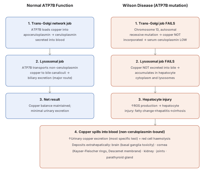
Dietary absorption and hepatic transport of copper are normal in Wilson’s disease — the defect is specifically in the ATP7B-mediated excretory step. The mutated ATP7B fails at both of its normal jobs simultaneously:
- Copper is not incorporated into apoceruloplasmin → serum ceruloplasmin is low.
- Copper is not excreted into bile → accumulates progressively in hepatocyte cytoplasm and lysosomes.
Accumulated hepatocellular copper increases reactive oxygen species (ROS) production, injuring hepatocytes (fatty change → hepatitis → cirrhosis). Once hepatocellular storage capacity is exceeded, non-ceruloplasmin-bound copper is released from injured hepatocytes into the circulation — causing red cell haemolysis and markedly increased urinary copper excretion — and deposits in extrahepatic tissues: brain (basal ganglia), cornea (Descemet membrane — Kayser-Fleischer rings), kidney, joints, and parathyroid glands. Copper deposition in the kidney does not produce clinically significant renal dysfunction, despite occurring.
Importantly, low ceruloplasmin is a hallmark finding but not itself the cause of disease — the pathogenesis is driven by hepatocellular copper accumulation and toxicity, not by ceruloplasmin deficiency per se.
Biochemical/Diagnostic Findings
- Decreased serum ceruloplasmin — from impaired apoceruloplasmin loading and defective copper mobilisation from hepatocellular lysosomes.
- Increased hepatic copper content (liver biopsy) — the most sensitive diagnostic test (>250 μg/g dry liver weight considered diagnostic, though only ~80% sensitive).
- Increased urinary copper excretion — the most specific diagnostic test.
- Serum copper level — of no diagnostic value; may be low, normal, or high depending on disease stage.
- Kayser-Fleischer rings — present in nearly all patients with neurologic involvement; supportive when hepatic copper is borderline.
- Genetic testing is complicated by the large number of distinct ATP7B mutations (unlike, say, HFE testing in haemochromatosis, where a limited number of variants make sequencing straightforward).
Morphology
Liver: highly variable, mimicking many other hepatic disease processes — mild-to-moderate fatty change with focal hepatocyte necrosis; acute fulminant hepatitis (can mimic acute viral hepatitis); chronic hepatitis with moderate-severe inflammation, hepatocyte necrosis, fatty change, and steatohepatitis features (ballooning, prominent Mallory bodies); advanced cases show macronodular cirrhosis. Copper is demonstrable in periportal hepatocytes by special stains (rubeanic acid, rhodamine, or copper-specific stains) as reddish cytoplasmic granules/coloration.
Brain: toxic injury to basal ganglia neurons.
Eye: greenish-brown copper deposits in Descemet’s membrane at the corneal periphery (Kayser-Fleischer rings).
Kidney: fatty and hydropic change (without significant functional impairment).
Treatment
Early recognition with long-term copper chelation therapy (D-penicillamine or trientine) or zinc-based therapy (inhibits intestinal copper uptake) has dramatically improved what was once an inexorably progressive disease. Patients presenting with hepatitis or advanced cirrhosis may require liver transplantation, which can be curative (since the underlying defect is hepatocyte-intrinsic).
Differential — Indian Childhood Cirrhosis
Indian childhood cirrhosis (ICC) is a distinct entity affecting children 6 months–3 years (rural, middle-class, Hindu populations in India and parts of South-East Asia/Middle East), with no viral aetiology — instead a combination of toxic effects and an inherited copper metabolism abnormality is implicated. Histology (type II, most common) shows hepatocyte injury, prominent Mallory bodies without fatty change, neutrophilic ± lymphocytic infiltrate, and creeping pericellular fibrosis progressing to micro-macronodular cirrhosis, with hepatic copper deposition often exceeding that seen in Wilson’s disease — largely from milk boiled and stored in copper vessels. Death typically occurs from hepatic failure within a year of diagnosis. ICC must be distinguished from Wilson’s disease despite the shared copper-overload picture.
Clinical Relevance
- The two-function ATP7B framework (trans-Golgi ceruloplasmin loading vs lysosomal biliary excretion) is the organising concept that explains both major biochemical findings at once — low ceruloplasmin (job 1 fails) and hepatic copper accumulation (job 2 fails) — rather than treating them as two unrelated lab abnormalities.
- Recognising that low ceruloplasmin is a marker, not the mechanism, of disease prevents the common misconception that Wilson’s disease is fundamentally “a ceruloplasmin deficiency” rather than a copper-excretion defect with ceruloplasmin synthesis as a secondary casualty.
- The sensitivity/specificity contrast between hepatic copper content (most sensitive) and urinary copper excretion (most specific) is directly relevant to diagnostic reasoning when Kayser-Fleischer rings are absent or ceruloplasmin is borderline.
- Distinguishing Wilson’s disease from Indian childhood cirrhosis matters clinically despite overlapping copper-overload histology — the epidemiology (age, cultural/dietary context), the absence of a genetic ATP7B defect in ICC, and markedly higher hepatic copper in ICC are the discriminating features, and the two require entirely different management approaches.
Diagram 1: Normal ATP7B Function Contrasted with Wilson’s Disease
Draw two parallel columns (Normal ATP7B function, Wilson’s Disease), each with three stacked stages matched step-for-step, converging into one shared wide box at the bottom for the extrahepatic consequences (Wilson’s column only).
- Trans-Golgi network job — loads copper into apoceruloplasmin, secreted as ceruloplasmin (normal) vs this job fails, serum ceruloplasmin low (Wilson’s).
- Lysosomal job — transports copper to bile for excretion, the major route (normal) vs this job fails too, copper accumulates in hepatocyte cytoplasm/lysosomes (Wilson’s).
- Net result — copper balance maintained (normal) vs ROS-driven hepatocyte injury: fatty change→hepatitis→cirrhosis (Wilson’s).
- Shared bottom box (Wilson’s only) — copper spills into blood → haemolysis + ↑urinary copper (most specific test) + extrahepatic deposition (brain, cornea, kidney, joints, parathyroid).
Labels required
- Both ATP7B jobs (trans-Golgi ceruloplasmin loading, lysosomal biliary excretion) must be shown as failing together in Wilson’s disease — a diagram showing only one failing loses the fact that both major biochemical abnormalities (low ceruloplasmin AND high hepatic copper) share one root cause.
- The bottom box must explicitly label urinary copper as the “most specific test” and (in the Wilson’s column, step 3) hepatic copper content as the pathway to the “most sensitive test” — these two labels are what make the diagram diagnostically useful, not just mechanistically descriptive.
- Kayser-Fleischer rings must be tied specifically to the Descemet membrane of the cornea, not just “the eye” generically.
Errors commonly made
- Treating low ceruloplasmin as the cause of Wilson’s disease rather than a downstream marker of the same ATP7B defect that separately causes hepatic copper accumulation — the two abnormalities are parallel consequences of one mutation, not cause-and-effect of each other.
- Omitting that copper deposition in the kidney, despite occurring, does not cause significant renal dysfunction — a detail easy to assume incorrectly by analogy with the liver/brain/eye damage.
- Confusing this diagram’s copper-overload mechanism with Indian childhood cirrhosis, a histologically overlapping but mechanistically and epidemiologically distinct entity covered separately in
notes.md.
Multiple Choice Questions (1)
My Sticky Notes
Personal revision notes, mnemonics and reminders.
