
Thrombosis
Definition
Solid mass from flowing blood constituents in intact CV system = thrombus. ≠ clotting (fibrinogen→fibrin only). Haemostatic plug = simplest/beneficial thrombosis. Pathologic thrombus → ischaemia or thromboembolism.
Virchow’s triad
-
Endothelial injury — intact endothelium: anti-thrombotic (heparin-like, thrombomodulin→protein C, ADPase/PGI2, tPA) + pro-thrombotic (tissue factor, vWF, PAF, PAI) factors. Injury exposes ECM. Dominant in ARTERIAL/cardiac thrombi (MI, myocarditis, prosthetic valves, ulcerated plaques, HTN, diabetes, smoking).
-
Platelets:
- Adhesion: GpIb + vWF (deficiency→vWD/Bernard-Soulier→bleeding)
- Release: dense bodies (ADP, Ca2+, serotonin) + alpha granules (fibrinogen, PDGF, PF4)
- Aggregation: ADP→secondary aggregation→temp plug; thrombin/fibrin/TXA2→stable plug
-
Coagulation system: Intrinsic (XII→XI→IX→VIII→X, +Ca+PF3) + Extrinsic (tissue factor+VII→X) → Common (Xa+Va+PF3+Ca→thrombin→fibrin, XIII polymerises). Regulators: antithrombin III, protein C, C1-inact, α1-AT, α2-M. Fibrinolysis: plasminogen→plasmin (via tPA/urokinase-PA)→FSP.
-
Altered blood flow — normal axial flow (RBC/WBC center, platelets laminar, plasma peripheral). Turbulence→endothelial injury→ARTERIAL/cardiac thrombi. Stasis alone→VENOUS thrombi.
-
Hypercoagulability (thrombophilia):
- Hereditary: antithrombin III def (AD, <50%) · protein C/S def (AD) · Factor V Leiden (AD, Arg506Gly, MOST COMMON cause) · fibrinolytic defects · ↑prothrombin/factor VIII
- Acquired: age, bed rest, immobilisation, smoking, obesity, heart/vascular disease, polycythaemia, nephrotic syndrome, cancer, shock, trauma, pregnancy, OCP/HRT
- APLA syndrome: lupus anticoagulant (→venous) vs anticardiolipin Ab (→arterial); + miscarriage, TIA, thrombocytopenia, ↑aPTT; linked to SLE
Sites
Cardiac (atrial appendages, valves — mural; ball-valve; agonal=fibrin-rich, pre-death) · Arterial (aorta, coronary, mesenteric, limb, renal, cerebral) · Venous (leg deep veins, popliteal/femoral/iliac, pulmonary, portal, IVC/SVC, mesenteric, renal) · Capillary (inflammation, vasculitis, DIC).
| Arterial | Venous | |
|---|---|---|
| Mechanism | Endothelial injury | Stasis |
| Development | Mural | Occlusive, casts vessel |
| Effect | Ischaemia→infarct | Thromboembolism, oedema, ulcers |
Morphology
Arterial=white/mural. Venous=red/occlusive. Lines of Zahn (alternating pale platelet-fibrin + dark RBC layers) = antemortem marker.
| Antemortem | Postmortem clot | |
|---|---|---|
| Gross | Dry, granular, firm, friable | Gelatinous, soft, rubbery |
| Wall | Adherent | Weakly attached |
| Shape | Independent of vessel | Casts vessel/bifurcation |
| Feature | Lines of Zahn | ”Chicken fat” over “currant jelly” |
Fate (4 outcomes)
- Resolution — fibrinolysis, complete dissolve, small venous thrombi; thrombolytics (urokinase/streptokinase) work best early (monomeric fibrin)
- Organisation — phagocytes clear debris, granulation tissue in → recanalisation or hyalinisation/calcification (phleboliths)
- Propagation — enlarges, may occlude
- Thromboembolism — friable/infected fragment detaches
Clinical effects by site
Cardiac→sudden death (obstruction/embolism). Arterial→infarct/gangrene. Venous (phlebothrombosis)→embolism, oedema, poor healing, ulcers, thrombophlebitis, phlegmasia alba dolens (postpartum), migratory thrombophlebitis (cancer). Capillary→DIC.
Clinical relevance
Factor V Leiden = #1 hereditary thrombophilia cause, first suspect in young unexplained recurrent VTE. Lupus anticoagulant(venous) vs anticardiolipin(arterial) = predicts vascular bed at risk, relevant in SLE. Antemortem vs postmortem clot distinction = real forensic skill. Thrombolysis works only while fibrin still monomeric = narrow treatment window.
Definition
Thrombosis is the formation of a solid mass from the constituents of flowing blood within the intact cardiovascular system; the mass itself is a thrombus. Thrombosis is often confused with “clotting,” but the two differ: thrombosis essentially involves platelet activation together with the coagulation cascade, whereas clotting refers narrowly to conversion of soluble fibrinogen to insoluble fibrin (including in vitro coagulation, e.g. in a test tube). A haemostatic plug — the clot that forms physiologically at a site of vessel injury to stop bleeding — is, in effect, the simplest and most beneficial form of thrombosis; a pathological thrombus forming in an unruptured vessel is instead harmful, causing ischaemic injury (by obstructing flow, potentially to the point of infarction) or thromboembolism (if part or all of it detaches and lodges elsewhere — see Embolism).
Virchow’s Triad
Rudolf Virchow described three primary predisposing events, still the organising framework for thrombogenesis:
1. Endothelial Injury
Intact endothelium normally protects flowing blood from the thrombogenic subendothelium and secretes both anti-thrombotic factors (heparin-like substance potentiating antithrombin III; thrombomodulin, activating protein C; ADPase and prostacyclin/PGI2 inhibiting platelet aggregation; tissue plasminogen activator promoting fibrinolysis) and pro-thrombotic factors (tissue factor/thromboplastin; von Willebrand factor; platelet-activating factor; plasminogen activator inhibitor). Injury exposes thrombogenic subendothelial ECM (collagen, elastin, fibronectin, laminin, glycosaminoglycans) and is the dominant mechanism in arterial and cardiac thrombus formation (myocardial infarction, myocarditis, cardiac surgery, prosthetic valves, ulcerated atherosclerotic plaques, hypertensive haemodynamic stress, diabetes, hypercholesterolaemia, endotoxins, cigarette smoke).
2. Role of Platelets
Following endothelial injury, platelets undergo a sequential response:
- Adhesion — platelet GpIb receptor recognises exposed subendothelial ECM; von Willebrand factor bridges GpIb to the ECM for firm adhesion (deficiency of either — von Willebrand disease, or Bernard-Soulier disease for GpIb — causes abnormal bleeding, confirming the step’s necessity).
- Release reaction — activated platelets release granule contents: dense bodies (ADP — a further platelet activator; ionic calcium — needed in coagulation; serotonin, histamine, epinephrine) and alpha granules (fibrinogen, fibronectin, PDGF, platelet factor 4, thrombospondin). Platelet factor 3 (a phospholipid complex) is also activated, feeding the intrinsic coagulation pathway.
- Aggregation — released ADP recruits further platelets (secondary aggregation), forming a temporary hameostatic plug; thrombin, fibrin and thromboxane A2 stabilise it into a permanent plug.
3. Role of the Coagulation System
The coagulation cascade converts soluble fibrinogen to insoluble fibrin via three linked pathways: the intrinsic (contact/blood) pathway — contact with an abnormal surface (e.g. exposed subendothelial ECM) sequentially activates factor XII, then XI, IX, VIII and X (with calcium and platelet factor 3); the extrinsic (tissue) pathway — tissue damage releases tissue factor, which with factor VII activates factor X; and the common pathway, where activated factor X, factor Va, platelet factor 3 and calcium activate prothrombin to thrombin, which converts fibrinogen to fibrin, polymerised to its insoluble form by factor XIII. Regulation is provided by protease inhibitors (antithrombin III, protein C, C1 inactivator, α1-antitrypsin, α2-macroglobulin) and the fibrinolytic system (plasminogen activators generate plasmin from plasminogen, which degrades fibrin to fibrin split products).
4. Alteration of Blood Flow
Normal axial flow keeps the fastest-moving central stream of red cells/leucocytes separated from a slower platelet-containing laminar layer and a peripheral, near-stationary plasma layer close to the endothelium. Turbulence (irregular flow) and stasis (slowed flow) disturb this, causing cells (including platelets) to marginate against the endothelium (margination/pavementing). Turbulence tends to injure endothelium directly, favouring arterial and cardiac thrombi; stasis alone, without endothelial injury, is sufficient to initiate venous thrombi.
5. Hypercoagulable States (Thrombophilia)
A group of conditions predisposing chiefly to venous thrombosis, hereditary or acquired (usually several factors coexist in any given patient):
- Hereditary: antithrombin III deficiency (autosomal dominant, <50% activity, recurrent venous thrombosis); protein C or S deficiency (autosomal dominant, lifelong deep-vein thrombosis risk); factor V Leiden mutation (autosomal dominant, arginine→glycine at position 506 — the single most common cause of thrombophilia); fibrinolytic defects (dysfibrinogenaemia, plasminogen disorders); elevated prothrombin/factor VIII from genetic mutation.
- Acquired: risk factors (advancing age, prolonged bed rest/immobilisation, smoking, obesity); predisposing conditions (heart disease, vascular disease, hypercoagulable states such as polycythaemia/myeloproliferative disease/dehydration/nephrotic syndrome/disseminated cancer, shock, tissue trauma, late pregnancy/puerperium, oral contraceptives/HRT); and antiphospholipid antibody (APLA) syndrome — lupus anticoagulant (predisposes to venous thrombosis) or anticardiolipin antibody (predisposes to arterial thrombosis), also causing recurrent miscarriage, TIAs, thrombocytopenia and prolonged aPTT; frequently coexpressed in SLE.
Sites of Origin
- Cardiac thrombi — any chamber or valve cusp; commonest in the atrial appendages (especially right atrium) and on mitral/aortic valves (infective and non-bacterial thrombotic endocarditis vegetations); typically mural (non-occlusive), as in large-vessel aneurysms; a large round obstructive thrombus in the mitral valve is a ball-valve thrombus; agonal thrombi, fibrin-rich, form shortly before death in either ventricle.
- Arterial thrombi — aorta (aneurysm, arteritis), coronary (atherosclerosis), mesenteric (atherosclerosis, arteritis), limb arteries (atherosclerosis, diabetes, Buerger’s/Raynaud’s disease), renal and cerebral arteries (atherosclerosis, vasculitis).
- Venous thrombi — leg deep veins/varicosities, popliteal/femoral/iliac veins (postoperative, postpartum), pulmonary veins (heart failure, pulmonary hypertension), hepatic/portal vein (portal hypertension), superior/inferior vena cava, mesenteric veins (volvulus, obstruction), renal vein (amyloidosis).
- Capillary thrombi — minute, red-cell-rich, in acute inflammation, vasculitis and DIC (see Disseminated Intravascular Coagulation).
| Feature | Arterial thrombi | Venous thrombi |
|---|---|---|
| Flow | Rapid | Slow |
| Mechanism | Endothelial injury (e.g. atherosclerosis) | Venous stasis |
| Development | Mural, may propagate | Occlusive, casts the vessel, may propagate both directions |
| Gross | Grey-white, friable, lines of Zahn | Red-blue, fibrin strands, lines of Zahn |
| Effect | Ischaemia → infarction | Thromboembolism, oedema, skin ulcers, poor healing |
Morphology
Grossly, thrombi vary by site — arterial thrombi are white and mural; venous thrombi are red and occlusive. Mixed/laminated thrombi show alternating pale (platelet/fibrin) and dark (red cell) bands, the lines of Zahn — a feature of antemortem thrombus formation under flow, distinguishing it from bland postmortem clotting. Red thrombi are soft and gelatinous; white thrombi firm and pale.
Antemortem thrombi vs postmortem clots (a frequently tested distinction):
| Feature | Antemortem thrombus | Postmortem clot |
|---|---|---|
| Gross | Dry, granular, firm, friable | Gelatinous, soft, rubbery |
| Vessel wall relation | Adherent | Weakly attached |
| Shape | May or may not fit vessel contour | Casts the vessel/bifurcation shape |
| Appearance | Lines of Zahn present | ”Chicken fat” yellow layer over “currant jelly” red clot |
Fate of Thrombus
- Resolution — fibrinolytic activation dissolves the thrombus completely; usual for small venous thrombi, less reliable for large ones. Thrombolytic drugs (urokinase, streptokinase) accelerate this, most effective early while fibrin is still monomeric (e.g. early acute MI thrombolysis).
- Organisation — if not resolved, phagocytes (neutrophils, macrophages) digest fibrin/debris while capillaries and fibroblasts grow in from the attachment site, forming fibrovascular granulation tissue that matures, becomes covered by endothelium, and incorporates into the vessel wall. New channels within it may re-establish flow — recanalisation. The organised thrombus may hyalinise and calcify (e.g. phleboliths in pelvic veins).
- Propagation — continued deposition from flowing blood enlarges the thrombus, potentially to the point of obstructing an important vessel.
- Thromboembolism — friable (especially early-stage or infected) thrombi can detach in part or whole, embolising to cause effects at their site of lodgement (see Embolism).
Clinical Effects by Site
- Cardiac thrombi — sudden death from mechanical obstruction or thromboembolism to vital organs.
- Arterial thrombi — ischaemic necrosis (infarct), potentially gangrene; sudden death with coronary thrombosis.
- Venous thrombi (phlebothrombosis) — thromboembolism, oedema of the drained area, poor wound healing, skin ulcers, painful thrombosed veins (thrombophlebitis), painful white leg (phlegmasia alba dolens, postpartum iliofemoral thrombosis), and migratory thrombophlebitis in malignancy.
- Capillary thrombi — can give rise to DIC.
Clinical Relevance
- Factor V Leiden being the single most common cause of thrombophilia is a specific, high-yield fact worth knowing by name and mutation site, since it is the first hereditary cause to consider in an otherwise unexplained recurrent venous thrombosis, particularly in a young patient.
- The distinction between lupus anticoagulant (venous thrombosis risk) and anticardiolipin antibody (arterial thrombosis risk) within antiphospholipid syndrome is directly clinically relevant to anticipating which vascular bed is at risk in a given patient, and its association with SLE makes it relevant whenever thrombosis occurs in a lupus patient.
- Distinguishing an antemortem thrombus from a postmortem clot at autopsy — using adherence to the vessel wall, lines of Zahn, and gross texture — is a genuinely applied forensic/pathological skill, not just a comparison exercise, since it determines whether a thrombotic event contributed to death.
- Recognising early thrombolytic therapy’s dependence on the thrombus still containing monomeric (not yet fully polymerised/organised) fibrin explains the narrow therapeutic time window for treatments like streptokinase in acute myocardial infarction.
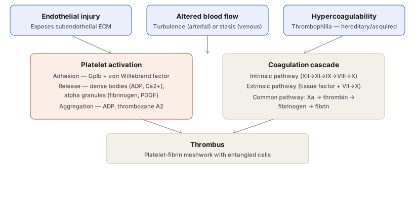
Diagram 1: Virchow’s Triad and Platelet Activation
Draw three top boxes (endothelial injury, altered blood flow, hypercoagulability) converging into two parallel process boxes (platelet activation, coagulation cascade), which both converge into a final thrombus box.
- Endothelial injury — exposes subendothelial ECM.
- Altered blood flow — turbulence (arterial) vs stasis (venous), explicitly labelled as favouring different thrombus types.
- Hypercoagulability — thrombophilia, hereditary/acquired.
- Platelet activation — three named sub-steps: adhesion (GpIb + vWF), release (dense bodies, alpha granules), aggregation (ADP, TXA2).
- Coagulation cascade — intrinsic and extrinsic pathways named with their key factors, converging on the common pathway (Xa → thrombin → fibrin).
Labels required
- Turbulence and stasis must each be labelled with the type of thrombus they favour (arterial/cardiac vs venous respectively), since this distinction is frequently tested on its own.
- The three platelet sub-steps (adhesion, release, aggregation) must appear in that order with their named receptors/mediators, since exam questions often ask for the sequence specifically.
Errors commonly made
- Treating Virchow’s triad as three independent, unrelated causes rather than three factors that converge on the same downstream platelet/coagulation machinery to produce a thrombus.
- Conflating platelet adhesion and aggregation — adhesion is the initial GpIb/vWF-mediated attachment to exposed ECM, aggregation is the later ADP/thromboxane-driven recruitment of additional platelets; the two involve different mediators and occur at different stages.
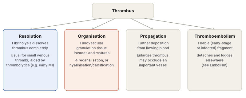
Diagram 2: Fate of a Thrombus
Draw a single “thrombus” box branching into four side-by-side outcome boxes.
- Resolution — fibrinolysis, complete dissolution, typical of small venous thrombi.
- Organisation — granulation tissue ingrowth, maturing to recanalisation or hyalinisation/calcification.
- Propagation — enlargement by further deposition.
- Thromboembolism — fragment detachment.
Labels required
- Recanalisation must be shown explicitly as a possible sub-outcome of organisation, not a separate fifth fate, since it is a specific structural consequence of the organisation process (new vascular channels within the organised thrombus).
- Each outcome’s typical clinical correlate (thrombolytic therapy for resolution; phleboliths for organisation; vessel occlusion for propagation) should be noted briefly under each box.
Errors commonly made
- Listing recanalisation as a standalone fifth fate of thrombus rather than correctly nesting it within organisation.
- Assuming all four fates are mutually exclusive single endpoints, when in practice a single thrombus can partially resolve, partially organise, and still propagate at its unresolved margin.
Short Essays (1)
Multiple Choice Questions (2)
My Sticky Notes
Personal revision notes, mnemonics and reminders.
