
Systemic Lupus Erythematosus and Autoimmunity
Tolerance mechanisms (general)
- Central deletion (thymus/marrow) — AIRE gene controls self-Ag expression in thymus; AIRE mutation → autoimmune polyglandular syndrome
- Receptor editing (B cells, 2nd chance before deletion)
- Treg (FoxP3-dependent) — CTLA-4, IL-2 competition, IL-10/TGF-β; FOXP3 mutation → IPEX
- Inhibitory receptors CTLA-4/PD-1 (checkpoint inhibitor drugs → autoimmunity as side effect)
- Fas/FasL-induced apoptosis of activated lymphocytes; FAS mutation → ALPS
Autoimmunity pathogenesis (general)
Genetic susceptibility (familial, twin concordance, weak HLA-DR/DQ links) + environmental triggers (infection → costimulator induction; molecular mimicry — best example = rheumatic heart disease; tissue injury exposing sequestered Ag; UV/smoking/hormones) → tolerance breakdown → epitope spreading (chronicity).
SLE overview
Systemic autoimmune/connective tissue disease. F:M 9:1 (age 17-55), 2:1 outside that range. Peak 20s-30s. Runs more frequent, more severe course in some non-white populations (e.g. African-American, Latin-American).
Autoantibodies
- ANA (4 groups: DNA, histone, RNA-bound nonhistone protein, nucleolar) — sensitive (95-100%) NOT specific
- Anti-dsDNA + anti-Sm — near-diagnostic for SLE, correlate with nephritis
- Anti-Ro (SS-A) — congenital heart block, neonatal lupus
- Antiphospholipid Ab (30-40%) — binds cardiolipin → false +VDRL/syphilis; prolongs PTT in vitro (“lupus anticoagulant”) but causes HYPERcoagulable state clinically (thrombosis, miscarriage, ischaemia)
Pathogenesis
Genetic: familial (MZ twin 25% vs DZ 1-3%), HLA-DR2/DR3 (RR2-3, ~5 if both), C1q/C2/C4 deficiency (10% — impairs immune complex + apoptotic debris clearance). Environmental: UV (apoptosis + immunogenic DNA + keratinocyte IL-1 → photosensitivity/flares), drugs (hydralazine, procainamide, D-penicillamine → SLE-like). Immunologic: B/T tolerance failure → autoAb; Type I IFN signature (IFN-α from plasmacytoid DC); TLR7(RNA)/TLR9(DNA) amplify B-cell autoAb production; BAFF↑ (→ anti-BAFF therapy).
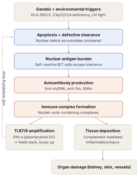
Model: susceptibility + UV → apoptosis + defective clearance → nuclear Ag burden → self-reactive lymphocytes → autoAb → immune complexes → TLR7/9 amplification loop (IFN-α) + tissue deposition → organ damage.
Shows BOTH Type II (cytopenias, Ab vs blood cell Ag) + Type III (visceral immune complex disease) hypersensitivity.
LE cell/bodies
ANA can’t enter intact cell; binds exposed damaged-cell nuclei → homogeneous LE body/haematoxylin body. LE cell = phagocyte that engulfed this. +ve in ~70% SLE (also RA, lupoid hepatitis, penicillin sensitivity). Historical test, superseded by ANA assays.
Morphology
Kidney (50%) — lupus nephritis classes I-VI:
- I minimal mesangial (deposits only, no LM change)
- II mesangial proliferative
- III focal proliferative (20-35%)
- IV diffuse proliferative (35-60%, most severe, wire-loop lesions, crescents)
- V membranous (10-15% → nephrotic syndrome)
- VI advanced sclerosing (>90% glomeruli)
Vessels — acute necrotising vasculitis, fibrinoid necrosis → chronic fibrous thickening. Heart — Libman-Sacks endocarditis: 1-3mm warty vegetations, either surface, any valve (vs bigger IE vegetations, vs smaller closure-line RHD verrucae).
Clinical features
Young woman, butterfly (malar) rash, fever, non-erosive arthritis, pleuritic chest pain, photosensitivity. ↑infection risk (disease + immunosuppression).
ACR criteria (1997) — need ≥4/11
Malar rash · discoid rash · photosensitivity · oral ulcers · arthritis (non-erosive, ≥2 joints) · serositis · renal (proteinuria >0.5g/d or casts) · neurologic (seizure/psychosis) · haematologic (haemolytic anaemia/leucopenia/lymphopenia/thrombocytopenia) · immunologic (anti-dsDNA/anti-Sm/antiphospholipid) · ANA.
Labs: ANA screen → anti-dsDNA/anti-Sm confirm; CBC, ESR, urinalysis; ↓C3/C4 = active disease marker.
Clinical relevance
False+ VDRL in young woman with joint pain/rash → think SLE/antiphospholipid syndrome. Wire-loop = proliferative (III/IV) class = worst prognosis, drives aggressive immunosuppression. ↓C3/C4 = flare (complement consumption). SLE = worked example of simultaneous Type II + Type III hypersensitivity.
Immunologic Tolerance and the General Basis of Autoimmunity
Autoimmune disease occurs when the immune system fails to distinguish self from non-self and mounts a sustained response against the body’s own antigens. Because healthy individuals do not normally react against self antigens — a state called immunologic tolerance — understanding autoimmunity begins with how tolerance is normally maintained:
- Central deletion. Immature T cells (in the thymus) and B cells (in the bone marrow) that recognise self antigen during development are eliminated (negative selection) or, for B cells, given a second chance via receptor editing before deletion. Expression of self antigens in the thymus depends on the protein AIRE; AIRE mutations cause autoimmune polyglandular syndrome because self antigens are never displayed centrally, so self-reactive T cells escape deletion.
- Regulatory T cells (Treg). CD4+ Tregs, dependent on the transcription factor FoxP3, actively suppress self-reactive lymphocytes — via CTLA-4 (removing costimulatory B7 from antigen-presenting cells), competing for IL-2, and secreting IL-10/TGF-β. FOXP3 mutations cause IPEX (immune dysregulation, polyendocrinopathy, enteropathy, X-linked).
- Inhibitory receptors. Activated T cells express CTLA-4 and PD-1, imposing checkpoints on continued activation — the mechanistic reason cancer patients treated with checkpoint inhibitors (which block these receptors) frequently develop autoimmune disease as a side effect.
- Activation-induced cell death. Fas/FasL co-expression on activated lymphocytes triggers their own apoptosis; FAS mutations cause autoimmune lymphoproliferative syndrome (ALPS).
Autoimmunity arises from combined genetic susceptibility and environmental triggers that together break down these safeguards:
- Genetic factors — familial clustering, higher monozygotic than dizygotic twin concordance, and HLA-DR/DQ associations (though most such associations carry only weak relative risk, and most people with a “susceptibility” HLA allele never develop disease).
- Environmental triggers — infections can induce costimulatory molecule expression on antigen-presenting cells, tipping self-reactive T cells into activation; or microbial antigens can share cross-reacting epitopes with self antigens (molecular mimicry — the clearest example being rheumatic heart disease, where anti-streptococcal antibody cross-reacts with cardiac antigen); tissue injury of any cause can expose normally sequestered self antigen (eye, testis) to the immune system; UV radiation, smoking, and hormonal factors (explaining the strong female predominance of most autoimmune disease) all modulate self-antigen display or immune activation.
- Epitope spreading — once an autoimmune response begins, the resulting tissue injury exposes further self epitopes, broadening the response over time and contributing to disease chronicity.
Systemic Lupus Erythematosus: Overview
SLE is the paradigm systemic autoimmune (connective tissue) disease, produced by autoantibodies against a wide range of self antigens with resulting immune-complex deposition and multi-organ injury. It predominantly affects women in the reproductive years (female:male ratio around 9:1 between ages 17–55, falling to about 2:1 outside that range), typically presenting in the twenties or thirties though onset can occur at any age. Ethnicity influences both prevalence and severity — the disease runs a more frequent and more severe course in some non-white populations (documented in African-American and Latin-American groups) than in white populations, a pattern relevant when assessing disease severity across different patient backgrounds.
Autoantibody Spectrum
The hallmark of SLE is autoantibody production, with both diagnostic and directly pathogenic significance (immune-complex glomerulonephritis being the clearest example of the latter):
- Antinuclear antibodies (ANAs), detected by indirect immunofluorescence, fall into four groups by target: DNA, histones, non-histone proteins bound to RNA, and nucleolar antigens. Generic ANA positivity is extremely sensitive (95–100% of SLE) but not specific, since it also occurs in other autoimmune diseases, chronic infection and cancer.
- Anti-double-stranded DNA and anti-Smith (Sm) antigen antibodies are near-diagnostic for SLE specifically (found in roughly 40–60% and 20–30% of patients respectively) and correlate with nephritis.
- Anti-Ro (SS-A) — associated with congenital heart block and neonatal lupus.
- Antiphospholipid antibodies, present in 30–40% of SLE patients, bind epitopes on plasma proteins complexed with phospholipid; because these same antibodies bind the cardiolipin antigen used in syphilis serology, SLE patients can have a false-positive VDRL/syphilis test. Despite prolonging the in vitro partial thromboplastin time (hence the older name “lupus anticoagulant”), these antibodies clinically cause a hypercoagulable state — venous/arterial thrombosis, recurrent miscarriage, and focal cerebral/ocular ischaemia (antiphospholipid antibody syndrome).
Pathogenesis
The fundamental defect is failure of self-tolerance, from combined genetic and environmental causes:
- Genetic factors: familial clustering (25% monozygotic twin concordance vs 1–3% dizygotic); HLA-DR2/DR3 association (relative risk 2–3, or ~5 with both haplotypes); genetic deficiency of early classical-pathway complement proteins (C1q, C2, C4) in about 10% of patients, which impairs clearance of immune complexes and apoptotic cell debris and disrupts B-cell tolerance; and polymorphisms in inhibitory Fc receptors contributing to unchecked B-cell activation.
- Environmental factors: UV light exposure exacerbates disease by inducing apoptosis and rendering DNA immunogenic (partly via enhanced TLR recognition), and by stimulating keratinocyte IL-1 production — the mechanistic basis for photosensitivity as a clinical feature and for SLE flares after sun exposure; certain drugs (hydralazine, procainamide, D-penicillamine) can induce an SLE-like syndrome.
- Immunologic mechanisms: failure of B-cell tolerance (defective receptor editing/peripheral tolerance) and escape of nucleosome-specific CD4+ helper T cells allow sustained high-affinity autoantibody production; a Type I interferon signature (excess IFN-α, chiefly from plasmacytoid dendritic cells) is characteristic; TLR9 (DNA-recognising) and TLR7 (RNA-recognising) engagement by nucleic-acid-containing immune complexes further stimulates self-reactive B cells; and BAFF (a B-cell survival cytokine) is often elevated, providing the rationale for BAFF-blocking therapy.
Putting this together as a model: defective lymphocyte tolerance allows self-reactive B and T cells to survive; UV light and other triggers drive cell apoptosis; inadequate clearance of the resulting nuclear debris creates a large burden of nuclear antigen; self-reactive lymphocytes respond to this antigen with autoantibody production; the resulting antigen–antibody complexes engage Fc receptors and TLRs on B cells and dendritic cells, further amplifying interferon and autoantibody production in a self-sustaining loop, while also depositing in tissues to cause the immune-complex-mediated inflammation responsible for organ damage (see Type III Hypersensitivity) — SLE in fact shows features of both Type II (cytopenias from antibody against blood cell surface antigens) and Type III (visceral immune-complex deposition) hypersensitivity simultaneously.
LE Cells and LE Bodies
ANAs cannot penetrate an intact cell, but once a cell’s nucleus is exposed by damage, ANAs bind it; the denatured, homogeneous nuclear remnant is an LE body (haematoxylin body). The LE cell is any phagocyte (neutrophil or macrophage) that has engulfed such a denatured nucleus, historically demonstrated in vitro by agitating a blood sample to damage nucleated cells and release nuclei for ANA binding. LE cells are positive in about 70% of SLE (and occasionally in rheumatoid arthritis, lupoid hepatitis, penicillin sensitivity) but the LE cell test has been superseded by direct ANA assays and is now of largely historical interest.
Morphology
Kidney (involved in ~50% of patients, one of the most prognostically important sites): DNA/anti-DNA immune complexes deposit in glomeruli, triggering endothelial, mesangial and/or epithelial cell proliferation. Six morphologic classes are recognised (none specific to SLE alone):
| Class | Pattern |
|---|---|
| I — Minimal mesangial | Mesangial immune deposits only, no light-microscopic structural change |
| II — Mesangial proliferative | Mesangial deposits with mild-moderate mesangial hypercellularity |
| III — Focal proliferative | 20–35% of patients; focal/segmental or global glomerular proliferation, fibrinoid necrosis, crescents, intracapillary thrombi |
| IV — Diffuse proliferative | Most severe, 35–60% of patients; >50% of glomeruli involved, cellular crescents, prominent subendothelial deposits producing “wire-loop” capillary wall thickening (also seen in class III) |
| V — Membranous | 10–15%; diffuse capillary wall thickening resembling idiopathic membranous GN → nephrotic syndrome |
| VI — Advanced sclerosing | Sclerosis of >90% of glomeruli |
Immunofluorescence shows granular immunoglobulin/complement deposits; electron microscopy shows electron-dense deposits in mesangial, subepithelial or subendothelial locations.
Blood vessels: acute necrotising vasculitis of small arteries/arterioles with fibrinoid necrosis, progressing to fibrous wall thickening and luminal narrowing in chronic disease.
Heart: any layer may be involved; Libman–Sacks endocarditis — single or multiple 1–3 mm warty (verrucous), non-bacterial vegetations on either surface of any valve leaflet — is characteristic (contrast with the larger vegetations of infective endocarditis and the smaller, closure-line verrucae of rheumatic heart disease, see Infective Endocarditis and Rheumatic Heart Disease).
Clinical Features
Multisystem disease with highly variable presentation; typical picture is a young woman with malar (butterfly) rash, fever, non-erosive/non-deforming polyarthralgia, pleuritic chest pain and photosensitivity. Immune dysfunction and immunosuppressive treatment both predispose SLE patients to infection.
Diagnostic Criteria (ACR, Revised 1997)
Any 4 or more of the following 11 criteria (serially or simultaneously) is diagnostic: malar rash; discoid rash; photosensitivity; oral/nasopharyngeal ulcers; non-erosive arthritis (≥2 peripheral joints); serositis (pleuritis/pericarditis); renal disorder (persistent proteinuria >0.5 g/day or cellular casts); neurologic disorder (seizures or psychosis without other cause); haematologic disorder (haemolytic anaemia, leucopenia <4000/mm³, lymphopenia <1500/mm³, or thrombocytopenia <100,000/mm³); immunologic disorder (anti-dsDNA, anti-Sm, or antiphospholipid antibodies); and antinuclear antibody.
Laboratory diagnosis serves three purposes — establishing/excluding diagnosis, following disease course, and monitoring treatment toxicity — and includes ANA screening (sensitive but non-specific), confirmatory anti-dsDNA/anti-Sm testing, complete blood count, ESR, urinalysis, and serum complement (C3/C4) levels, which fall during active disease from complement consumption and thus serve as a disease-activity marker.
Clinical Relevance
- A false-positive syphilis serology (VDRL/RPR) in a young woman with joint pain and rash should immediately raise suspicion of SLE and antiphospholipid antibody syndrome rather than syphilis, since the antiphospholipid antibody cross-reacts with the same cardiolipin antigen used in that test.
- Recognising wire-loop lesions specifically as a marker of proliferative (class III/IV) rather than membranous (class V) lupus nephritis has direct prognostic value, since the proliferative classes carry the highest risk of progression to renal failure and drive the choice of aggressive immunosuppression.
- Falling serum complement (C3/C4) in a known SLE patient indicates active immune-complex consumption — i.e. a flare — and should be interpreted the same way the equivalent finding is interpreted in any Type III hypersensitivity process.
- SLE’s genuinely dual hypersensitivity mechanism (Type II cytopenias plus Type III visceral immune-complex disease) is a useful worked example when a question asks a student to apply the four-type hypersensitivity classification to a single real disease rather than to isolated textbook examples.
Diagram 1: Model for the Pathogenesis of SLE
Draw a single downward column of five stages, then branch into two boxes (TLR amplification, tissue deposition), with a dashed feedback line running from the TLR box back up to the “apoptosis and defective clearance” stage to show the self-amplifying loop, before both branches converge into a final organ-damage box.
- Genetic + environmental triggers — name specific examples: HLA-DR2/DR3, C1q/C2/C4 deficiency, UV light.
- Apoptosis + defective clearance — nuclear debris accumulates because it isn’t cleared.
- Nuclear antigen burden — self-reactive B/T cells, which have escaped tolerance, respond to it.
- Autoantibody production — anti-dsDNA, anti-Sm, ANAs.
- Immune complex formation — nucleic-acid-containing complexes.
- Branch: TLR7/9 amplification (IFN-α from plasmacytoid dendritic cells, feeding back to stage 2) and tissue deposition (complement-mediated inflammation).
- Converge: organ damage — kidney, skin, vessels.
Labels required
- The feedback loop from TLR7/9 activation back to stage 2 must be drawn explicitly as a loop, not a one-way arrow, since the self-amplifying nature of this cycle is the reason SLE is chronic and relapsing rather than self-limited.
- Each stage must carry at least one named, specific factor (a gene, a cell type, a cytokine) rather than a generic label — this diagram is frequently tested by asking which specific factor acts at which stage.
Errors commonly made
- Treating SLE pathogenesis as a single linear pathway ending in tissue damage, omitting the amplification loop that is what actually distinguishes this model from a simple one-shot immune reaction.
- Placing complement deficiency (C1q/C2/C4) at the tissue-injury end of the pathway rather than correctly at the clearance-failure stage — complement deficiency in SLE causes disease by impairing debris clearance, not by directly causing more inflammation.
Multiple Choice Questions (1)
My Sticky Notes
Personal revision notes, mnemonics and reminders.
