
Reversible and Irreversible Cell Injury
Definition
Cell injury is the change produced in a cell by a harmful stimulus. Outcome depends on host factors (cell type, adaptability) and agent factors (type, duration, severity). Mild and brief injury is reversible; severe and sustained injury becomes irreversible and ends in cell death.
Causes
Hypoxia/ischaemia (leading cause) · physical agents (trauma, thermal, radiation) · chemical agents and drugs · microbial agents · immunological agents · nutritional derangements · ageing · genetic causes · iatrogenic and idiopathic disease. Tissue tolerance varies sharply: skeletal muscle withstands hypoxia comparatively long; cardiac muscle sustains irreversible injury after ~20 min of total coronary occlusion (basis for urgency of reperfusion in MI).
Reversible injury — sequence of change
- Falling ATP: ischaemia cuts off both oxygen and glucose supply together, so injury is faster than hypoxia from other causes, where anaerobic glycolysis can continue. Myocardium, proximal renal tubular cells, and CNS neurons depend almost entirely on aerobic respiration and are affected earliest.
- Anaerobic glycolysis depletes glycogen, accumulates lactic acid, and lowers intracellular pH, causing nuclear chromatin to clump.
- Membrane pump failure: loss of Na⁺-K⁺ ATPase activity lets sodium accumulate and potassium leak out, drawing water into the cell (hydropic swelling); loss of calcium regulation lets calcium enter the mitochondria, causing mitochondrial swelling.
- Ribosomes detach from the rough endoplasmic reticulum, reducing protein synthesis.
Ultrastructural features: membrane blebbing and blunting with loss of microvilli, mitochondrial swelling with small amorphous densities, dilated endoplasmic reticulum with detached polysomes, disaggregated nuclear chromatin, and myelin figures.
Irreversible injury — sequence of change
- Massive calcium influx, most marked on reperfusion, collects in the mitochondria and disables them; amorphous calcium deposits and vacuoles enlarge.
- Calcium-activated phospholipases degrade membrane phospholipid, while falling ATP slows its replacement.
- Activated proteases degrade the cytoskeleton, adding to membrane injury.
- Activated endonucleases damage the nucleus, producing pyknosis, karyorrhexis, and karyolysis; DNA damage can also activate proapoptotic proteins.
- Lysosomal membranes rupture, releasing hydrolytic enzymes that digest the cell and leak into the serum, where they serve as diagnostic markers of cell death (troponin, CK-MB, LDH, AST after MI; lipase/amylase after pancreatic injury).
The two features that define irreversibility
Mitochondrial dysfunction that cannot be restored even on reoxygenation, and plasma membrane damage that persists.
Ultrastructural comparison
| Structure | Reversible | Irreversible |
|---|---|---|
| Plasma membrane | Blebbing, blunting, loss of microvilli | Discontinuities in membrane |
| Mitochondria | Swelling, small amorphous densities | Marked dilatation, large amorphous (calcium) densities |
| Endoplasmic reticulum | Dilated, detached polysomes | Swollen and fragmented |
| Nucleus | Chromatin disaggregation | Pyknosis, karyorrhexis, karyolysis |
Calcium as a common final pathway
Cytosolic calcium is normally low, sequestered in mitochondria and endoplasmic reticulum. Ischaemia and toxins raise it, first by release from these stores, then by influx across the damaged membrane. Elevated calcium activates phospholipases, endonucleases, proteases, and caspases, linking the necrotic pathway of cell injury to apoptosis when the calcium overload is severe.
Morphology of reversible injury — three forms
| Form | Recognised by |
|---|---|
| Hydropic change | Clear cytoplasmic vacuoles, pale nucleus; earliest and commonest change |
| Hyaline change | Glassy, pink, homogeneous material — Russell bodies, Mallory’s hyaline, Zenker’s degeneration, hyaline arteriolosclerosis |
| Mucoid (myxoid) change | Mucin deposition — epithelial mucin (PAS-positive) or connective tissue mucin (colloidal iron-positive) |
Gross swelling is often easier to see than LM swelling (fluid lost in processing); progression to necrosis shows increasing eosinophilia on H&E.
Definition
Cell injury is the change produced within a cell’s internal and external environment when it encounters a harmful stimulus. The outcome depends on two variables: factors belonging to the host cell — the type of tissue and its capacity to adapt — and factors belonging to the injurious agent — its type, duration, and severity. A mild or brief stimulus produces a state the cell can recover from once the stimulus is withdrawn, termed reversible cell injury. A severe or sustained stimulus carries the cell past a point of no return into irreversible cell injury, culminating in cell death.
Causes of Cell Injury
| Category | Examples |
|---|---|
| Hypoxia and ischaemia | The single most common cause; results from interrupted blood flow (ischaemia) or from impaired oxygen delivery by other routes, such as anaemia, carbon monoxide poisoning, or cardiac and pulmonary disease |
| Physical agents | Mechanical trauma, thermal injury, electricity, ionising or ultraviolet radiation, sudden changes in atmospheric pressure |
| Chemical agents and drugs | Poisons, strong acids and alkalis, environmental pollutants, pesticides, high-concentration oxygen, hypertonic solutions, alcohol, therapeutic drugs |
| Microbial agents | Bacteria, viruses, fungi, protozoa, and other parasites |
| Immunological agents | Hypersensitivity reactions, anaphylaxis, autoimmune disease |
| Nutritional derangements | Deficiency states, such as starvation and protein-calorie malnutrition, or excess, contributing to obesity-related disease |
| Ageing | Cellular senescence, with impaired capacity for replication and repair |
| Genetic causes | Inherited abnormalities |
| Other | Psychogenic stress leading to organic disease, iatrogenic injury, idiopathic disease |
More than one of these factors often contributes to a given episode of injury. Hypoxic-ischaemic injury has been worked out in the greatest molecular detail and is described below, since interruption of blood supply and impaired oxygen delivery are the forms of injury encountered most often in practice — and the same tissue hierarchy of tolerance applies throughout: skeletal muscle withstands hypoxic injury for a comparatively long period, whereas cardiac muscle sustains irreversible injury after roughly twenty minutes of total coronary occlusion, reflecting its near-total dependence on aerobic respiration for ATP.
Pathogenesis of Hypoxic-Ischaemic Injury
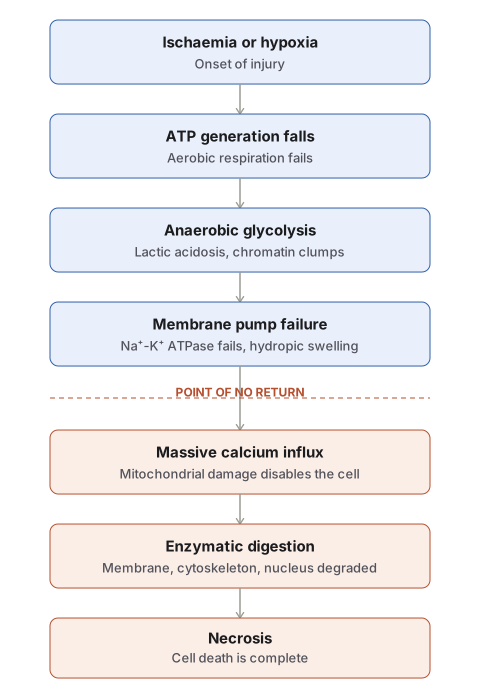
Reversible Cell Injury
When ischaemia or hypoxia is brief, the cell recovers fully once circulation is restored, as in coronary occlusion where prompt reperfusion restores contractility, metabolism, and ultrastructure. The sequence of changes is as follows.
- Fall in cellular ATP. Cells generate ATP by aerobic oxidative phosphorylation in the mitochondria, and can shift to anaerobic glycolysis when oxygen is scarce. In true ischaemia, both oxygen and the glucose needed for glycolysis are cut off together, so injury develops faster and more severely than in hypoxia arising from other causes, where glycolysis can continue for a time.
- Intracellular lactic acidosis. Once aerobic respiration fails, the cell turns to anaerobic glycolysis, rapidly depleting glycogen and accumulating lactic acid. The resulting fall in intracellular pH causes clumping of nuclear chromatin.
- Damage to membrane ion pumps. Falling ATP impairs the phospholipid synthesis needed for continuous membrane repair, disabling the pumps that regulate sodium, potassium, and calcium.
- Failure of the Na⁺-K⁺ ATPase allows sodium to accumulate within the cell and potassium to leak out; the resulting osmotic shift draws water into the cell, producing hydropic (cloudy) swelling.
- Failure of calcium regulation allows calcium to enter the cell, particularly into the mitochondria, causing mitochondrial swelling and deposition of amorphous phospholipid-rich densities.
- Reduced protein synthesis. Membranes of the endoplasmic reticulum and Golgi apparatus swell, and ribosomes detach from the rough endoplasmic reticulum, breaking down into inactive monosomes and reducing protein synthesis.
Up to this point, removal of the injurious stimulus restores the cell to normal.
Irreversible Cell Injury
Persistent ischaemia or hypoxia eventually crosses into irreversibility. Two features distinguish irreversible from reversible injury: the cell can no longer reverse mitochondrial dysfunction even after reperfusion, and disturbance of plasma membrane function persists. Beyond these, there is further ATP depletion, continued protein loss, a further fall in intracellular pH, and leakage of lysosomal enzymes into the plasma.
- Calcium influx and mitochondrial damage. A large influx of calcium into the cytosol — most marked after reperfusion of an already irreversibly injured cell — collects in the mitochondria and disables their function. Vacuoles and amorphous calcium deposits appear within the mitochondrial matrix, now larger and more numerous than in reversible injury.
- Activated phospholipases and membrane damage. Rising cytosolic calcium activates endogenous phospholipases, which progressively degrade membrane phospholipids, the main constituent of the lipid bilayer. Reduced ATP simultaneously slows replacement synthesis of these phospholipids, and an activated ATPase adds to the loss of ATP.
- Activated proteases and cytoskeletal damage. The cytoskeleton — microfilaments, microtubules, and intermediate filaments anchoring the cell membrane — is degraded by activated intracellular proteases and by the physical stress of cell swelling, contributing further to irreversible membrane injury.
- Activated endonucleases and nuclear damage. DNA and nucleoproteins are attacked by activated lysosomal proteases and endonucleases, producing pyknosis, karyorrhexis, and karyolysis. DNA damage of this kind also activates proapoptotic proteins, so a severely injured cell may be driven toward apoptosis as well as necrosis.
- Lysosomal enzyme release. Ruptured lysosomal membranes release hydrolytic enzymes — hydrolase, RNase, DNase, protease, glycosidase, phosphatase, lipase, amylase, and cathepsin — that digest the cell’s own components. The dead cell is eventually replaced by masses of phospholipid known as myelin figures, either phagocytosed by macrophages or converted into calcium soaps. These same enzymes leak across the damaged membrane into the serum, which is the mechanistic basis for using their levels clinically to detect cell death — troponin, creatine kinase-MB, lactate dehydrogenase, and aspartate/alanine aminotransferases after myocardial infarction, and lipase/amylase after pancreatic injury.
Ischaemia-Reperfusion Injury
Restoring blood flow after ischaemia has three possible outcomes depending on how long the ischaemia lasted:
- Short ischaemia → reversible injury. Reperfusion fully restores structure and function.
- Very prolonged ischaemia → injury already irreversible. Cell death occurred during the ischaemic period itself; reperfusion neither helps nor triggers additional injury. Death here is not attributable to reactive oxygen species but to persistent membrane damage with further sodium and calcium accumulation.
- Intermediate-duration ischaemia → true reperfusion injury. Cells still viable at the point of reperfusion paradoxically sustain additional injury from the restored blood flow itself, chiefly through generation of reactive oxygen species — meaning reperfusion, while necessary to rescue reversibly injured cells, can itself add to the injury burden in a subset of borderline cells.
Calcium as a Common Final Pathway
Cytosolic calcium is normally kept very low, most of it sequestered in the mitochondria and endoplasmic reticulum. Ischaemia and certain toxins raise cytosolic calcium, initially by release from these intracellular stores and later by influx across the damaged cell membrane. This rise in calcium activates a set of damaging enzymes — phospholipases, endonucleases, and proteases — as well as caspases, so that severe calcium overload can push a cell toward apoptosis as well as toward the necrotic pathway described above. Membrane damage in cell injury therefore arises through several converging mechanisms: indirect damage from free radical–mediated lipid peroxidation, reduced phospholipid synthesis secondary to ATP depletion, calcium-activated phospholipase degradation of existing phospholipid, and calcium-activated protease damage to the cytoskeleton, together with direct damage from bacterial toxins, viral proteins, or physical and chemical agents acting on the membrane itself. Damage to the mitochondrial membrane opens the mitochondrial permeability transition pore, further lowering ATP and releasing proteins that trigger apoptosis; damage to the plasma membrane leads to loss of the ionic and metabolic gradients the cell depends on for survival.
Morphology
Gross
Organs affected by widespread reversible injury (kidney, liver, pancreas, cardiac muscle) appear enlarged, with a bulging, slightly opaque cut surface — the visible correlate of cellular swelling, which is frequently easier to appreciate on gross examination than under the light microscope, since intracellular fluid is largely extracted during tissue processing.
Light Microscopy
The older term “degeneration” has been replaced by “reversible cell injury” or “retrogressive change,” since it describes non-lethal injury without implying anything about the underlying mechanism. Recognised light-microscopic forms:
| Form | Feature |
|---|---|
| Hydropic change (cloudy swelling, vacuolar degeneration) | The earliest and commonest form of cell injury from almost any cause. Tubular epithelial cells, classically in the kidney, show small clear cytoplasmic vacuoles (distended endoplasmic reticulum cisternae), pale nuclei, and compressed interstitial capillaries. |
| Hyaline change | Glassy, homogeneous, eosinophilic material, intracellular or extracellular, seen in varied settings — hyaline droplets in proteinuria, Zenker’s degeneration of muscle in typhoid fever, Mallory’s hyaline in alcoholic liver injury, Russell bodies in plasma cells, hyalinised arterioles in hypertension and diabetes mellitus. |
| Mucoid (myxoid) change | Deposition of mucin-like material — epithelial mucin in catarrhal inflammation, mucocele formation, and mucin-secreting tumours; connective tissue (myxoid) mucin in myxomas, neurofibromas, and the aortic medial degeneration of Marfan syndrome. |
Progression toward necrosis is signalled by the cytoplasm becoming progressively more eosinophilic (redder on H&E).
Electron Microscopy (Ultrastructural)
Ultrastructural change precedes light-microscopic change and gives the clearest evidence of where an injured cell sits on the reversible-irreversible continuum:
| Structure | Reversible injury | Irreversible injury |
|---|---|---|
| Plasma membrane | Blebbing, blunting, loss of microvilli | Discontinuities in plasma and organelle membranes |
| Mitochondria | Swelling, small amorphous densities | Marked dilatation, large amorphous densities of precipitated calcium, aggregates of denatured protein |
| Endoplasmic reticulum | Dilatation, detachment of polysomes | Swelling and fragmentation |
| Myelin figures | May be present | Usually present |
| Nucleus | Disaggregation of granular and fibrillar chromatin | Pyknosis, karyorrhexis, karyolysis |
The Point of No Return
There is no single sharp biochemical marker separating reversible from irreversible injury, but two features together define the transition: the cell can no longer restore mitochondrial function even when oxygen is resupplied, and plasma membrane integrity remains permanently compromised. Everything downstream of this point — calcium overload, enzymatic digestion of membranes, the cytoskeleton, and the nucleus, followed by lysosomal enzyme release — culminates in necrosis. Recognising the reversible morphologies (hydropic, hyaline, mucoid change) on biopsy is what signals that the underlying insult is still correctable, in contrast to necrosis, which represents completed cell death.
Diagram 1: Sequence of Change in Hypoxic-Ischaemic Cell Injury
Draw a single horizontal timeline with a normal cell at the far left and a necrotic cell at the far right, with two intermediate cell stages in between, connected by arrows, as one continuous sequence.
Stage 1 — normal cell: intact organelles, smooth membrane, central round nucleus.
Stage 2 — early reversible injury: small clear cytoplasmic vacuoles (hydropic swelling), mildly swollen mitochondria, ribosomes shown detaching from the rough endoplasmic reticulum and scattering as dots in the cytoplasm, a few small membrane blebs.
Stage 3 — advanced reversible injury: more pronounced swelling, myelin figures drawn as whorled membrane loops in the cytoplasm, early chromatin clumping at the nuclear margin, mitochondria more swollen with a few amorphous densities.
Stage 4 — irreversible injury (cell death): ruptured plasma membrane, mitochondria with visible calcium deposits (small dark granules) and vacuoles, nucleus shown with the three changes in separate small insets — pyknotic, karyorrhectic, karyolytic — a disorganised cytoskeleton, and ruptured lysosomes with enzyme symbols escaping.
Labels required
- An arrow between Stage 3 and Stage 4 labelled: point of no return — irreversible mitochondrial dysfunction and persistent membrane damage.
- The relevant biochemical driver written beneath each stage — falling ATP under Stage 2, calcium influx under Stage 3–4, lysosomal enzyme release under Stage 4.
- All three nuclear changes individually labelled in Stage 4, not grouped as a single “nuclear damage” label.
Errors commonly made
- Placing hydropic swelling in the irreversible stage rather than the reversible stage, where it is the earliest change.
- Omitting the point-of-no-return label, leaving a continuous progression without stating where reversibility ends.
- Showing only one type of nuclear change in Stage 4 instead of all three.
- Omitting myelin figures, a specifically asked-for ultrastructural feature.
Diagram 2: Membrane Pump Failure
Draw a small cross-section of the cell membrane with two ion pumps side by side.
Na⁺-K⁺ ATPase: working state on the left half (arrows moving Na⁺ out and K⁺ in, an ATP symbol being consumed); failed state on the right half (pump inactive, Na⁺ accumulating inside the cell with water following it inward, cell outline bulging).
Calcium pump: same format — working state regulating Ca²⁺ efflux; failed state showing Ca²⁺ flooding in, with an arrow directing it toward a nearby mitochondrion.
Labels required
- Name both pumps explicitly: Na⁺-K⁺ ATPase and calcium pump.
- Label the water-influx arrow as producing hydropic swelling through an iso-osmotic water shift.
- Label the destination of the calcium arrow as the mitochondrial matrix.
Errors commonly made
- Showing water entering the cell without connecting it to sodium accumulation, when the iso-osmotic mechanism is the point being tested.
- Drawing calcium influx without directing it specifically into the mitochondria, which omits the link to mitochondrial damage in irreversible injury.
Short Essays (1)
Multiple Choice Questions (2)
My Sticky Notes
Personal revision notes, mnemonics and reminders.
