
Neoplasia Nomenclature Classification and Anaplasia
Definition
New growth = neoplasm/tumour = “abnormal, excessive, uncoordinated, autonomous, purposeless proliferation… even after cessation of stimulus.” Benign (slow, localised) vs malignant (=cancer, rapid, spreads, fatal). Oncology = study of tumours.
Structure
Parenchyma (tumour cells, determines behaviour) + stroma (CT+vessels, support). Monoclonal origin (even mixed tumours = single clone, divergent differentiation).
Nomenclature
-oma = benign. Carcinoma = malignant EPITHELIAL (any germ layer) — adenocarcinoma(glandular)/SCC(squamous). Sarcoma = malignant MESENCHYMAL (named by cell type).
Exceptions (memorise): melanoma, hepatoma, lymphoma, seminoma, mesothelioma = ALL MALIGNANT despite no carcinoma/sarcoma suffix. Leukaemia = blood cell cancer.
Special categories
- Mixed tumours — adenosquamous Ca, adenoacanthoma, carcinosarcoma, collision tumour, pleomorphic adenoma (benign, salivary)
- Teratoma — totipotent cells, all 3 germ layers, gonadal>extragonadal(midline). Benign/mature(ovarian) vs malignant/immature(testicular)
- Blastoma — embryonal cells, children <5yo: neuroblastoma, Wilms/nephroblastoma, hepatoblastoma, retinoblastoma, medulloblastoma, pulmonary blastoma
- Hamartoma — disorganised MATURE native tissue (e.g. lung: cartilage+muscle+epithelium). Modern view: clonal (acquired somatic mutation) = unusual BENIGN neoplasm, not just malformation
- Choristoma — ectopic NORMAL tissue island (heterotopia). NOT a true tumour despite “-oma”
Classification table (examples)
| Origin | Benign | Malignant |
|---|---|---|
| Squamous epi | Papilloma | SCC |
| Glandular epi | Adenoma | Adenocarcinoma |
| Melanocyte | Naevus | Melanoma |
| Hepatocyte | Adenoma | Hepatoma |
| Fat | Lipoma | Liposarcoma |
| Cartilage | Chondroma | Chondrosarcoma |
| Bone | Osteoma | Osteosarcoma |
| Smooth muscle | Leiomyoma | Leiomyosarcoma |
| Blood vessel | Haemangioma | Angiosarcoma |
| Haematopoietic | — | Leukaemia |
| Lymphoid | — | Lymphoma |
Benign vs malignant
| Benign | Malignant | |
|---|---|---|
| Boundary | Encapsulated | Poorly circumscribed |
| Surrounding tissue | Compressed | Invaded |
| Growth | Slow | Rapid |
| Metastasis | Absent | Frequent |
Growth rate depends on: (1) cell production vs loss balance (malignant = shortened doubling time + evades death, growth=production>loss) (2) degree of differentiation (poorly differentiated=aggressive). Rare spontaneous regression→later mets: choriocarcinoma, melanoma.
Differentiation and anaplasia
Differentiation = resemblance to normal cell. Anaplasia (“backward formation”) = lack of differentiation = malignancy indicator, degree correlates with grade.
Anaplasia features (10 — HIGH YIELD)
- Loss of polarity — nuclei leave basal position (selectin/CAM-dependent normally)
- Pleomorphism — variable size/shape
- ↑N:C ratio — normal 1:5 → 1:1
- Anisonucleosis — nuclear size/shape variation
- Hyperchromatism — coarse clumped chromatin, dark nuclei
- Nucleolar changes — prominent nucleoli (AgNOR silver stain)
- Mitotic figures — ↑number; ABNORMAL (tripolar/quadripolar/multipolar) = more significant than normal mitoses seen in benign/proliferating tissue
- Tumour giant cells — multinucleate or single bizarre nucleus
- Functional changes — quantitative (product amount ↑↓) vs qualitative (product WRONG, e.g. myeloma abnormal Ig) vs both. Ectopic hormone production = tumour secretes hormone unrelated to its tissue (oat cell lung Ca→ACTH/ADH; RCC/HCC/cerebellar haemangioblastoma→erythropoietin)
- Chromosomal abnormalities — DNA aneuploidy (usually ↑number→↑nuclear size). Philadelphia chromosome t(9;22) = 95% CML. Also: Burkitt lymphoma, ALL, myeloma, retinoblastoma, small cell lung Ca, Wilms tumour
Angiogenesis and stroma
Angiogenesis (VEGF-driven) feeds growth; microvascular density = grade marker; outgrowing supply → central necrosis. Stroma: scanty→soft/fleshy (sarcoma, lymphoma); abundant/desmoplastic (bFGF-driven)→hard/scirrhous (infiltrating duct Ca breast, linitis plastica). Nearly-pure parenchyma = “medullary” (breast/thyroid).
Inflammatory reaction (host immune response, sometimes improves prognosis): seminoma, melanoma, lymphoepithelioma, medullary breast Ca, choriocarcinoma, Warthin tumour.
Clinical relevance
Nomenclature exceptions must be memorised as a set — “melanoma” sounds benign by suffix but is always malignant. Abnormal (not just increased) mitoses = pathology report emphasis. Philadelphia chromosome = diagnostic AND therapeutic target (imatinib, BCR-ABL). Ectopic hormone production ≠ metastatic spread — distinct mechanism, relevant to Paraneoplastic Syndromes.
Definition
Neoplasia means “new growth,” and the new growth produced is a neoplasm or tumour — but not every new growth of tissue is neoplastic (embryogenesis, regeneration, hyperplasia and hormonal stimulation are all controlled proliferation, not neoplasia). A satisfactory definition: a neoplasm is “a mass of tissue formed as a result of abnormal, excessive, uncoordinated, autonomous and purposeless proliferation of cells, even after cessation of the stimulus that caused it.” This autonomy — persistence of growth beyond the original stimulus — is the defining biological feature. Oncology is the study of neoplasms. Neoplasms are benign (slow-growing, localised, rarely troublesome) or malignant (rapidly proliferative, capable of spread, potentially fatal) — the collective term for malignant tumours is cancer, from the Greek karkinos (coined by Hippocrates for breast cancer — “crab,” reflecting how the tumour “sticks to the part stubbornly like a crab”).
Basic Structure
Every tumour, benign or malignant, has two components: the parenchyma (proliferating tumour cells, which determines the tumour’s nature and behaviour) and the supportive stroma (connective tissue and blood vessels providing the structural/nutritive framework). Evidence indicates cancer cells originate by clonal proliferation from a single transformed progenitor cell (monoclonality) — even in “mixed” tumours, the clonal progenitor simply retains capacity to differentiate along more than one lineage.
Nomenclature
Tumours are named for their parenchymal component. The suffix “-oma” denotes a benign tumour. Malignant tumours of epithelial origin are carcinomas (subdivided by pattern — adenocarcinoma if glandular, squamous cell carcinoma if squamous — regardless of which germ layer the epithelium derived from: renal tubular, cutaneous and gut-lining carcinomas are all “carcinomas” despite mesodermal/ectodermal/endodermal origin respectively); malignant tumours of mesenchymal origin are sarcomas (named by principal cell type — liposarcoma, chondrosarcoma). Highly undifferentiated malignant tumours are simply termed undifferentiated.
Firmly entrenched exceptions to this logic (worth memorising as exceptions, not deriving): melanoma (malignant melanocyte tumour), hepatoma (hepatocellular carcinoma), lymphoma (malignant lymphoid tumour), seminoma (malignant testicular tumour), mesothelioma — all malignant despite lacking “carcinoma/sarcoma.” Leukaemia denotes cancer of blood-forming cells.
Special Categories of Tumours
- Mixed tumours — combine two tumour types: adenosquamous carcinoma (adeno + squamous, endometrium), adenoacanthoma (adenocarcinoma + benign squamous elements, endometrium), carcinosarcoma (carcinoma + sarcoma, e.g. thyroid), collision tumour (two distinct, non-mixing cancers in one organ), and pleomorphic adenoma/mixed salivary tumour (benign, epithelial + mesenchymal elements together).
- Teratomas — arise from totipotent cells of all three germ layers (ectoderm, mesoderm, endoderm), chiefly gonadal (ovary, testis) but also midline extragonadal sites (head/neck, mediastinum, retroperitoneum, sacrococcygeal region); may be benign/mature (most ovarian) or malignant/immature (most testicular).
- Blastomas (embryomas) — malignant tumours from embryonal/blastemal cells, characteristically in infants/young children (<5 years): neuroblastoma, nephroblastoma (Wilms tumour), hepatoblastoma, retinoblastoma, medulloblastoma, pulmonary blastoma.
- Hamartoma — a mass of mature but disorganised tissue native to the organ (e.g. pulmonary hamartoma: mature cartilage, smooth muscle and epithelium jumbled together). Traditionally regarded as a developmental malformation rather than a true neoplasm, but modern evidence of clonal chromosomal aberrations from acquired somatic mutation means hamartomas are now better considered unusual benign neoplasms.
- Choristoma — an ectopic island of otherwise normal tissue (heterotopia); despite the “-oma” suffix implying neoplasia, it is not a true tumour.
Classification
Tumours are classified by histogenesis (cell of origin) and anticipated behaviour (benign/malignant), summarised for the major tissue categories:
| Tissue of origin | Benign | Malignant |
|---|---|---|
| Squamous epithelium | Squamous papilloma | Squamous cell carcinoma |
| Glandular epithelium | Adenoma | Adenocarcinoma |
| Transitional epithelium | Transitional cell papilloma | Transitional cell carcinoma |
| Basal cell layer | — | Basal cell carcinoma |
| Neuroectoderm (melanocyte) | Naevus | Melanoma |
| Hepatocytes | Liver cell adenoma | Hepatoma |
| Adipose tissue | Lipoma | Liposarcoma |
| Fibrous tissue | Fibroma | Fibrosarcoma |
| Cartilage | Chondroma | Chondrosarcoma |
| Bone | Osteoma | Osteosarcoma |
| Smooth muscle | Leiomyoma | Leiomyosarcoma |
| Skeletal muscle | Rhabdomyoma | Rhabdomyosarcoma |
| Blood vessels | Haemangioma | Angiosarcoma |
| Mesothelium | — | Mesothelioma |
| Meninges | Meningioma | Invasive meningioma |
| Haematopoietic cells | — | Leukaemia |
| Lymphoid tissue | — | Malignant lymphoma |
| Nerve sheath | Neurilemmoma, neurofibroma | Neurogenic sarcoma |
| Totipotent germ cells | Mature teratoma | Immature teratoma |
Characteristics: Benign vs Malignant
| Feature | Benign | Malignant |
|---|---|---|
| Boundaries | Encapsulated/well-circumscribed | Poorly circumscribed, irregular |
| Surrounding tissue | Compressed | Invaded |
| Size | Usually small | Often larger |
| Growth rate | Usually slow | Usually rapid |
| Local invasion | Compresses without infiltrating | Infiltrates/invades |
| Metastasis | Absent | Frequently present |
| Prognosis | Local complications | Death by local/metastatic complications |
Rate of growth depends on two factors: (1) the balance of cell production, growth fraction (proportion of cells actively in the proliferative pool) and cell loss — malignant cells have a shortened doubling time and evade normal death controls (relative immortality), so tumour growth reflects production exceeding loss (loss occurring via apoptosis or shedding when nutrient supply cannot keep pace); (2) degree of differentiation — poorly differentiated tumours grow more aggressively than well-differentiated ones, and some tumours acquire a sudden aggressive growth spurt from emergence of a more malignant subclone. Rarely, a malignant tumour (choriocarcinoma, melanoma) can spontaneously regress at its primary site from immune-mediated necrosis, only to recur as metastases.
Differentiation and Anaplasia
Differentiation is the extent of morphological/functional resemblance between tumour parenchymal cells and their normal counterpart; minimal deviation = well-differentiated (most benign, low-grade malignant tumours); marked deviation = poorly differentiated/undifferentiated/dedifferentiated. Anaplasia (literally “backward formation”) is lack of differentiation — a reliable indicator of malignancy — and its degree correlates with the tumour’s grade of malignancy.
The cytomorphologic features of anaplasia, best seen at high magnification:
- Loss of polarity — normal epithelial nuclei align along the basement membrane (basal polarity, dependent on cell adhesion molecules including selectins); anaplastic cells lose this orientation.
- Pleomorphism — variation in cell size and shape, correlating with degree of anaplasia; cells are often larger than normal but occasionally smaller.
- Increased nucleocytoplasmic (N:C) ratio — disproportionate nuclear enlargement shifts the normal ~1:5 ratio toward 1:1.
- Anisonucleosis — variation in nuclear size and shape, paralleling cellular pleomorphism.
- Hyperchromatism — increased, coarsely clumped nuclear chromatin (increased nucleoprotein) producing dark-staining nuclei, with irregular nuclear membranes and chromatin clumped peripherally.
- Nucleolar changes — prominent nucleoli reflecting increased nucleoprotein synthesis, demonstrable as AgNOR material by silver staining of the nucleolar organiser region.
- Mitotic figures — increased numbers in poorly differentiated tumours; normal mitotic figures also occur in proliferating non-neoplastic tissue (marrow, gut epithelium) and low-grade tumours, whereas abnormal/atypical mitotic figures (tripolar, quadripolar, multipolar spindles) are the more significant, malignancy-associated finding.
- Tumour giant cells — multinucleate cells, or cells with a single large bizarre nucleus sharing the surrounding tumour cells’ nuclear characteristics.
- Functional (cytoplasmic) changes — quantitative or qualitative. Better-differentiated tumours usually function qualitatively normally with only quantitative product change (variable collagen from fibrous tumours, keratin in well-differentiated squamous carcinoma); more anaplastic tumours typically show quantitative loss of product (absent keratin in anaplastic squamous carcinoma). Some tumours show both abnormalities (multiple myeloma producing abnormal immunoglobulin in excess). Endocrine tumours may over-produce their hormone; ectopic hormone production — hormone secretion by a tumour unrelated to any endocrine gland — is a distinct phenomenon (e.g. oat cell/small cell lung carcinoma secreting ACTH or ADH, occasionally gonadotropin, thyrotropin, parathormone, calcitonin, growth hormone; ectopic erythropoietin from renal cell carcinoma, hepatocellular carcinoma, cerebellar haemangioblastoma — see Paraneoplastic Syndromes).
- Chromosomal abnormalities — more marked in more malignant tumours, both numerical (DNA aneuploidy, usually increased chromosome number, reflected in enlarged nuclei) and structural. The classical example is the Philadelphia chromosome — t(9;22) — present in 95% of chronic myeloid leukaemia; other tumours with characteristic chromosomal abnormalities include Burkitt lymphoma, acute lymphoblastic leukaemia, multiple myeloma, retinoblastoma, small cell lung carcinoma, and Wilms tumour.
Tumour Angiogenesis and Stroma
New vessel formation from pre-existing vasculature (angiogenesis, driven by tumour-elaborated VEGF and other factors) supplies growing tumour tissue; microvascular density is used as a marker of growth rate/grade, while tumours that outgrow their blood supply (or fail to induce adequate angiogenesis) develop central ischaemic necrosis. Stromal collagen content varies: scanty stroma produces soft, fleshy tumours (sarcomas, lymphomas); abundant stroma (desmoplasia, driven by tumour-elaborated basic FGF) produces hard, gritty (“scirrhous”) tumours such as infiltrating ductal breast carcinoma or linitis plastica of the stomach. A tumour almost entirely parenchymal (little stroma) is termed medullary (medullary carcinoma of breast/thyroid).
Inflammatory reaction in/around a tumour may reflect secondary infection at an ulcerated surface, or — in the absence of ulceration — a host cell-mediated immune response against the tumour (lymphocytes, plasma cells, macrophages, occasionally granulomatous), sometimes associated with improved prognosis: seen in seminoma, malignant melanoma, lymphoepithelioma of the throat, medullary breast carcinoma, choriocarcinoma, Warthin tumour.
Clinical Relevance
- The nomenclature exceptions (melanoma, hepatoma, lymphoma, seminoma, mesothelioma) are worth deliberately memorising as a set, since applying the “-oma = benign” rule to any of them is a classic student error with real clinical stakes — mistaking “melanoma” for a benign lesion because of its suffix would be a dangerous misreading.
- Recognising abnormal (tripolar/multipolar) mitotic figures as more diagnostically significant than mere mitotic count is the reason a pathology report specifically comments on atypical mitoses, not simply “increased mitotic activity.”
- The Philadelphia chromosome’s near-universal presence in CML (95%) makes it both a diagnostic marker and, via the resulting BCR-ABL fusion protein, the direct target of imatinib and related tyrosine kinase inhibitor therapy — a rare case where a classical cytogenetic finding translates directly into targeted treatment.
- Recognising ectopic hormone production (e.g. ACTH/ADH from small cell lung carcinoma) as a tumour-related but non-metastatic phenomenon is essential to avoid misattributing new hormonal symptoms in a cancer patient purely to disease spread — see Paraneoplastic Syndromes for the full clinical picture.
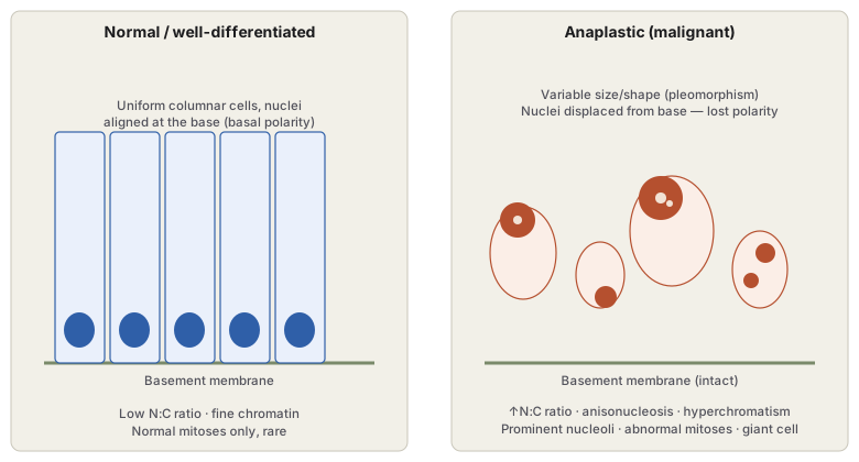
Diagram 1: Cytomorphology of Anaplasia vs Normal Epithelium
Draw two side-by-side panels over a common basement membrane baseline.
- Left panel (normal/well-differentiated): a row of uniform columnar cells, each with a small, uniformly sized nucleus aligned at the base of the cell (basal polarity), low N:C ratio, fine chromatin.
- Right panel (anaplastic): cells of markedly varying size and shape (pleomorphism), with nuclei displaced away from the base (loss of polarity), showing variable size (anisonucleosis), dark clumped chromatin (hyperchromatism), and one or more visible nucleoli; include at least one cell with two nuclei to represent a tumour giant cell.
Labels required
- Each of the ten anaplasia features named in the notes should be attributable to a specific visual element in the right panel (e.g. the double-nucleus cell = tumour giant cell; the variably sized dark nuclei = anisonucleosis + hyperchromatism), not left as a disconnected list beside a generic “abnormal cells” drawing.
- The intact basement membrane must be shown under both panels, since loss of polarity is specifically about nuclear position relative to an intact basement membrane — invasion through the membrane is a separate, later event (see Invasion and Metastasis), not part of anaplasia itself.
Errors commonly made
- Drawing the anaplastic panel with cells breaching the basement membrane, conflating anaplasia (a cytological feature) with invasion (a behavioural/architectural feature) — a tumour can be cytologically anaplastic (e.g. carcinoma in situ) while the basement membrane remains completely intact.
- Showing uniform enlargement of all anaplastic nuclei rather than variable enlargement — anisonucleosis specifically requires nucleus-to-nucleus variation, not just generalised nuclear enlargement.
Long Essays (1)
Multiple Choice Questions (2)
My Sticky Notes
Personal revision notes, mnemonics and reminders.
