
Molecular Basis of Cancer
General concepts
Monoclonality (single transformed cell — myeloma Ig spike, leiomyoma G6PD isotype) · field effect (only some cells in exposed organ progress) · multistep (multi-hit, many generations) · genetic theory (inherited or induced mutation, transmitted to progeny).
4 classes of regulatory genes
- Proto-oncogenes — growth-promoting
- Anti-oncogenes — growth-suppressing
- Apoptosis regulatory genes — control programmed death
- DNA repair genes — fix mitotic DNA damage (incl. damage to genes 1-3)
Cancer = oncogene activation (dominant, one allele) + tumour suppressor gene inactivation (recessive, both alleles) + abnormal apoptosis-regulating gene (either allele) + DNA repair gene failure.
Oncogenes (mutant proto-oncogene; product=oncoprotein)
3 activation mechanisms: point mutation (RAS — 1/3 of ALL tumours, colon/lung/pancreas, GTPase-resistant) · translocation (Philadelphia t(9;22) ABL-BCR=95% CML; t(8;14) MYC=75% Burkitt) · amplification (N-MYC=neuroblastoma; HER2=breast Ca).
5 categories:
| Category | Gene | Tumour |
|---|---|---|
| Growth factors | SIS(PDGF-β) | Glioma/sarcoma |
| GF receptors | ERBB1/HER1(EGFR) | Lung SCC 80%, glioblastoma |
| ERBB2/HER2 | Breast 25%, ovary, stomach, lung | |
| c-KIT | GIST | |
| RET | MEN2A/2B, medullary thyroid Ca | |
| Cytoplasmic signal | RAS | 1/3 tumours |
| ABL-BCR | CML | |
| Nuclear TF | C-MYC | Burkitt |
| N-MYC | Neuroblastoma, SCLC | |
| Cell cycle | Cyclin D | Mantle cell lymphoma |
| CDK4 | Melanoma, glioblastoma, sarcoma |
Anti-oncogenes (tumour suppressors, RECESSIVE — both alleles needed)
- RB (13q) — FIRST TSG identified. pRB blocks G1→S via E2F binding; hyperphosphorylation (by cyclin-CDK) inactivates it. Retinoblastoma + osteosarcoma.
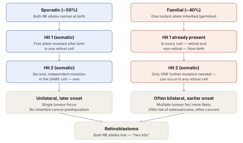
- Knudson’s 2-hit hypothesis: Sporadic (50%, unilateral) = 2 somatic hits same cell, rare, later onset. Familial (40%, often bilateral) = 1 germline hit (every cell) + 1 somatic hit anywhere = earlier, + 200x risk of 2nd cancers (osteosarcoma, breast, colon, lung).
- p53/TP53 (17p) — “guardian of genome.” Low baseline, ↑after DNA damage. Functions: (a) G1 arrest for repair (b) apoptosis via BAX (works with RB). >70% of cancers = homozygous p53 loss (lung, H&N, colon, breast). Germline mutation = Li-Fraumeni syndrome (breast, bone, brain, sarcoma).
- TGF-β + receptor — activates CDKIs, suppresses MYC/CDK/cyclin. Mutated: pancreas, colon, stomach, endometrium.
- APC/β-catenin — APC normally degrades β-catenin. APC loss → β-catenin accumulates → unchecked mitosis. Colon cancer. Germline APC mutation = FAP (polyps early, malignant transformation after 20yo when 2nd allele lost).
- BRCA1(17q21)/BRCA2(13q12-13) — breast Ca susceptibility. BRCA1: 85% breast Ca risk, 40% ovarian. Inherited breast Ca = 5-10%, younger, bilateral.
- VHL(3p) — VHL disease, 60% of RCC inactivated.
- WT1/WT2(chr11) — hereditary Wilms.
- NF1/NF2 — neurofibromatosis type 1/2.
Apoptosis evasion
CD95 death receptor + pro-apoptotic (BAD, BAX, BID, p53) vs anti-apoptotic (BCL2, BCL-X).
- BCL2 — t(14;18), first in B-cell lymphoma, also breast/thyroid/prostate. Removes apoptotic brake. MYC(proliferate)+BCL2(no death)=powerful combo.
- Mutant p53 → no BAX activation → ↓apoptosis
- CD95 depletion → hepatocellular carcinoma escapes apoptosis
Telomerase (avoiding senescence)
Normal cells: telomere shortening each division → senescence after 60-70 divisions. Telomerase active in stem cells only (normally). Cancer: telomerase UPREGULATED → immortalisation.
Angiogenesis
Pro: VEGF, bFGF. Anti: thrombospondin-1, angiostatin, endostatin, vasculostatin. p53 loss → ↓thrombospondin-1 → unchecked angiogenesis.
Clinical relevance
2-hit hypothesis explains hereditary cancer syndromes’ earlier/bilateral/multifocal presentation (1 hit already done). Oncogene→drug mapping: BCR-ABL→imatinib, HER2→trastuzumab, EGFR→gefitinib/erlotinib, c-KIT→imatinib(GIST) — molecular Dx guides Rx directly. p53 = single most important TSG (frequency + pathway convergence). Telomerase = near-universal cancer target, active drug development area.
General Concepts
Several converging lines of evidence underpin the molecular understanding of cancer: monoclonality — most human cancers arise from a single transformed cell (demonstrated by, e.g., the single immunoglobulin spike in multiple myeloma, or the single G6PD isoenzyme type found throughout a uterine leiomyoma despite the surrounding normal myometrium being a mosaic of both types); the field effect — within an organ, only a limited subset of cells under carcinogenic influence actually progress to cancer; the multistep nature of carcinogenesis — a gradual, multi-hit process across many cell generations, producing progressively transformed, phenotypically malignant cells; and the genetic theory of cancer — abnormal cell growth reflects either genetic damage or normal genes with abnormal expression, arising from inherited or induced (chemical, viral, radiation) mutation, transmitted to progeny.
Four Classes of Regulatory Genes
Normal cell growth is governed by four classes of genes, and cancer results from damage to one or more of them:
- Proto-oncogenes — growth-promoting genes encoding the cell proliferation pathway.
- Anti-oncogenes (tumour suppressor genes) — growth-inhibiting/suppressing genes.
- Apoptosis regulatory genes — control programmed cell death.
- DNA repair genes — regulate repair of DNA damage occurring during mitosis, including damage to the other three gene classes.
Corresponding abnormalities producing cancer: activation of proto-oncogenes to oncogenes (dominant — active despite normal alleles being present); inactivation of anti-oncogenes (recessive — both alleles must be lost/damaged for effect); abnormal apoptosis regulatory genes (may behave dominantly or recessively); and failure of DNA repair genes, permitting mutations to accumulate unchecked.
Molecular Hallmarks of Cancer
The genetic properties of cancer cells, corresponding to these gene classes and their downstream effects:
1. Excessive Autonomous Growth — Growth-Promoting Oncogenes
A mutant proto-oncogene is an oncogene; its product is an oncoprotein. Proto-oncogene → oncogene conversion occurs by three mechanisms:
- Point mutation — single base alteration; the classic example is RAS, mutated in about a third of all human tumours (colon, lung, pancreatic cancer) — normal RAS is inactivated by its own GTPase activity after signalling, but mutant RAS is GTPase-resistant and signals continuously.
- Chromosomal translocation — a proto-oncogene moves to a new chromosomal location, escaping normal growth control: the Philadelphia chromosome t(9;22) fuses c-ABL (chromosome 9) with BCR (chromosome 22), seen in 95% of CML; t(8;14) translocates c-MYC in 75% of Burkitt lymphoma.
- Gene amplification — increased copy number, raising oncoprotein output: N-MYC amplification in neuroblastoma; ERBB2/HER2 amplification in breast cancer.
Oncogenes are classified by which component of the growth-signalling cascade they encode:
| Class | Example gene(s) | Mechanism | Associated tumour |
|---|---|---|---|
| Growth factors | SIS (PDGF-β) | Overexpression | Glioma, sarcoma |
| HST-1/INT-2 (FGF) | Overexpression/amplification | Bowel, breast cancer | |
| GF receptors | ERBB1/HER1 (EGFR) | Overexpression | Lung SCC (80%), glioblastoma |
| ERBB2/HER2/neu | Amplification | Breast (25%), ovarian, gastric, lung cancer | |
| c-KIT | Point mutation | GIST | |
| RET | Point mutation | MEN 2A/2B, medullary thyroid carcinoma | |
| Cytoplasmic signal transduction | RAS | Point mutation | ~1/3 of all tumours; colon, lung, pancreas |
| ABL-BCR | Translocation | CML, some acute leukaemias | |
| Nuclear transcription factors | C-MYC | Translocation t(8;14) | Burkitt lymphoma |
| N-MYC | Amplification | Neuroblastoma, small cell lung Ca | |
| Cell cycle regulators | Cyclin D | Translocation | Mantle cell lymphoma |
| CDK4 | Amplification | Melanoma, glioblastoma, sarcoma |
2. Refractoriness to Growth Inhibition — Anti-Oncogenes
Mutated anti-oncogenes remove the normal “brake” on proliferation, behaving functionally like oncogenes — but require loss/damage of both alleles to manifest (recessive), typically via deletion, point mutation, or loss of a chromosomal segment.
- RB gene (13q) — the first tumour suppressor gene identified. Its product, pRB, blocks G1→S transition by binding transcription factor E2F; hyperphosphorylation by cyclin-CDK complexes inactivates pRB, releasing the brake. Mutant RB underlies retinoblastoma (the most common childhood intraocular tumour) and osteosarcoma. This is the basis of Knudson’s two-hit hypothesis: sporadic retinoblastoma (~50% of cases, unilateral) requires two independent somatic mutations in the same retinal cell after birth; familial retinoblastoma (~40%, often bilateral) involves one germline-inherited mutant allele present in every cell from birth, so only a single additional somatic hit is needed — explaining both its earlier onset and its association with a markedly increased (200-fold) risk of later second cancers (osteosarcoma, breast, colon, lung).
- p53 (TP53) (17p) — the “guardian/protector of the genome.” Normally present at low levels, accumulating after DNA damage; acts by (a) transiently arresting the cycle at G1 (via CDK/cyclin inhibition) to allow DNA repair, and (b) directing cells with irreparable damage to apoptosis (via BAX activation), working alongside RB. Homozygous p53 loss occurs in >70% of human cancers (lung, head/neck, colon, breast among the commonest), and is seen across the hyperplasia→carcinoma-in-situ→invasive-carcinoma sequence. Germline p53 mutation (one inherited defective allele, second hit acquired) causes Li-Fraumeni syndrome — multi-organ cancer susceptibility (breast, bone, brain, sarcoma).
- TGF-β and its receptor — normally inhibits proliferation (activates CDK inhibitors, suppresses MYC/cyclins/CDKs); mutated in pancreatic, colon, gastric, endometrial cancer.
- APC gene / β-catenin — APC normally promotes β-catenin degradation, restraining its dual role (E-cadherin binding, proliferation signalling); APC loss (colon cancer) allows β-catenin accumulation and unchecked mitotic signalling. Germline APC mutation causes early florid colonic polyposis, with near-universal malignant transformation once the second allele is lost after age 20 (familial adenomatous polyposis).
- Other named anti-oncogenes: BRCA1 (17q21) and BRCA2 (13q12-13) — breast cancer susceptibility genes; BRCA1 germline mutation confers ~85% lifetime breast cancer risk and ~40% ovarian cancer risk, with inherited breast cancer (5–10% of cases) presenting younger and more often bilaterally. VHL (3p) — von Hippel-Lindau disease, multi-organ benign/malignant tumours, inactivated in 60% of renal cell carcinoma. WT1/WT2 (chromosome 11) — hereditary Wilms tumour. NF1/NF2 — neurofibromatosis type 1 and 2.
3. Escaping Apoptosis
Normal apoptosis is triggered via death receptor CD95, pro-apoptotic factors (BAD, BAX, BID, p53) and restrained by anti-apoptotic factors (BCL2, BCL-X). Cancer-relevant mutations: BCL2 — first described with t(14;18) in B-cell lymphoma, also seen in breast/thyroid/prostate cancer — removes apoptotic restraint on damaged cells (MYC drives proliferation while BCL2 blocks death, a particularly effective combination for tumour cells); mutant/absent p53 fails to activate BAX, reducing apoptosis; CD95 receptor depletion in hepatocellular carcinoma allows apoptosis evasion.
4. Avoiding Cellular Ageing — Telomerase
Normal somatic cells progressively shorten their telomeres with each division, ceasing mitosis after 60–70 divisions (replicative senescence); telomerase (which maintains telomere length) is active in stem cells but not normal somatic cells. Most cancers markedly upregulate telomerase, preserving telomere length and effectively immortalising the malignant clone.
5. Continued Perfusion — Tumour Angiogenesis
Neovascularisation, essential for tumour growth beyond a small size, is driven by VEGF and basic FGF (pro-angiogenic) balanced against thrombospondin-1, angiostatin, endostatin, vasculostatin (anti-angiogenic); loss of p53 removes thrombospondin-1’s restraint, favouring unchecked angiogenesis (see Invasion and Metastasis for the full mechanics of tumour spread this supports).
Clinical Relevance
- Knudson’s two-hit hypothesis is the direct explanation for why hereditary cancer syndromes (retinoblastoma, BRCA-associated breast/ovarian cancer, Li-Fraumeni syndrome) present earlier, more often bilaterally/multifocally, and with higher lifetime risk than their sporadic counterparts — the inherited first hit means only one further somatic event, rather than two independent ones, is needed.
- The specific oncogene-to-drug mapping in current practice — BCR-ABL and imatinib in CML, HER2 and trastuzumab in breast cancer, EGFR and gefitinib/erlotinib in lung cancer, c-KIT and imatinib in GIST — makes this molecular framework directly therapeutic, not just diagnostic, and is why identifying the specific oncogenic driver in a tumour now routinely guides treatment selection.
- p53’s central, repeatedly-cited role (guardian of the genome, mutated in >70% of cancers, germline mutation causing Li-Fraumeni syndrome) makes it the single most important tumour suppressor gene to know in depth, both for its own frequent appearance in questions and because so many other pathways (apoptosis, angiogenesis, cell cycle arrest) converge on it.
- Telomerase upregulation as a near-universal cancer feature (present in the great majority of malignancies but essentially absent from normal somatic cells) has made it an active target for anticancer drug development, distinct from its diagnostic significance.
Diagram 1: Knudson’s Two-Hit Hypothesis in Retinoblastoma
Draw two parallel columns (sporadic, familial), each with three stages, converging into a shared final “retinoblastoma” box.
- Sporadic column: both alleles normal at birth → first somatic hit in one retinal cell → second, independent somatic hit in that same cell (statistically rare) → unilateral, later-onset disease, no inherited predisposition.
- Familial column: one mutant allele already present in every cell (germline) at birth → only one further somatic hit needed, in any retinal cell → often bilateral, earlier-onset disease, plus markedly increased risk of second cancers (osteosarcoma etc.).
- Convergence: both pathways end in the same outcome — both RB alleles lost in a retinal cell — but reached by a different number of required post-birth events.
Labels required
- The column each hit occurs in (“somatic” vs “germline, present from birth”) must be explicit, since the entire logic of the hypothesis rests on this distinction, not on the hits being otherwise different in nature.
- The downstream consequences (unilateral/no predisposition vs bilateral/200x second-cancer risk) must be attached to the correct column, since this is the clinically testable payoff of the genetic explanation.
Errors commonly made
- Describing familial retinoblastoma as requiring “no mutation” or being present at birth in complete form, rather than correctly requiring one further somatic hit after the inherited germline hit.
- Losing the reason sporadic disease is rarer per-eye and later-onset — because it requires two independent chance somatic events in the same cell, versus only one for familial disease — and instead treating the two forms as differing only in family history.
Long Essays (1)
Short Essays (1)
Short Answers (2)
Multiple Choice Questions (4)
My Sticky Notes
Personal revision notes, mnemonics and reminders.
