
Klinefelter Syndrome
Overview
Male hypogonadism + ≥2 X chromosomes + ≥1 Y chromosome. One of MOST COMMON causes male hypogonadism. Sex chromosome trisomies > autosomal trisomies (general rule) in frequency.
Karyotype
Most: 47,XXY — meiotic nondisjunction. Maternal+paternal contribute EQUALLY (≠ Down syndrome’s maternal-age-dominant pattern). ~15% mosaic (46,XY/47,XXY, 47,XXY/48,XXXY, variants) — substantial 46,XY line = MILDER phenotype.
Pathogenesis
Extra X → seminiferous tubule dysgenesis → hyalinisation (“ghost-like” tubules histologically); Leydig cells prominent (hyperplasia OR relative increase as tubules lost).
→ 2 parallel effects:
- Hormonal/germinal: ↓testosterone + impaired spermatogenesis, often complete azoospermia. Fertility RARE (only in mosaics with large 46,XY proportion)
- Feedback loss: ↑urinary gonadotropins (esp. FSH) — testicular atrophy removes normal pituitary-gonadal negative feedback
= Hypergonadotropic hypogonadism pattern (↓testosterone + ↑FSH) → localises defect to TESTIS itself, not pituitary/hypothalamus.
Clinical features
Variable — some ONLY hypogonadism. Most: eunuchoid habitus (↑sole-to-pubis length, elongated body), ↓facial/body/pubic hair, gynaecomastia, small testes (down to ~2cm). ↓Serum testosterone, ↑urinary FSH.
Cognition: average to below-average, modest VERBAL skill deficit specifically.
Comorbidities
- Type 2 DM + metabolic syndrome (insulin resistance)
- Congenital heart disease — mitral valve prolapse ~50% of adults
- 20-30x ↑extragonadal germ cell tumours (mostly MEDIASTINAL teratoma)
- ↑Breast cancer
- ↑Autoimmune disease (e.g. SLE)
Physical attributes VARIABLE overall — hypogonadism = only consistent finding (same teaching point as Turner syndrome).
Clinical relevance
Cascade (nondisjunction→tubule dysgenesis→parallel ↓testosterone+↑FSH→phenotype) explains hypergonadotropic hypogonadism pattern = testicular-level defect (not pituitary/hypothalamic). Equal maternal/paternal nondisjunction = contrast point vs Down’s maternal-age-dominant pattern (no strong maternal-age association here). Mediastinal teratoma 20-30x risk = specific testable fact → consider Klinefelter when young male has extragonadal germ cell tumour. Shared teaching point with Turner: “hypogonadism = only universally consistent finding” — textbook phenotype = composite picture, not fixed always-present feature set.
Overview
Klinefelter syndrome is defined by male hypogonadism in an individual carrying at least two X chromosomes and one or more Y chromosomes. It is one of the most common causes of male hypogonadism, and sex chromosome trisomies of this kind are, in general, more common than autosomal trisomies.
Karyotype
Most affected patients have a 47,XXY karyotype, arising from meiotic nondisjunction of sex chromosomes — maternal and paternal nondisjunction contribute equally to Klinefelter syndrome (a notable contrast with Down syndrome, where maternal nondisjunction dominates). Approximately 15% of patients show mosaic patterns — e.g. 46,XY/47,XXY, 47,XXY/48,XXXY, and variants — and the presence of a substantial 46,XY cell line in mosaics is usually associated with a milder clinical phenotype.
Pathogenesis
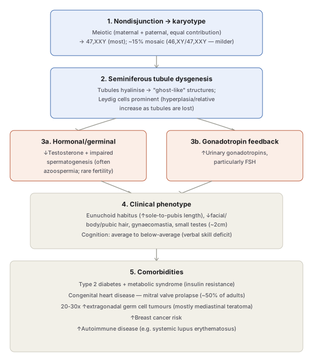
The extra X chromosome drives seminiferous tubule dysgenesis — tubules progressively undergo hyalinisation, appearing as “ghost-like” structures histologically, while Leydig cells appear prominent (from true hyperplasia, or simply relative increase as surrounding tubular tissue is lost). This produces two parallel downstream effects:
- Hormonal/germinal failure: reduced testosterone with impaired spermatogenesis, often complete azoospermia. Fertility is rare and, when present, is thought to occur only in mosaics with a large proportion of 46,XY cells.
- Loss of negative feedback: elevated urinary gonadotropins, particularly FSH — testicular atrophy removes the normal inhibitory feedback on the pituitary-gonadal axis.
Clinical Features
Variable overall — in some individuals, hypogonadism alone is noted, but most patients show a distinctive eunuchoid body habitus: increased length between the soles and the pubic bone, producing an elongated body appearance. Also frequent: reduced facial, body, and pubic hair; gynaecomastia; and markedly small testes, sometimes as little as 2 cm in greatest dimension. Consistent with testicular atrophy: low serum testosterone, elevated urinary FSH.
Cognition: ranges from average to below average, with a modest deficit specifically in verbal skills.
Comorbidities:
- Type 2 diabetes and metabolic syndrome (insulin resistance).
- Congenital heart disease, particularly mitral valve prolapse — seen in roughly 50% of adults.
- 20- to 30-fold increased risk of extragonadal germ cell tumours, mostly mediastinal teratomas.
- Increased incidence of breast cancer.
- Increased incidence of autoimmune disease (e.g. systemic lupus erythematosus).
As with Turner syndrome, the described physical attributes are quite variable — hypogonadism is the only consistently present finding.
Clinical Relevance
- The single-column pathogenetic cascade (nondisjunction → tubule dysgenesis → parallel hormonal failure + gonadotropin elevation → clinical phenotype) is the organising structure that explains why Klinefelter patients simultaneously show low testosterone AND high FSH — a pattern of hypergonadotropic hypogonadism that localises the defect to the testis itself, not the pituitary or hypothalamus.
- Recognising equal maternal/paternal contribution to nondisjunction (unlike Down syndrome’s maternal-age-dominant pattern) is a useful contrast point — Klinefelter syndrome does not show the same strong maternal-age association seen in trisomy 21.
- The mediastinal teratoma association (20–30× increased risk) is a specific, testable fact that should prompt consideration of Klinefelter syndrome when an extragonadal germ cell tumour is encountered, particularly in a young male.
- Both Klinefelter and Turner syndromes share the teaching point that hypogonadism is the only universally consistent finding despite a “classic” described phenotype — a reminder that the textbook description represents a composite picture, not a fixed, always-present set of features in every affected individual.
Diagram 1: Testicular Pathogenetic Cascade and Comorbidities
Draw a single downward column: karyotype → tubule dysgenesis → splitting into two parallel boxes (hormonal/germinal failure, gonadotropin elevation) → converging into one clinical phenotype box → one comorbidities box.
- Nondisjunction → karyotype — equal maternal/paternal contribution, 47,XXY (most) or mosaic (~15%, milder with substantial 46,XY line).
- Seminiferous tubule dysgenesis — hyalinised “ghost-like” tubules, prominent Leydig cells. 3a. Hormonal/germinal — ↓testosterone, impaired spermatogenesis, rare fertility. 3b. Gonadotropin feedback — ↑FSH from loss of tubular feedback.
- Clinical phenotype — eunuchoid habitus, gynaecomastia, small testes, verbal-skill cognitive deficit.
- Comorbidities — type 2 diabetes/metabolic syndrome, mitral valve prolapse (~50%), 20-30x mediastinal teratoma risk, breast cancer, autoimmune disease.
Labels required
- Both step 3 branches (↓testosterone and ↑FSH) must appear together as parallel outputs of the same tubule dysgenesis — this pairing is what demonstrates the hypergonadotropic hypogonadism pattern, the key physiological signature that localises the defect to the testis.
- “Equal maternal/paternal contribution” in step 1 must be explicit — it is a specific, testable point of contrast with Down syndrome’s maternal-age-dominant nondisjunction pattern.
- The comorbidities box must carry the specific numbers (50% mitral valve prolapse, 20-30x teratoma risk) rather than vague qualifiers like “increased risk” — the numbers themselves are what get tested.
Errors commonly made
- Describing only low testosterone without also naming elevated FSH — the combination (not either alone) is what defines the hypergonadotropic hypogonadism pattern and localises pathology to the testis.
- Assuming Klinefelter syndrome shows the same maternal-age association as Down syndrome — it does not; nondisjunction here is maternal/paternal in roughly equal measure.
- Treating mediastinal teratoma as a random association rather than a specific, numerically quantified (20-30x) risk elevation worth remembering as a distinct fact from the syndrome’s more commonly recalled hypogonadal features.
My Sticky Notes
Personal revision notes, mnemonics and reminders.
