
Chemical Mediators of Inflammation
General properties
Cell-derived (preformed or synthesised) vs plasma-derived (liver, inactive precursors, activated by cleavage). Stimulus-triggered, act on self/other cells, short lifespan (built-in brake).
I. Cell-derived
Vasoactive amines
- Histamine (mast cells > basophils, platelets) — H1 receptor → vasodilatation, ↑ permeability, itching/pain
- Serotonin — weaker, similar action (platelets, GIT, carcinoid tumour source)
- Neuropeptides (substance P) — ↑ permeability, pain, mast cell degranulation
Arachidonic acid metabolites
AA —PLA2⇒ released, then:
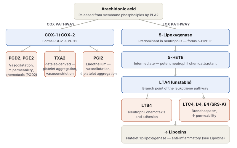
- COX-1/COX-2 → PGG2 → PGH2 → PGD2/PGE2 (vasodilatation, ↑permeability), TXA2 (platelet — aggregation, vasoconstriction), PGI2 (endothelium — vasodilatation, ↓aggregation)
- 5-Lipoxygenase (neutrophils) → 5-HPETE → 5-HETE (chemotactic) + Leukotrienes: LTA4 → LTB4 (chemotaxis/adhesion) or LTC4/D4/E4 = SRS-A (bronchospasm, vasoconstriction, ↑permeability)
- → Lipoxins (platelet 12-LOX on neutrophil LTA4) — anti-inflammatory, see Lipoxins
Drugs: NSAIDs/aspirin (COX inhibitors) · COX-2 selective (↑CV risk — spares platelet TXA2, blocks endothelial PGI2) · corticosteroids (block COX-2, PLA2, cytokines transcriptionally) · zileuton (LOX inhibitor) · zafirlukast (LT receptor antagonist) — asthma drugs.
Lysosomal components — neutrophil granules: primary/azurophil (MPO, hydrolases, defensins, elastase), secondary/specific (lactoferrin, collagenase, lysozyme), tertiary (gelatinase). Checked by α1-antitrypsin, α2-macroglobulin.
PAF — mast cells/leukocytes/endothelium/platelets → ↑permeability, vasodilatation(low)/constriction(high), bronchoconstriction, adhesion, chemotaxis, platelet aggregation.
Cytokines — TNF-α + IL-1 central: endothelial adhesion molecule expression, leukocyte activation, acute-phase response (fever, shock at high TNF). IL-6 (acute phase). IL-8 (chemokine). MCP-1/eotaxin/PF-4 (specific chemoattractants). IL-12/IL-17 = chronic inflammation cytokines. TNF antagonists → RA treatment but ↑TB risk.
Free radicals — O2 metabolites (endothelial damage, protease activation) + NO (vasodilatation, antiplatelet, microbicidal).
II. Plasma protein-derived (4 interlinked systems via Factor XIIa)
- Kinin: XIIa → kallikrein → bradykinin (permeability, pain, smooth muscle contraction, vasodilatation)
- Clotting: XIIa → fibrin + fibrinopeptides (↑permeability, chemotaxis)
- Fibrinolytic: plasminogen activator → plasmin → activates XII, cleaves C3→C3a, fibrin split products (chemotactic)
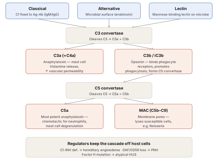
- Complement: 3 pathways (classical=Ag-Ab/C1; alternative=microbial surface; lectin=MBL-mannose) → C3 convertase → C3a/C3b → C5 convertase → C5a + MAC(C5b-9)
- C3a/C4a/C5a = anaphylatoxins (histamine release, permeability); C5a also chemotactic
- C3b/iC3b = opsonin
- MAC = lysis (e.g. Neisseria)
- Regulators: C1-INH (def→hereditary angioedema), DAF/CD59 (loss→PNH), Factor H (mutation→atypical HUS)
Regulation of inflammation
Acute-phase reactants (liver — CRP=opsonin, clinical marker) · endogenous glucocorticoids · soluble cytokine receptors · lipoxins/resolvins/PGI2 (active resolution, not passive fading).
Clinical relevance
Drug classes map directly onto mediator pathway points (NSAIDs/COX-2i/steroids/LT-antagonists/anti-TNF). CRP = bedside marker driven by IL-1/IL-6/TNF. Complement regulator deficiencies = named diseases (angioedema, PNH, aHUS).
Definition and General Properties
Chemical mediators of inflammation are the endogenous substances that translate recognition of an offending agent into the visible vascular and cellular events of acute inflammation. They share a set of properties worth knowing as a group, since several are tested directly:
- They are either cell-derived, released from preformed intracellular stores (e.g. histamine from mast cell granules) or synthesised anew on stimulation (e.g. prostaglandins, cytokines), or plasma-derived, produced mainly by the liver as inactive precursors that are activated at the site of inflammation by proteolytic cleavage (e.g. complement, kinin, clotting and fibrinolytic system products).
- They are produced only in response to a stimulus — microbial products or material released from necrotic cells — which restricts inflammation to where and when it is actually needed.
- A given mediator may act on the same cell that produced it, or on other cells; and one mediator’s release commonly stimulates or opposes the release of another, so that the mediators of a full inflammatory reaction interact as a network rather than acting independently.
- Their range of action covers vasodilatation, increased vascular permeability, chemotaxis, fever, pain and tissue damage — the mediators are, collectively, the mechanistic explanation for every cardinal sign (see Acute Inflammation).
- They have a deliberately short lifespan: they decay spontaneously, are enzymatically inactivated, or are removed by antioxidants and regulatory proteins, providing an inbuilt brake on the reaction once the stimulus is cleared.
I. Cell-Derived Mediators
Vasoactive Amines
Histamine, the major vasoactive amine, is stored preformed in the granules of mast cells (its richest source), basophils and platelets, which is why it is among the very first mediators released in an inflammatory reaction. Release is triggered by physical injury (heat, cold, trauma), by the anaphylatoxins C3a and C5a, by cytokines (IL-1, IL-8), and by binding of IgE to mast cells in immediate hypersensitivity reactions. Acting chiefly through H1 receptors on microvascular endothelium — the target of antihistamine drugs — histamine causes arteriolar dilatation and increased venular permeability, and also produces itching and pain.
Serotonin (5-hydroxytryptamine), stored in platelets, GIT chromaffin cells and mast cells, produces effects qualitatively similar to but weaker than histamine; carcinoid tumours are notable as serotonin-secreting neoplasms.
Neuropeptides such as substance P, neurokinin A and vasoactive intestinal polypeptide are produced by the nervous system and contribute to increased vascular permeability, pain transmission and mast cell degranulation.
Arachidonic Acid Metabolites (Eicosanoids)
Arachidonic acid, a 20-carbon polyunsaturated fatty acid, is released from membrane phospholipids by phospholipase A2 in response to inflammatory stimuli and metabolised along two enzymatic routes, making eicosanoids arguably the most potent mediator class in acute inflammation.
Cyclo-oxygenase (COX) pathway. COX exists as two isoforms — COX-1, constitutively expressed and important for tissue homeostasis (renal fluid/electrolyte balance, gastric cytoprotection), and COX-2, induced specifically by inflammatory stimuli — acting on arachidonic acid to form PGG2 and then PGH2, from which three principal products arise:
- PGD2 and PGE2 — vasodilatation and increased venular permeability; PGD2 is also chemotactic for neutrophils.
- Thromboxane A2 (TXA2) — formed via thromboxane synthase, present in platelets; causes platelet aggregation and vasoconstriction.
- Prostacyclin (PGI2) — formed via prostacyclin synthase in endothelium; a vasodilator and potent inhibitor of platelet aggregation, opposing TXA2. A thromboxane–prostacyclin imbalance is implicated in early coronary and cerebral thrombosis.
Prostaglandins also mediate the pain and fever of the systemic acute-phase response.
Lipo-oxygenase pathway. 5-lipoxygenase, the predominant enzyme in neutrophils, converts arachidonic acid to 5-HPETE and then to two further products:
- 5-HETE, an intermediate, is itself a potent neutrophil chemoattractant.
- Leukotrienes: unstable LTA4 gives rise to LTB4 (chemotactic and neutrophil-activating) and to LTC4/LTD4/LTE4, collectively the slow-reacting substances of anaphylaxis, which cause smooth muscle contraction, bronchospasm, vasoconstriction and increased vascular permeability.
Lipoxins, formed when neutrophil-derived LTA4 is taken up transcellularly by platelet 12-lipoxygenase, are the counter-regulatory branch of this same pathway: they inhibit neutrophil chemotaxis and leucocyte adhesion, actively terminating rather than propagating the reaction (full mechanism in Lipoxins). Resolvins, another anti-inflammatory derivative, are the mechanism by which aspirin — which inhibits COX activity — also promotes resolution.
Because this pathway is the direct target of major drug classes, its pharmacology is worth fixing alongside the biochemistry: aspirin and other NSAIDs inhibit both COX isoforms (aspirin irreversibly), blocking prostaglandin synthesis and thereby treating pain and fever; selective COX-2 inhibitors spare COX-1-mediated gastric and platelet effects but can increase cardiovascular risk by sparing endothelial thromboxane while suppressing protective prostacyclin; corticosteroids act further upstream, reducing transcription of COX-2, phospholipase A2 and pro-inflammatory cytokines; and leukotriene-pathway drugs (zileuton, a lipoxygenase inhibitor, and zafirlukast, a leukotriene receptor antagonist) are used specifically in asthma, reflecting the leukotrienes’ potency in bronchospasm.
Lysosomal Components
Neutrophils carry three granule types, released on degranulation: primary (azurophil) granules contain myeloperoxidase, acid hydrolases, defensins, elastase, cathepsin G and other proteases; secondary (specific) granules contain lactoferrin, collagenase, gelatinase and lysozyme; tertiary granules contain gelatinase and acid hydrolases. Myeloperoxidase generates oxidative lysis via free radicals, acid hydrolases destroy ingested bacteria within the phagolysosome, and the proteases attack extracellular matrix components (basement membrane, collagen, elastin) — tissue-damaging activity that is normally kept in check by circulating antiproteases such as α1-antitrypsin and α2-macroglobulin. Monocyte and macrophage granules release similar mediators but contribute more to chronic than to acute inflammation.
Platelet-Activating Factor (PAF)
Released from IgE-sensitised mast cells and basophils, other leucocytes, endothelium and platelets, PAF causes increased vascular permeability, vasodilatation at low concentration (vasoconstriction otherwise), bronchoconstriction, leucocyte adhesion to endothelium and chemotaxis, in addition to its eponymous role in platelet aggregation.
Cytokines
Cytokines are the polypeptide products of activated lymphocytes (lymphokines) and monocytes/macrophages (monokines); chemokines are the specific chemoattractant subfamily. TNF-α and IL-1 are the two cytokines most central to acute inflammation, sharing largely overlapping actions: both increase endothelial expression of adhesion molecules (chiefly E- and P-selectins and integrin ligands) to drive leukocyte recruitment, both activate leukocytes and other cells (TNF augments neutrophil responses and macrophage microbicidal activity; IL-1 activates fibroblasts and synovial cells), and both drive the systemic acute-phase response — fever, hepatic acute-phase protein synthesis, and, at high concentration, the hypotension and vascular collapse of shock. IL-6 similarly drives hepatic acute-phase protein production and lymphocyte differentiation. IL-8 is itself a chemokine, inducing neutrophil, macrophage and T-cell migration and stimulating basophil histamine release. Other named chemokines with more restricted roles include MCP-1 (monocyte/T-cell/NK-cell chemoattractant), eotaxin (eosinophil/basophil chemoattractant, implicated in allergic pulmonary disease) and PF-4 (fibroblast chemoattractant, released by platelets). IL-12 and IL-17, by contrast, are chiefly cytokines of chronic rather than acute inflammation. TNF antagonists are now important therapy for chronic inflammatory diseases such as rheumatoid arthritis, at the cost of impairing macrophage killing of intracellular organisms such as mycobacteria.
Free Radicals: Oxygen Metabolites and Nitric Oxide
Released from activated neutrophils and macrophages, oxygen-derived free radicals (superoxide, hydrogen peroxide, hydroxyl radical) damage endothelium (increasing permeability), activate proteases while inactivating antiproteases (causing matrix damage), and injure other cells directly; their action is normally opposed by tissue and serum antioxidants. Nitric oxide, generated from arginine by NO synthase in activated macrophages, contributes vasodilatation, anti-platelet activity and probable microbicidal action; it was originally identified as an endothelium-derived vascular relaxation factor.
II. Plasma Protein–Derived Mediators (Plasma Proteases)
Four interlinked plasma protease systems — kinin, clotting, fibrinolytic and complement — generate the plasma-derived mediators of inflammation, each with its own inhibitors and accelerators providing negative and positive feedback. Factor XII (Hageman factor) of the clotting system is the key point of interaction between the four: its activation, triggered in vivo by contact with exposed basement membrane or bacterial endotoxin leaking through injured endothelium, sets off the clotting, fibrinolytic and kinin cascades simultaneously, whose products in turn activate complement, whose permeability-increasing products further activate clotting — a self-amplifying loop.
- The kinin system. Factor XIIa generates kallikrein from plasma prekallikrein, and kallikrein then cleaves high-molecular-weight kininogen to release bradykinin. Acting early in inflammation, bradykinin causes smooth muscle contraction, vasodilatation, increased vascular permeability and pain.
- The clotting system. Factor XIIa initiates the cascade converting fibrinogen to fibrin (via thrombin) with release of fibrinopeptides, which themselves increase vascular permeability, are chemotactic for leukocytes, and have anticoagulant activity.
- The fibrinolytic system. Plasminogen activator (from kallikrein, endothelium and leukocytes) converts plasminogen to plasmin, which activates factor XII (feeding back into the kinin system), cleaves complement C3 to the permeability factor C3a, and degrades fibrin into split products that are themselves chemotactic and permeability-increasing.
- The complement system. Activation proceeds through the classical pathway (C1 fixed to antigen–antibody complexes, IgM or IgG), the alternative pathway (triggered directly by microbial surfaces — endotoxin, complex polysaccharides — without antibody), or the lectin pathway (plasma mannose-binding lectin bound to microbial mannose, again antibody-independent). All three converge on cleavage of C3 by C3 convertase into C3a and C3b, and progress via a C5 convertase to C5a and the membrane attack complex (C5b–C9). The functional consequences are: C3a, C4a and C5a (anaphylatoxins) trigger mast cell histamine release and are directly chemotactic (C5a) for leukocytes; C3b and iC3b act as opsonins, binding microbial surfaces to promote phagocytosis; and the membrane attack complex inserts into lipid membranes, lysing susceptible cells such as Neisseria. Regulatory proteins — C1 inhibitor, decay-accelerating factor, CD59, Factor H — normally keep this cascade confined to its target and off host cells; their deficiency states are clinically important in their own right (hereditary angioedema with C1 inhibitor deficiency; paroxysmal nocturnal haemoglobinuria with loss of the GPI-anchored regulators DAF and CD59; atypical haemolytic uraemic syndrome with Factor H mutation).
Regulation of Inflammation
Because an unchecked inflammatory response is itself capable of tissue damage — as seen in hypersensitivity states — the host maintains regulatory mechanisms that keep mediator activity in check and allow the reaction to resolve:
- Acute-phase reactant proteins, synthesised mainly by the liver, include cellular protection factors (α1-antitrypsin, α2-antiplasmin) that limit proteolytic damage, coagulation proteins that replace factors consumed in clotting, transport proteins (ceruloplasmin, haptoglobin), and immune agents such as C-reactive protein, an opsonin whose serum level is a widely used clinical marker of ongoing inflammation.
- Endogenous glucocorticoids, raised during infection and trauma, act as natural anti-inflammatory agents.
- Soluble cytokine receptors, shed into serum, correlate with disease activity and buffer free cytokine levels.
- Anti-inflammatory lipid mediators — lipoxins and resolvins, and the dual pro-/anti-inflammatory action of prostacyclin — actively terminate the reaction rather than simply allowing it to fade passively.
Clinical Relevance
- Almost every major class of anti-inflammatory drug in clinical use targets a specific point on this mediator map — NSAIDs and COX inhibitors block prostaglandin synthesis, leukotriene antagonists block the lipoxygenase arm, corticosteroids act broadly upstream on gene transcription, and TNF antagonists block a cytokine central to both leukocyte recruitment and the systemic acute-phase response — which is why this topic is examined as much through pharmacology as through pure biochemistry.
- C-reactive protein, an acute-phase reactant activated through this same mediator cascade, is used at the bedside as a quantitative marker of inflammatory activity precisely because its hepatic synthesis is driven by IL-1, IL-6 and TNF-α — the same cytokines responsible for fever and leucocytosis.
- Recognising the complement regulatory-protein deficiency states (hereditary angioedema, paroxysmal nocturnal haemoglobinuria, atypical HUS) as direct consequences of losing a specific brake on this cascade is a common way the complement system is tested beyond the basic classical/alternative/lectin pathway distinction.
Diagram 1: Arachidonic Acid Metabolite Cascade
Draw a single starting box, “Arachidonic acid,” branching into two labelled columns.
- COX pathway column: COX-1/COX-2 → PGG2/PGH2 → three end-product boxes side by side: PGD2/PGE2 (vasodilatation, permeability), TXA2 (platelet aggregation, vasoconstriction), PGI2 (vasodilatation, anti-aggregation).
- LOX pathway column: 5-lipoxygenase → 5-HETE (chemotactic) → LTA4 → branches into LTB4 (chemotaxis/adhesion) and LTC4/D4/E4 (bronchospasm, vasoconstriction, permeability) → a final box for lipoxins, noting the platelet 12-lipoxygenase step.
Labels required
- Both branches must originate visibly from the single arachidonic acid box, since the shared precursor is the point most often tested (students otherwise learn the two pathways as unrelated lists).
- TXA2 and PGI2 must be labelled with their opposing platelet actions (aggregation vs anti-aggregation) since their balance, not either alone, is the clinically relevant point.
- The LTA4 branch point must be shown explicitly, since LTB4 and LTC4/D4/E4 have very different actions (chemotaxis vs bronchospasm) despite sharing this one precursor.
Errors commonly made
- Describing prostaglandins and leukotrienes as produced by unrelated enzymes without showing that both trace back to the same arachidonic acid.
- Omitting the lipoxin end-point, leaving the impression that the lipoxygenase pathway is purely pro-inflammatory.
Diagram 2: Complement Activation Pathways
Draw three boxes at the top, one per activation pathway, converging onto a single “C3 convertase” box, which branches into a C3a box and a C3b box; the C3b box continues down into a “C5 convertase” box, which branches again into a C5a box and a membrane attack complex (MAC) box. A final note box lists the regulatory-protein deficiency states.
- Classical — C1 fixed to antigen–antibody complex (IgM/IgG).
- Alternative — direct microbial surface trigger (endotoxin), no antibody needed.
- Lectin — mannose-binding lectin bound to microbial mannose, no antibody needed.
- C3a — anaphylatoxin: mast cell histamine release, increased permeability.
- C3b — opsonin, and the component that forms C5 convertase.
- C5a — the most potent anaphylatoxin, also directly chemotactic for neutrophils.
- MAC (C5b–C9) — membrane pore formation and cell lysis.
Labels required
- All three pathways must be shown converging on the same C3 convertase step, since the exam-relevant point is that they differ only in how C3 is triggered, not in what happens after.
- C3b must be shown continuing on to form C5 convertase, distinguishing its downstream role from C3a’s immediate local action.
Errors commonly made
- Treating C3a and C5a as functionally identical rather than noting C5a is the more potent chemotactic anaphylatoxin.
- Forgetting that C3b’s opsonin function and its role in forming C5 convertase are two separate, both examinable, actions of the same fragment.
Short Essays (1)
Multiple Choice Questions (3)
My Sticky Notes
Personal revision notes, mnemonics and reminders.
