
Basic Haematology Laboratory Techniques
Overview
Core haematology bench techniques: anticoagulants, CBC, Hb estimation, PCV/haematocrit, reticulocyte count, ESR. See Haemostasis Screening Tests (coagulation), Blood Groups and Transfusion Medicine (transfusion testing).
Collection/anticoagulants
Vacutainer colours: Red=none (serum), Purple/lavender=EDTA (routine CBC), Light blue=sodium citrate (coagulation), Grey=fluoride (glucose), Green=heparin (osmotic fragility, enzyme studies), Black=citrate (ESR). Haemolysed samples unsuitable (narrow needle, excess suction, water contamination).
CBC/Hemogram
Hb, TLC, RBC count, platelet count, DLC, PCV, reticulocyte count, PBF (morphology, haemoparasites, abnormal cells). Automated: adds MCV/MCH/MCHC/RDW + MPV — cross-checks manual params.
Hb estimation
Colorimetric (Sahli’s/alkaline haematin — visual; cyanmethemoglobin/oxyHb — photoelectric), physical (specific gravity), chemical (iron content), gasometric (O2-combining capacity), autoanalyser (lysate colour vs standard). Cyanmethemoglobin = most widely used.
PCV/Haematocrit
= RBC volume : whole blood ratio. ↓anaemia, ↑polycythaemia. Wintrobe tube (macromethod): 11cm, 2.5mm bore, 1mL, dual-calibrated (ascending=ESR, descending=PCV).
Centrifuged blood → 3 layers: Lower=packed RBCs, Middle=buffy coat (WBC+platelets), Upper=plasma.
Normal: males 38-47%, females 36-46%, infants (cord) 45-70%. ↑PV vera/secondary polycythaemia; ↓anaemia.
Uses: anaemia assessment, polycythaemia detection, red cell indices (MCV/MCHC), Hb cross-check (Hb g/dL ×3 ≈ Hct).
Buffy coat: 0.1mm ≈ 1000 WBC/cu mm. ↑thickness = marked leucocytosis (CML/CLL/acute leukaemia); absent = leucopenia. Subleukaemic leukaemia: buffy-coat smear concentrates WBCs, eases abnormal cell ID.
Reticulocyte count
Immature non-nucleated RBCs, slightly larger, still synthesising Hb. Supravital stain (new methylene blue/brilliant cresyl blue) on live blood — RNA precipitates as dye-RNP complex (dark blue reticulum). Romanowsky stain → polychromatic (“polychromatophil”).
Normal: 0.5-2.5% (newborn 1-7%).
↑ count: haemolytic anaemias/crisis, haemorrhage, treatment response (iron/folate/B12 — peaks day 6-7, confirms marrow response). ↓ count: aplastic anaemia, aplastic crisis (parvovirus in HS/sickle cell), pure red cell aplasia, Fanconi anaemia, myelofibrosis.
ESR
Nonspecific inflammation marker (CRP now often preferred). Principle: anticoagulated blood, vertical tube, RBCs settle 1hr under gravity (mm). RBCs net negative surface charge → mutual repulsion; positive plasma proteins counteract this → plasma protein composition drives ESR.
3 stages: (1) aggregation/rouleaux (first 10min, minimal sedimentation), (2) settling (~40min, constant rate), (3) packing (final 10min).
Factors:
- Plasma: ↑fibrinogen/globulins/cholesterol → accelerate (↑positive charge); albumin/lecithin → retard
- RBC: rate ∝ aggregate weight, ∝1/surface area. Microcytes sediment slower than macrocytes. Rouleaux ACCELERATES ESR; abnormal shape (sickle cells, spherocytes) CAN’T rouleaux → LOW ESR despite active disease
- Numerical: anaemia↑ESR, polycythaemia↓ESR
- Technical: tube must stay vertical
Methods: Westergren (preferred, more accurate) — 30cm glass pipette, 0-200mm, 3.8% trisodium citrate, blood:anticoagulant 4:1. Wintrobe — same tube as PCV, ascending scale.
Clinical relevance
Hb×3≈Hct cross-check = simple useful consistency test — violation should prompt re-checking sample/analyser, not blind acceptance. Sickle cells/spherocytes → LOW ESR despite active haemolysis = frequently missed counter-intuitive point — abnormal SHAPE prevents rouleaux, suppressing ESR independent of actual disease activity. Reticulocyte peak day 6-7 of haematinic Rx = specific testable timepoint confirming marrow response — checked too early, looks falsely unresponsive. Buffy coat thickness as visual WBC proxy (+ subleukaemic leukaemia smear trick) = simple technique still practically useful, not just historical.
Overview
This note collects the core haematology bench techniques referenced throughout the haematopathology topics — anticoagulant choice, the complete blood count, haemoglobin estimation, PCV/haematocrit, reticulocyte count, and ESR — into one reference. See Haemostasis Screening Tests for coagulation-specific tests and Blood Groups and Transfusion Medicine for transfusion-related testing.
Blood Collection and Anticoagulants
Venous blood is collected by syringe or vacuum tubes (vacutainers), colour-coded by anticoagulant:
| Tube colour | Anticoagulant | Use |
|---|---|---|
| Red | None (plain) | Serum studies |
| Purple/lavender | EDTA | Routine haematology (CBC) |
| Light blue | Sodium citrate | Coagulation studies |
| Grey | Fluoride | Glucose estimation |
| Green | Heparin | Osmotic fragility test, red cell enzyme studies |
| Black | Citrate | ESR |
Capillary blood (finger/heel prick) and arterial blood (blood gas analysis) are collected separately as needed. Haemolysed samples are unsuitable for testing — haemolysis results from narrow-bore needles, excessive suction, or syringe contamination with water.
Complete Blood Count (CBC/Hemogram)
Standard components: haemoglobin, total leucocyte count, RBC count, platelet count, differential leucocyte count, haematocrit (PCV), reticulocyte count, and peripheral blood smear (RBC/platelet morphology, haemoparasites, abnormal cells). Automated analysers additionally derive absolute values — MCV, MCH, MCHC, RDW — plus mean platelet volume (MPV), useful for cross-checking manually-derived parameters.
Haemoglobin Estimation
| Method | Principle |
|---|---|
| Colorimetric — Sahli’s/acid haematin, alkaline haematin | Visual colour matching |
| Cyanmethemoglobin (HiCN), oxyhaemoglobin | Photoelectric/photocolorimetric |
| Physical | Specific gravity |
| Chemical | Iron content |
| Gasometric | Oxygen-combining capacity |
| Cell counter/autoanalyser | RBC lysate colour matched to standard (most use cyanide-free reagent) |
Cyanmethemoglobin is the most widely used routine method.
PCV / Haematocrit
Haematocrit — the ratio of red cell volume to whole blood volume — indicates relative red cell mass (reduced in anaemia, increased in polycythaemia). Estimated by macromethod (Wintrobe tube), micromethod (capillary tube), or automated analyser.
Wintrobe tube: thick-walled glass, 11 cm long, 2.5 mm bore, 1 mL capacity; calibrated ascending (0→100+) for ESR and descending (100→0) for PCV on the same tube.
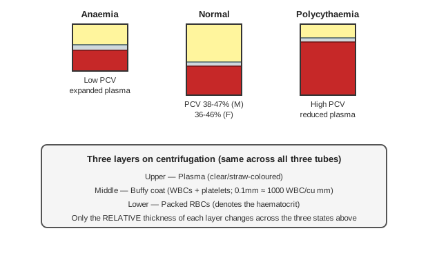
Anticoagulated whole blood, centrifuged, separates into three layers:
- Lower — packed RBCs.
- Middle — the buffy coat (thin grey-red layer of WBCs and platelets).
- Upper — plasma (clear/straw-coloured).
Normal PCV: adult males 38–47%, adult females 36–46%, infants (cord blood) 45–70%. Increased in polycythaemia vera and secondary polycythaemia; decreased in anaemia.
Uses of Wintrobe/haematocrit: assessing anaemia (presence, severity, treatment response), detecting polycythaemia, deriving red cell indices (MCV, MCHC), and cross-checking haemoglobin accuracy (Hb in g/dL × 3 ≈ haematocrit — a quick internal consistency check).
Buffy coat: normally 0.1 mm of this layer ≈ 1000 WBC/cu mm. Thickness increases with marked leucocytosis (CML, CLL, acute leukaemias); absent/minimal buffy coat implies leucopenia. In subleukaemic leukaemia, a smear made directly from the buffy coat concentrates WBCs, making abnormal cells easier to identify.
Reticulocyte Count
Reticulocytes are immature, non-nucleated RBCs recently released from marrow, slightly larger than mature RBCs, still synthesising haemoglobin. Stained by supravital staining (new methylene blue/brilliant cresyl blue) on live, unfixed blood — ribosomal RNA precipitates as a dye-ribonucleoprotein complex, visible as a dark blue reticular network. On Romanowsky stains, reticulocytes appear polychromatic (“polychromatophils”).
Normal range: 0.5–2.5% of total red cells (newborn/cord blood: 1–7%).
| Increased reticulocyte count | Reduced reticulocyte count |
|---|---|
| Haemolytic anaemias, haemolytic crisis, haemorrhage | Aplastic anaemia |
| Response to treatment (iron/folate/B12 deficiency) — peaks day 6–7, confirms marrow response | Aplastic crisis (parvovirus, in hereditary spherocytosis/sickle cell disease) |
| — | Pure red cell aplasia, Fanconi anaemia, myelofibrosis |
Erythrocyte Sedimentation Rate (ESR)
A commonly used but nonspecific marker of underlying inflammation (higher-sensitivity markers like CRP are now often preferred for monitoring conditions such as cardiovascular disease).
Principle: anticoagulated blood in a vertical tube — RBCs settle under gravity over 1 hour, measured as the length (mm) of the sedimented column. RBCs carry a net negative surface charge that promotes mutual repulsion; this repulsion is partially counteracted by positively-charged plasma proteins, which is why plasma protein composition drives ESR.
Three stages: (1) aggregation/rouleaux formation — first 10 minutes, minimal sedimentation while rouleaux form; (2) settling — ~40 minutes, constant-rate sedimentation; (3) packing — final 10 minutes.
Factors affecting ESR:
- Plasma factors — raised fibrinogen, globulins, cholesterol accelerate ESR (increase plasma’s positive charge); albumin and lecithin retard it.
- Red cell factors — sedimentation rate is directly proportional to aggregate weight, inversely proportional to surface area. Microcytes sediment slower than macrocytes. Rouleaux formation accelerates ESR; cells with abnormal/irregular shape (sickle cells, spherocytes) cannot form rouleaux and therefore have a low ESR despite potentially active disease.
- Numerical — anaemia increases ESR, polycythaemia decreases it.
- Technical — the ESR tube must be kept strictly vertical, or results become inaccurate.
Methods: Westergren (preferred, more accurate) — a straight glass pipette, 30 cm, 0–200 mm calibration, 3.8% trisodium citrate anticoagulant, blood:anticoagulant ratio 4:1. Wintrobe method — uses the same Wintrobe tube as PCV estimation (ascending scale).
Clinical Relevance
- The haematocrit-to-haemoglobin cross-check (Hb × 3 ≈ Hct) is a simple, genuinely useful internal consistency test — a result that violates this ratio should prompt re-checking the sample or the analyser, rather than being accepted at face value.
- Sickle cells and spherocytes producing a low ESR despite being markers of active haemolytic disease is a frequently missed, counter-intuitive point — it is the abnormal cell shape itself (preventing rouleaux formation) that suppresses ESR, independent of how much inflammation or disease activity is actually present.
- The reticulocyte count’s peak on day 6–7 of haematinic treatment is the specific, testable timepoint that confirms genuine marrow response to iron/folate/B12 therapy — a reticulocyte count checked too early can appear falsely unresponsive even in a patient who is about to respond.
- Buffy coat thickness as a rough visual proxy for leucocyte count (and its practical use in subleukaemic leukaemia to concentrate WBCs for smear examination) is a simple bedside-adjacent technique worth remembering as more than a historical curiosity — it directly aids morphologic diagnosis when the peripheral count itself is low.
Diagram 1: PCV Layer Separation — Anaemia vs Normal vs Polycythaemia
Draw three side-by-side tubes (Anaemia / Normal / Polycythaemia), each with the same three layers in the same order — plasma on top, a thin buffy coat band, packed RBCs at the bottom — varying only the relative height of each band across the three tubes.
Labels required
- All three tubes must show the same three-layer order — this is a fixed anatomy of the test; only the proportions change.
- The buffy coat must stay visually thin and consistently labelled (WBCs + platelets) across all three tubes, since its own thickness is a separate diagnostic signal (leucocytosis) from PCV itself and should not be conflated with the RBC layer’s changes.
Errors commonly made
- Assuming the layer order changes between disease states — it never does; only the relative volume of plasma versus packed RBCs shifts.
- Reading buffy coat thickness as if it tracked PCV — it doesn’t; buffy coat thickness reflects white cell/platelet mass, which is an independent parameter from the red cell mass that PCV actually measures.
My Sticky Notes
Personal revision notes, mnemonics and reminders.
