
Infective Endocarditis
Definition
Infection, usually bacterial, of the valvular or mural endocardium, producing bulky, friable, infected vegetations composed of thrombotic material, inflammatory cells, and organisms.
Acute vs subacute
| Acute | Subacute | |
|---|---|---|
| Course | Fulminant, death by ~6 weeks if untreated | Insidious, weeks to months |
| Organism | Highly virulent — S. aureus | Low virulence — S. viridans |
| Valve | Previously normal | Previously damaged |
| Lesion | Invasive, destructive, suppurative | Not invasive/suppurative |
| Clinical picture | Acute sepsis | Splenomegaly, clubbing, petechiae |
Etiology
90% streptococci and staphylococci. Acute: S. aureus (IV drug users). Subacute: S. viridans (oral commensal), also S. bovis, S. epidermidis (prosthetic valve endocarditis), HACEK group.
Predisposing factors: transient bacteraemia (dental/GU/GI procedures, IV drug use) + underlying heart disease (rheumatic, congenital, MVP, degenerative calcific, prosthetic valve) + impaired host defence (diabetes, leukaemia/lymphoma, cytotoxic therapy, neutropenia).
Pathogenesis
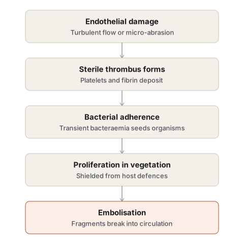
Endocardial damage/denudation (turbulent flow, micro-abrasion) → sterile platelet-fibrin thrombus forms → transient bacteraemia seeds organisms onto the thrombus → organisms proliferate, protected under further fibrin/platelet deposition → vegetation enlarges → fragments embolise.
Morphology
Gross: mitral > aortic > combined mitral+aortic (left heart); tricuspid in IV drug users. Same anatomical sites as rheumatic vegetations (atrial surface of AV valves, ventricular surface of semilunar valves) but larger, grey-tawny to greenish, irregular, and friable (vs rheumatic: small, warty, firmly attached). Acute-disease vegetations bulkier, more destructive — ulceration, perforation, myocardial abscess.
Microscopy — three zones: outer eosinophilic fibrin/platelet cap → basophilic bacterial colony zone → deep inflammatory zone (necrosis/abscess in acute; granulation tissue/healing in subacute).
Complications
Cardiac: valvular stenosis/insufficiency · perforation/rupture/leaflet aneurysm · ring abscess · myocardial abscess · suppurative pericarditis · cardiac failure.
Extracardiac (from friable emboli):
- Left-heart emboli → systemic → septic infarcts/abscesses, mycotic aneurysms (spleen, kidney, brain).
- Right-heart emboli → pulmonary abscess.
- Immunologic: glomerulonephritis (immune complex) · Osler nodes (tender, subacute) · Janeway lesions (painless macules, acute) · Roth spots (retinal haemorrhage, pale centre) · splinter/subungual haemorrhages, petechiae.
Diagnosis — modified Duke criteria
Definite IE = 2 major, or 1 major + 3 minor, or 5 minor.
Major: positive blood cultures (typical organism, ≥2 samples, or persistently positive) · echocardiographic evidence (vegetation, abscess, new prosthetic dehiscence) · new valvular regurgitation.
Minor: fever ≥38°C · predisposing lesion or IV drug use · vascular phenomena (emboli, mycotic aneurysm, Janeway lesions) · immunologic phenomena (glomerulonephritis, Osler nodes, Roth spots) · microbiological evidence not meeting major criteria.
Noninfected vegetations
Non-bacterial thrombotic (marantic) endocarditis: small, sterile, bland vegetations on the line of closure of a normal valve; associated with hypercoagulable states (DIC, hyperoestrogenic states, mucinous adenocarcinoma); does not destroy the valve, but can seed infective endocarditis.
Libman-Sacks endocarditis: ~10% of SLE patients; small-medium sterile vegetations, can occur on either leaflet surface or on cords/mural endocardium (unlike other types); immune complex-mediated, fibrinoid necrosis; may leave chronic deformity resembling rheumatic disease.
Clinical relevance
Any unexplained fever with a prosthetic valve, known rheumatic/congenital heart disease, or IV drug use history should raise suspicion for infective endocarditis. Splinter haemorrhage, Osler node, or Roth spot in a febrile patient point to embolisation from a vegetation.
Definition
Infective endocarditis is an infection, usually bacterial, of the valvular or mural endocardium, in which microorganisms colonise the endothelial surface and provoke the formation of bulky, friable, infected vegetations composed of thrombotic material, inflammatory cells, and organisms. Left untreated, it runs a destructive and often fatal course; even with treatment, it can leave permanent valvular damage and produce serious complications elsewhere in the body through embolisation of its friable vegetations.
Classification by Clinical Course
Infective endocarditis has traditionally been divided into acute and subacute forms, distinguished chiefly by the virulence of the organism and the condition of the valve it infects. Antibiotic treatment has blurred this distinction clinically, but the underlying pathological contrast remains a useful teaching framework.
| Feature | Acute | Subacute |
|---|---|---|
| Onset and course | Fulminant; death within about six weeks if untreated | Insidious; a protracted course of weeks to months, occasionally years |
| Typical organism | Highly virulent — chiefly Staphylococcus aureus | Low virulence — chiefly Streptococcus viridans |
| Valve affected | Usually previously normal | Usually previously damaged |
| Effect on the valve | Invasive, destructive, suppurative | Usually not invasive or suppurative |
| Clinical picture | Features of acute systemic infection | Splenomegaly, clubbing, petechiae |
Etiology
More than ninety percent of cases are caused by streptococci and staphylococci.
- Acute disease: predominantly Staphylococcus aureus, a highly virulent organism capable of infecting either a normal or an already deformed valve, and the leading cause of endocarditis in intravenous drug users; other organisms include pneumococci, gonococci, and enterococci.
- Subacute disease: predominantly Streptococcus viridans, a low-virulence commensal of the mouth and pharynx that typically requires a previously abnormal valve to establish infection; other organisms include Streptococcus bovis (a gut commensal), coagulase-negative staphylococci such as S. epidermidis (the leading cause of prosthetic valve endocarditis), gram-negative enteric bacilli, and the fastidious HACEK group of oral commensals.
Predisposing Factors
Three categories of predisposing condition act together to produce disease.
- Transient bacteraemia, arising from everyday sources such as dental procedures, genitourinary instrumentation or delivery, gastrointestinal or biliary surgery, skin infections, respiratory infections, or intravenous drug use.
- Underlying heart disease, which provides an abnormal surface for organisms to colonise: chronic rheumatic valvular disease (historically the leading substrate), congenital heart disease, mitral valve prolapse, degenerative calcific valve disease in the elderly, a bicuspid aortic valve, and prosthetic valves or indwelling catheters.
- Impaired host defence, as in diabetes mellitus, leukaemia and lymphoma, cytotoxic therapy, or neutropenia.
Pathogenesis
Development of infective endocarditis on a previously abnormal valve follows a predictable sequence, though the mechanism by which virulent organisms occasionally infect a completely normal valve is less well understood.
- Endocardial damage and denudation. Turbulent or high-velocity blood flow across a deformed valve, or micro-abrasion from repeated intravenous injection, damages the endothelial surface.
- Formation of a sterile thrombus. The exposed extracellular matrix attracts platelets and fibrin, which aggregate into a small, sterile vegetation — essentially a form of non-bacterial thrombotic endocarditis.
- Bacterial adherence. Transient bacteraemia delivers circulating organisms, which possess surface adhesion molecules that let them attach to the sterile thrombus.
- Proliferation within the vegetation. Further platelets and fibrin are deposited over the adherent organisms, sealing them off from the bloodstream and from host defences, allowing the colony to expand.
- Embolisation. Fragments of the enlarging, friable vegetation may detach and travel in the circulation, seeding infection or infarction at distant sites.
Morphology
Gross
The valves of the left heart — mitral, followed by aortic, then combined mitral and aortic — are affected far more often than those of the right heart, though right-sided (typically tricuspid) involvement is characteristic of intravenous drug users. Vegetations in subacute disease more often develop on a previously diseased valve, while those of acute disease more often appear on a previously normal one; in both, the vegetations occupy the same anatomical locations as rheumatic vegetations — the atrial surface of the atrioventricular valves and the ventricular surface of the semilunar valves — beginning at the line of closure and sometimes extending onto adjacent structures. Compared with the small, firmly attached, warty vegetations of rheumatic disease, the vegetations of infective endocarditis are typically larger, grey-tawny to greenish, irregular, single or multiple, and friable — a fragility that directly explains their tendency to embolise. Acute-disease vegetations tend to be bulkier and more destructive, capable of ulcerating or perforating the underlying leaflet or producing a myocardial abscess.
Microscopy
A vegetation of infective endocarditis has three histological zones: an outer eosinophilic cap of fibrin and platelets; a basophilic zone beneath it containing colonies of bacteria (which may be scanty or absent in previously treated cases); and a deeper zone of non-specific inflammatory reaction within the valve tissue itself, showing frank tissue necrosis and abscess formation in acute disease, or evidence of healing by granulation tissue in subacute disease.
Complications
Cardiac
- Valvular stenosis or insufficiency from destruction of the leaflet.
- Perforation, rupture, or aneurysm formation of a valve leaflet.
- Ring abscess, where the infection erodes into the underlying myocardium at the valve annulus.
- Myocardial abscess.
- Suppurative pericarditis.
- Cardiac failure resulting from any of the above.
Extracardiac
Because the vegetations are typically friable, they readily fragment and embolise, and most of the serious extracardiac complications follow directly from this.
- Septic emboli from the left heart enter the systemic circulation, producing infarcts, abscesses, and mycotic aneurysms in organs such as the spleen, kidney, and brain.
- Septic emboli from the right heart enter the pulmonary circulation and produce pulmonary abscesses.
- Immunologic phenomena, arising from sustained bacteraemia stimulating both humoral and cellular immunity: focal (or diffuse) glomerulonephritis from immune complex deposition; Osler nodes, small, tender, subcutaneous nodules on the finger pulps, more typical of subacute disease; Janeway lesions, painless, non-tender, erythematous macules on the palms and soles, more typical of acute disease; and Roth spots, retinal haemorrhages with pale fibrin-rich centres. Small emboli or toxic capillary damage may also produce splinter or subungual haemorrhages and petechiae of the skin and conjunctiva.
Diagnosis
Diagnosis rests on a combination of blood culture, echocardiographic evidence of endocardial involvement, and supporting clinical features, formalised in the modified Duke criteria, which require either two major criteria, one major plus three minor criteria, or five minor criteria for a definite diagnosis.
| Major criteria | Minor criteria |
|---|---|
| Positive blood cultures with a typical organism from two separate samples, or persistently positive cultures | Fever ≥ 38°C |
| Echocardiographic evidence of a vegetation, an abscess, or new dehiscence of a prosthetic valve | Predisposing heart lesion or intravenous drug use |
| New valvular regurgitation | Vascular phenomena — arterial emboli, septic pulmonary infarcts, mycotic aneurysm, intracranial or conjunctival haemorrhage, Janeway lesions |
| Immunologic phenomena — glomerulonephritis, Osler nodes, Roth spots | |
| Microbiological evidence not meeting a major criterion |
Noninfected Vegetations
Not every vegetation on a heart valve is infected; a small group of conditions produce sterile vegetations through mechanisms unrelated to microbial colonisation, and distinguishing them from infective endocarditis is a common source of confusion.
- Non-bacterial thrombotic endocarditis (also called marantic endocarditis) consists of small, sterile, bland vegetations attached to the line of valve closure, typically on a previously normal valve, occurring in patients with an underlying hypercoagulable state — chronic disseminated intravascular coagulation, hyperoestrogenic states, or an associated mucin-producing adenocarcinoma. Unlike infective vegetations, these do not destroy the underlying valve, though they can themselves embolise, and — because a sterile thrombus is the first step in the pathogenesis of true infective endocarditis — they can act as a nidus for subsequent bacterial colonisation.
- Libman-Sacks endocarditis occurs in roughly a tenth of patients with systemic lupus erythematosus, producing small to medium sterile vegetations that, unlike other forms, may appear on either surface of the leaflet or even on the cords and mural endocardium, arising from immune complex deposition; the adjacent valve tissue shows fibrinoid necrosis, and subsequent fibrosis can produce a chronic deformity resembling rheumatic heart disease.
- Atypical verrucous (Libman-Sacks-type) lesions more broadly can be seen in other collagen vascular diseases, again as small, granular, multiple vegetations.
Clinical Relevance
- The traditional acute/subacute distinction, though softened by modern antibiotic use, remains clinically useful as a first approximation: a virulent organism attacking a normal valve behaves very differently from an indolent organism attacking an already damaged one, and this shapes both urgency of treatment and expected complications.
- Any unexplained fever in a patient with a prosthetic valve, a history of rheumatic or congenital heart disease, or a history of intravenous drug use should prompt consideration of infective endocarditis, since the predisposing lesion is frequently already known before the infection develops.
- The friability of the vegetation, rather than its infective nature alone, is what drives the extracardiac disease pattern — recognising a fresh splinter haemorrhage, an Osler node, or a Roth spot in a febrile patient should immediately raise the possibility of endocarditis as the source of embolisation.
Diagram 1: Pathogenesis of Vegetation Formation (mechanism flow)
Draw a horizontal sequence of five small panels, each a close-up of a valve leaflet edge.
- Normal, smooth endothelial surface of the leaflet, with a small arrow above it labelled “turbulent/high-velocity flow” pointing at one spot.
- The endothelium at that spot shown disrupted, with the underlying extracellular matrix exposed.
- A small pale mass of platelets and fibrin deposited at the site of exposure, labelled “sterile thrombus.”
- Small round bacteria drawn adhering to and embedded within the sterile thrombus, with an arrow labelled “transient bacteraemia” bringing them in from a nearby vessel.
- The mass now enlarged, with a visible layered structure — an outer pale cap and a darker interior packed with more bacteria — and a small fragment shown breaking off with an arrow labelled “embolisation.”
Labels required
- Each of the five steps must be individually captioned: endothelial damage, sterile thrombus formation, bacterial adherence, proliferation within the vegetation, embolisation.
- The mechanism of endothelial damage must be specified (turbulent flow across a deformed valve, or micro-abrasion from intravenous injection), not left as a generic arrow.
- The final panel must show the vegetation as layered rather than uniform, foreshadowing the three histological zones seen on microscopy.
Errors commonly made
- Skipping the sterile-thrombus stage and drawing bacteria adhering directly to bare endothelium — the platelet-fibrin thrombus is the necessary intermediate step and is frequently asked about by name.
- Omitting the embolisation step, which is what links this mechanism diagram to the extracardiac complications topic.
Diagram 2: Rheumatic vs Infective Vegetations (comparative valve diagram)
Draw two mitral valves side by side, both viewed from the atrial surface, both showing vegetations along the line of leaflet closure.
Left — rheumatic: numerous small, uniform, closely spaced warty nodules forming an almost continuous rough ridge along the closure line, drawn firmly seated onto the leaflet edge.
Right — infective endocarditis: fewer, larger, irregular, bulky vegetations of varying size, drawn with a ragged, friable outline, one shown partly detached with a small fragment breaking away into the atrial chamber.
Labels required
- Size, number, and attachment must each be labelled with a comparative descriptor: small/multiple/firmly attached (rheumatic) vs large/irregular/friable (infective).
- The detaching fragment on the infective side must be explicitly labelled “embolus,” tying the drawing to the friability that drives extracardiac complications.
Errors commonly made
- Drawing infective vegetations as small and uniform like the rheumatic ones — the size and friability contrast is the entire point of the comparison and is commonly lost.
- Omitting the embolic fragment, which is the visual explanation for why infective endocarditis, unlike rheumatic valvulitis, produces systemic embolic complications.
Diagram 3: Sites of Extracardiac Complications (body map)
Draw a simplified human body outline with the heart at the centre, and arrows radiating outward from the left side of the heart to labelled organs, plus a separate arrow from the right side of the heart to the lungs.
From the left heart (systemic circulation): arrows to the spleen (labelled “septic infarct”), kidney (labelled “septic infarct / glomerulonephritis”), brain (labelled “septic infarct / mycotic aneurysm”), skin of the fingers and palms (labelled “Osler nodes / Janeway lesions”), and the eye (labelled “Roth spots”).
From the right heart (pulmonary circulation): an arrow to the lungs, labelled “pulmonary abscess.”
Labels required
- Each destination organ must carry its specific complication, not a generic “embolism” label.
- The diagram must visually separate left-heart (systemic) and right-heart (pulmonary) embolic routes, since which valve is infected determines which circulation receives the emboli.
- Osler nodes, Janeway lesions, and Roth spots must each be named individually rather than grouped as “skin findings.”
Errors commonly made
- Sending all emboli to the same set of organs regardless of whether the infection is left- or right-sided — this conflates two distinct embolic routes that are commonly tested as a pair.
- Mislabelling Osler nodes and Janeway lesions as interchangeable — they differ in tenderness and in their more typical association with subacute versus acute disease respectively, and mixing them up is a frequent error.
Multiple Choice Questions (2)
My Sticky Notes
Personal revision notes, mnemonics and reminders.
