
Atherosclerosis
Definition
Progressive intimal disease of large and medium elastic/muscular arteries producing lipid-rich fibrofatty plaques (atheromas). Leading cause of ischaemic cardiovascular, cerebrovascular, and peripheral vascular disease.
Risk factors
Modifiable major: dyslipidaemia, hypertension, smoking, diabetes. Constitutional: age, male sex, genetic/family factors. Emerging: inflammation/CRP, hyperhomocysteinaemia, lipoprotein(a), metabolic syndrome, procoagulant state, inactivity, stress, obesity.
LDL = atherogenic (“bad”); HDL = protective, mobilises cholesterol out of lesions for hepatic excretion. Combining hyperlipidaemia + hypertension + smoking raises risk ~sevenfold.
Pathogenesis — response-to-injury hypothesis
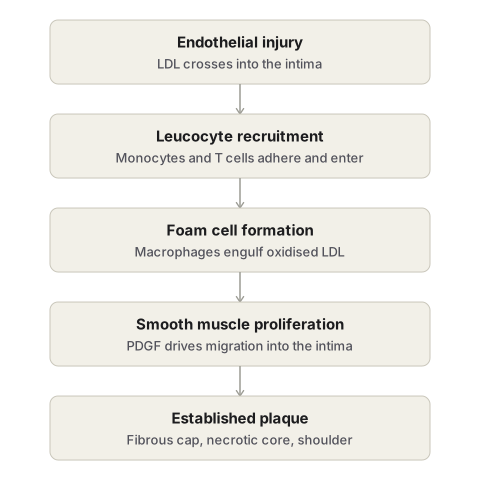
- Endothelial injury/dysfunction (disturbed flow at branch points, hyperlipidaemia, hypertension, smoking toxins, glycation end-products) → ↑permeability, leucocyte and platelet adhesion, LDL entry.
- Monocytes/T cells adhere and migrate into intima → monocytes become macrophages.
- LDL entering the intima is oxidised (by macrophage- and endothelium-derived free radicals) — oxidised LDL is cytotoxic.
- Macrophages engulf oxidised LDL via scavenger receptors → foam cells; apoptotic foam cells release lipid → necrotic core. Activated macrophages release cytokines, chemokines, growth factors, more free radicals.
- PDGF and other growth factors recruit smooth muscle cells from media into intima → proliferation + ECM synthesis (collagen, elastin, proteoglycans) → fibrous cap. Smooth muscle cells can also become foam cells.
- Lipid accumulates both intra- and extracellularly; HDL counteracts by removing cholesterol.
(Older/rival theories: Virchow’s insudation/lipid theory, Rokitansky’s encrustation/thrombogenic theory, monoclonal hypothesis — folded into or superseded by response-to-injury.)
Morphology — sequence
Fatty streaks: earliest lesion, present even in children; flat/raised yellow dots or streaks; foam cells under intact endothelium; harmless, may not progress.
Atheromatous plaque — three zones:
- Fibrous cap: smooth muscle + collagen + ECM, covered by endothelium.
- Necrotic core: cholesterol clefts, cell debris, foam cells, ± thrombus.
- Shoulder: cellular margin — macrophages, smooth muscle, T cells, neovascularisation.
Gross: white-yellow (red-brown if thrombosed), eccentric, focal. Site preference (descending frequency): abdominal aorta > coronary > popliteal > internal carotid > circle of Willis.
Stable vs vulnerable plaque
| Stable | Vulnerable | |
|---|---|---|
| Cap | Thick, collagenous | Thin |
| Inflammation | Minimal | Marked |
| Lipid core | Small | Large |
| Rupture risk | Low | High |
Cap integrity = balance of collagen synthesis (smooth muscle cells) vs degradation (MMPs from inflammatory cells).
Complications
Rupture/ulceration/erosion · haemorrhage into plaque · superimposed thrombosis (occlusion or thromboembolism) · atheroembolism · dystrophic calcification of the necrotic core (distinct from Mönckeberg’s medial calcification) · aneurysmal dilatation from medial/adventitial weakening.
Clinical consequences
Heart: angina, MI. Brain: TIA, stroke. Limbs: claudication, gangrene. Gut: mesenteric ischaemia. Aorta: aneurysm. Critical stenosis ≈ 70% cross-sectional narrowing — threshold for exertional symptoms.
Clinical relevance
Plaque biology (vulnerable vs stable), not size alone, determines risk of an acute event. Statins lower LDL and independently stabilise plaques by reducing inflammation.
Definition
Atherosclerosis is a progressive disease of the intima of large and medium-sized elastic and muscular arteries, marked by focal, lipid-rich intimal lesions called atheromas or atheromatous plaques, which thicken and stiffen the arterial wall and can encroach on the lumen. The name derives from the Greek for porridge, describing the soft, lipid-rich material found at the centre of a mature plaque, combined with the fibrous scarring that forms around it. It is the single most important disease of arteries, since the ischaemic consequences of plaques in the coronary, cerebral, and peripheral circulations account for the majority of cardiovascular deaths worldwide.
Risk Factors
Risk rises sharply as risk factors accumulate rather than simply adding together — two factors raise risk roughly fourfold, and the combination of hyperlipidaemia, hypertension, and smoking raises it about sevenfold.
| Category | Factors |
|---|---|
| Modifiable major | Dyslipidaemia, hypertension, cigarette smoking, diabetes mellitus |
| Constitutional (non-modifiable) | Increasing age, male sex, genetic factors, family history |
| Additional/emerging | Inflammation and elevated C-reactive protein, hyperhomocysteinaemia, elevated lipoprotein(a), metabolic syndrome, raised procoagulant levels, physical inactivity, stressful lifestyle, obesity, certain infections |
Dyslipidaemia is the most firmly established of the major risk factors. Low-density lipoprotein (LDL) delivers cholesterol to peripheral tissue and is directly atherogenic, while high-density lipoprotein (HDL) mobilises cholesterol away from the periphery, including from existing atheroma, for excretion by the liver, and is therefore protective. Hypercholesterolaemia can initiate atherosclerotic lesions even without any other risk factor present, and lowering LDL — whether by diet, exercise, or statin therapy, which inhibits the rate-limiting hepatic enzyme HMG-CoA reductase — reduces that risk.
Hypertension doubles the risk of cardiovascular disease overall, acting largely through mechanical injury to the arterial wall, and treating it reduces the risk of both myocardial infarction and stroke.
Cigarette smoking is the single most important avoidable cause; risk rises with the dose of exposure and falls again once smoking stops.
Diabetes mellitus accelerates atherosclerosis through hypercholesterolaemia, endothelial dysfunction, and a prothrombotic state, and substantially raises the risk of myocardial infarction, stroke, and peripheral gangrene.
Pathogenesis: The Response-to-Injury Hypothesis
The currently accepted model treats atherosclerosis as a chronic inflammatory and fibroproliferative response of the arterial wall to sustained or repeated injury of the endothelium, incorporating elements of two earlier historical theories — Virchow’s proposal that lipid accumulates within the intima, and Rokitansky’s proposal that thrombotic material becomes incorporated into the wall. The process unfolds as a sequence of interacting steps.
- Endothelial injury and dysfunction. Disturbed blood flow at vessel branch points and ostia, together with risk factors such as hyperlipidaemia, hypertension, cigarette toxins, and advanced glycation end-products in diabetes, injures or activates the endothelium. This increases vascular permeability, promotes leucocyte adhesion, favours platelet adhesion, and allows LDL to cross into the intima.
- Monocyte migration into the intima. Monocytes and T lymphocytes adhere to the dysfunctional endothelium and, drawn by locally produced chemokines, migrate into the intima, where the monocytes differentiate into macrophages.
- Lipid accumulation and oxidation. LDL entering the intima through the leaky endothelium is oxidised, chiefly by free radicals generated by the infiltrating macrophages and the dysfunctional endothelium itself. Oxidised LDL is directly cytotoxic to endothelial and smooth muscle cells.
- Foam cell formation. Macrophages take up native and oxidised LDL through scavenger receptors, becoming lipid-laden foam cells. Activated macrophages release cytokines that recruit further leucocytes, growth factors that drive smooth muscle proliferation, and more free radicals that perpetuate LDL oxidation. Foam cells that die by apoptosis release their lipid, contributing to the necrotic lipid core of the maturing plaque.
- Smooth muscle recruitment and proliferation. Growth factors — chiefly platelet-derived growth factor, released by activated platelets, macrophages, and endothelium — draw smooth muscle cells from the media into the intima, where they proliferate and synthesise extracellular matrix (collagen, elastic fibres, proteoglycans), building the fibrous component of the plaque. Smooth muscle cells may themselves take up oxidised LDL and become foam cells.
- Lipid accumulation continues both within cells and extracellularly, from ongoing insudation of plasma lipid and from the debris of dying foam cells, while HDL works in the opposite direction, mobilising cholesterol out of the lesion.
A rival monoclonal hypothesis, based on the observation that smooth muscle cells within a plaque often share a single glucose-6-phosphate dehydrogenase isoenzyme, proposes that the smooth muscle proliferation is itself a clonal, neoplasm-like event triggered by mutagenic exposures such as cigarette smoke or lipid metabolites; this concept has been folded into, rather than replacing, the response-to-injury model.
Morphology
Atherosclerotic lesions evolve through a recognisable sequence, from harmless precursor lesions to fully developed plaques capable of producing disease.
Fatty Streaks
The earliest lesion, present even in children, composed of lipid-laden foam cells lying beneath an intact endothelium. Grossly they appear as small, flat or slightly raised yellow dots or elongated streaks, most prominent in the aorta; they do not themselves obstruct flow and, in many individuals, do not progress further, though they can be the precursor of a true plaque.
The Atheromatous Plaque
A fully developed lesion — also called a fibrous or fibrofatty plaque — with three structural zones.
- Fibrous cap: the superficial, luminal layer, composed of smooth muscle cells, dense collagen, and extracellular matrix, covered by endothelium.
- Necrotic core: deep to the cap, containing extracellular lipid (seen as needle-shaped cholesterol clefts on routine staining), debris from dead cells, foam cells, and sometimes fibrin or organised thrombus.
- Shoulder region: the more cellular margin beneath and to the side of the cap, containing macrophages, smooth muscle cells, and T lymphocytes, often with small new blood vessels (neovascularisation).
Grossly, plaques are white to yellow, becoming red-brown if a thrombus is superimposed, ranging from a few millimetres up to several centimetres, with a soft, yellow, grumous core beneath a firm white cap. They are distributed focally and eccentrically rather than uniformly around the vessel circumference, and show a strong preference for particular sites in descending order of frequency: the abdominal aorta (more than the thoracic aorta), the coronary arteries, the popliteal arteries, the internal carotid arteries, and the vessels of the circle of Willis.
Stable versus Vulnerable Plaques
The clinical behaviour of a plaque depends less on its size than on its structural stability.
| Feature | Stable plaque | Vulnerable plaque |
|---|---|---|
| Fibrous cap | Thick, densely collagenous | Thin |
| Inflammation | Minimal | Marked, with abundant inflammatory cells |
| Lipid core | Small or negligible | Large, rich in foam cells and extracellular lipid |
| Smooth muscle content | Preserved | Sparse |
| Risk of rupture | Low | High |
The strength of the fibrous cap reflects a balance between ongoing collagen synthesis by smooth muscle cells and collagen degradation by matrix metalloproteinases from activated inflammatory cells; when degradation outpaces synthesis, the cap thins and becomes prone to rupture.
Complications of the Plaque
A once-stable plaque can undergo a series of superimposed changes, several of which are the immediate mechanical cause of acute clinical events.
- Rupture, ulceration, or erosion of the surface, exposing highly thrombogenic material to the bloodstream.
- Haemorrhage into the plaque, from rupture of the cap or of fragile neovascular vessels, abruptly enlarging the lesion.
- Superimposed thrombosis, which may partially or completely occlude the lumen, or fragment to produce distal thromboembolism.
- Atheroembolism, the discharge of atheromatous debris itself into the circulation after plaque rupture.
- Calcification, occurring in the necrotic core of advanced plaques, giving the intima a brittle, egg-shell-like quality on incision — distinct from the medial calcification of Mönckeberg’s sclerosis, which spares the intima entirely.
- Aneurysmal dilatation, from secondary weakening, atrophy, and thinning of the underlying media and adventitia beneath a severe plaque.
Clinical Consequences
The effect of atherosclerosis at any site depends on the degree of luminal narrowing and the metabolic demands of the tissue supplied. Critical stenosis — most often defined as roughly seventy percent reduction in cross-sectional area — is the threshold beyond which fixed obstruction typically produces symptoms on exertion, such as stable angina in the coronary circulation. The major clinical syndromes are:
- Heart: angina and myocardial infarction.
- Brain: transient ischaemic attacks and cerebral infarction (stroke).
- Peripheral circulation: intermittent claudication and gangrene of the extremities, most often the lower limb.
- Mesenteric circulation: bowel ischaemia.
- Aorta: aneurysmal dilatation, particularly of the abdominal aorta.
Clinical Relevance
- The distinction between a stable and a vulnerable plaque explains why a coronary lesion causing only moderate, unremarkable narrowing on angiography can still be the site of a fatal acute event — the danger lies in the plaque’s biology, not simply its size.
- Statins reduce cardiovascular risk through two separate mechanisms: lowering circulating LDL, and independently stabilising existing plaques by reducing the inflammatory activity that thins the fibrous cap.
- Recognising the strong site preference of atherosclerosis — abdominal aorta, coronary, popliteal, internal carotid, and circle of Willis arteries, in that order — helps anticipate where clinical disease is most likely to first declare itself in a given patient.
Diagram 1: Response-to-Injury Pathogenesis (mechanism sequence)
Draw a cross-section of an arterial wall (intima, media, adventitia labelled) as a sequence of five panels left to right, each showing the same cross-section progressing further.
Panel 1 — endothelial injury: intact media and adventitia; endothelial cells drawn as a continuous line except for one region shown disrupted or dysfunctional, with small arrows showing LDL particles crossing into the intima at that point.
Panel 2 — leucocyte recruitment: monocytes and lymphocytes shown adhering to the dysfunctional endothelium and squeezing between endothelial cells into the intima.
Panel 3 — foam cell formation: monocytes now drawn as macrophages within the intima, several containing multiple small round vacuoles (lipid) to represent foam cells; a few oxidised LDL particles shown nearby with a jagged outline to distinguish them from native LDL.
Panel 4 — smooth muscle migration and proliferation: smooth muscle cells shown migrating from the media through a gap in the internal elastic lamina into the intima, with several shown dividing.
Panel 5 — established plaque: the full three-zone structure — a fibrous cap of smooth muscle and collagen at the top, a necrotic lipid core with needle-shaped clefts beneath it, and a cellular shoulder region at the margins containing macrophages, smooth muscle cells, and lymphocytes.
Labels required
- Each panel individually titled with the step it represents.
- Panel 3 must distinguish native LDL from oxidised LDL by label, since the oxidation step is a specific examinable point.
- Panel 5 must label all three zones (fibrous cap, necrotic core, shoulder) by name.
- PDGF should be labelled as the growth factor driving the panel 4 transition.
Errors commonly made
- Skipping the oxidation of LDL and drawing foam cells forming directly from native LDL — oxidation is the step that makes LDL recognisable to macrophage scavenger receptors and is frequently asked about by name.
- Drawing the necrotic core in the wrong position, superficial to the fibrous cap instead of deep to it.
- Omitting the shoulder region entirely and drawing only cap and core — the shoulder is where plaque rupture most often originates and is a specifically testable structure.
Diagram 2: Stable vs Vulnerable Plaque (comparative cross-section)
Draw two arterial cross-sections side by side, both narrowing the lumen to a similar degree, so the visual contrast is purely about internal structure rather than size.
Left — stable plaque: thick, densely hatched fibrous cap; small, barely visible lipid core; only a few scattered inflammatory cells at the shoulder.
Right — vulnerable plaque: thin, minimally hatched fibrous cap; large lipid core filling most of the plaque, drawn with dense cholesterol-cleft markings; numerous inflammatory cells clustered at the shoulder and beneath the cap; a small tear drawn partway through the thin cap to foreshadow rupture.
Labels required
- Cap thickness, core size, and inflammatory cell density must each be labelled with a comparative note (thick vs thin, small vs large, sparse vs marked) rather than left to the drawing alone.
- A caption beneath each plaque stating its clinical behaviour: “low rupture risk” vs “high rupture risk.”
- A note that both plaques can produce the same degree of luminal narrowing, since this is the specific point the diagram is meant to illustrate — that stenosis severity alone does not predict acute risk.
Errors commonly made
- Drawing the vulnerable plaque as more severely stenotic than the stable one, which defeats the purpose of the comparison — the two should look similar in the degree of narrowing and differ only in internal composition.
- Omitting the small tear/fissure in the vulnerable cap, which is the visual cue tying this diagram to the mechanism of acute plaque change.
Short Essays (1)
Multiple Choice Questions (1)
My Sticky Notes
Personal revision notes, mnemonics and reminders.
