
Wound Healing
Definition
Healing = regeneration (parenchymal cell proliferation, complete restoration) + repair (connective tissue replacement → fibrosis/scar). Often both together.
Cell types (regenerative capacity)
- Labile — always dividing (epidermis, GI/resp/urinary epithelium, endometrium, marrow/lymphoid)
- Stable — G0, can re-enter cycle (liver, kidney, pancreas, adrenal, thyroid, fibroblasts, endothelium, bone, cartilage)
- Permanent — never regenerate (neurons, skeletal muscle, cardiac muscle) → repair only
Regeneration = margin proliferation + migration → differentiation/maturation. Driven by growth factors (EGF, FGF, PDGF, TGF-β) → cyclins/CDKs.
Granulation tissue formation (3 phases)
- Inflammation (<24h) — clot, exudation (plasma, neutrophils, monocytes)
- Clearance — neutrophil proteases + macrophage phagocytosis clear debris
- Ingrowth:
- Angiogenesis — endothelial budding from severed vessel margins, initially leaky (oedema) → arterioles/venules/capillaries. Driven by VEGF (macrophage-derived, hypoxia/HIF-triggered) + PDGF/TGF-β/bFGF/integrins
- Fibrogenesis — myofibroblasts, collagen from day 6 (fibronectin cross-links) → ↑collagen ↓cells/vessels = cicatrisation
Skin wound healing
Primary union (1° intention) — clean, sutured, minimal loss:
- Haemorrhage/clot seals wound
- Polymorphs (24h) → macrophages (d3)
- Epithelial spurs, covered by 48h, multilayered by d5
- Fibroblasts (d3) → collagen (d5) → mature scar (4wk)
- Suture tracks = mini-wounds; removed d7 → risk stitch abscess/implantation cyst
Secondary union (2° intention) — open, large defect, ± infected, unsutured: Same steps but bridges larger gap (base-up + margin-in) → slower, bigger scar. Extra features: exuberant granulation tissue; wound contraction (myofibroblasts, unique to 2° union, →1/3-1/4 original size, starts d2-3, complete by d14); adnexa (hair/sweat glands) NOT regenerated; infection risk higher.
| Primary | Secondary | |
|---|---|---|
| Wound | Clean | Unclean/infected |
| Sutures | Yes | No |
| Granulation | Scanty | Exuberant |
| Scar | Neat linear | Contracted, irregular |
Complications
Infection · implantation cyst · pigmentation (haemosiderin) · deficient scar · incisional hernia/dehiscence · hypertrophied scar (within wound margins) vs keloid (beyond margins, tumour-like, more in Blacks) · contracture (Dupuytren’s, plantar, Peyronie’s) · neoplasia (Marjolin’s ulcer = SCC in burn scar).
ECM (5 components)
Collagen (I/III/V fibrillar = scar; IV = basement membrane) · adhesive glycoproteins (fibronectin-plasma/tissue, tenascin @48h then disappears, thrombospondin) · basement membrane (PAS+, type IV + laminin) · elastic fibres (recoil, not strength; elastase-degraded) · proteoglycans (chondroitin/heparan/dermatan/keratan sulphate, hyaluronic acid — deposited BEFORE collagen).
Wound strength: 10% @d7 (suture removal) → 80% @3mo.
Factors affecting healing
Local: infection (most important) · poor blood supply · foreign bodies/sutures · movement · radiation (delays) · UV (helps) · injury type/size/site. Systemic: age · nutrition (protein, vit C-scurvy, vit A, zinc deficiency) · systemic infection · glucocorticoids (slow healing) · diabetes · neutrophil defects/neutropenia/bleeding disorders.
Fracture healing
Primary union — surgical apposition (plates/clamps) → medullary callus only, no periosteal callus, early ambulation but more necrosis, slower.
Secondary union (plaster cast — more common), 3 stages:
- Procallus: haematoma → inflammation/clearance (macrophages+osteoclasts) → granulation ingrowth (periosteum/endosteum) → woven bone ± cartilage callus (fusiform, “provisional callus”)
- Osseous callus: osteoclasts clear woven bone/cartilage → osteoblasts lay lamellar bone (Haversian systems)
- Remodelling: osteoblast+osteoclast reshaping → external callus cleared, cortex reforms, marrow cavity reforms internally
Complications: fibrous union (± pseudo-arthrosis) · non-union (soft tissue interposed) · delayed union (infection, poor supply/nutrition, movement, age).
Nervous tissue healing
CNS: neurons = permanent, no regeneration; axons don’t regenerate; astrocyte proliferation = gliosis (glial scar). PNS: limited but real regeneration.
Clinical relevance
Labile/stable/permanent classification predicts regeneration vs scar outcome (MI/neuron = permanent scar; skin/gut = full restoration). Wound contraction specific to 2° union. Hypertrophied scar vs keloid = real clinical distinction. VEGF/hypoxia link = same biology behind pro-angiogenic (ischaemic heart) and anti-angiogenic (tumour, wet AMD) therapy.
Definition
Healing is the body’s attempt to restore normal structure and function after injury, and proceeds by two processes that frequently occur together: regeneration, restoration of the original tissue by proliferation of parenchymal cells, and repair, replacement of lost tissue by connective tissue, resulting in fibrosis and scarring.
Regenerative Capacity of Tissues
Whether regeneration is even possible for a given tissue depends on the proliferative capacity of its parenchymal cells, which fall into three groups defined by their relationship to the cell cycle:
- Labile cells — continuously dividing throughout life, remaining actively in the cell cycle from one mitosis to the next: surface epithelium of epidermis, alimentary/respiratory/urinary tract, vagina, cervix, uterine endometrium, and the haematopoietic cells of bone marrow, lymph nodes and spleen.
- Stable cells — normally quiescent (resting in G0) after adolescence but retain the capacity to re-enter the cycle and proliferate when stimulated: parenchymal cells of liver, pancreas, kidney, adrenal and thyroid, and mesenchymal cells (smooth muscle, fibroblasts, vascular endothelium, bone and cartilage).
- Permanent cells — lose proliferative capacity around the time of birth and, once destroyed, are never replaced: neurons, skeletal muscle, cardiac muscle. Injury here can only be repaired, never regenerated.
Regeneration of any parenchymal cell type requires two coupled processes: proliferation of surviving cells at the margin of injury with migration to cover the defect, and subsequent differentiation/maturation of the migrated cells to reconstitute the original tissue architecture. Mitosis itself is driven by cyclins (A, B, E) activating cyclin-dependent kinases (CDKs), with growth factors (EGF, FGF, PDGF, TGF-β and others) providing the upstream regulatory signal that pushes stable cells out of G0 and into cycle.
Repair: Granulation Tissue Formation
Repair proceeds through granulation tissue formation followed by wound contraction, involving mesenchymal cells, endothelial cells, macrophages, platelets and the parenchymal cells of the injured organ. Granulation tissue takes its name from the granular, pink appearance produced by numerous small new blood vessels lifted slightly above the surface by a thin covering of fibroblasts and young collagen. Its formation proceeds through three phases:
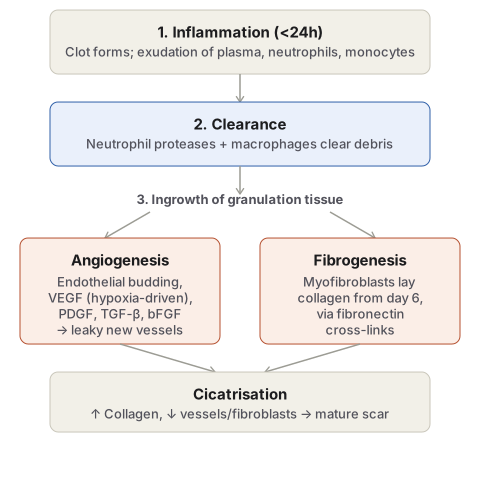
- Phase of inflammation — blood clots at the injury site within minutes; an acute inflammatory response follows within 24 hours, with exudation of plasma, neutrophils and some monocytes.
- Phase of clearance — proteolytic enzymes from neutrophils, autolytic enzymes from dead cells, and macrophage phagocytosis together clear necrotic tissue, debris and red cells.
- Phase of ingrowth of granulation tissue — combining two parallel processes:
- Angiogenesis, new vessel formation by proliferation of endothelial cells from the margins of severed vessels — initially solid buds, developing a lumen and blood flow within hours; these early vessels are more leaky, accounting for the oedematous look of fresh granulation tissue, before differentiating into arterioles, venules and true capillaries. Angiogenesis is triggered by proteolytic destruction of the basement membrane and driven chiefly by vascular endothelial growth factor (VEGF) — produced by macrophages and other mesenchymal cells, often under hypoxia-inducible-factor control in response to local hypoxia, and acting on endothelium-restricted VEGF receptors to stimulate endothelial migration and proliferation — with PDGF, TGF-β, basic FGF and surface integrins also contributing to cellular proliferation.
- Fibrogenesis, in which new fibroblasts (intermediate in character between fibroblasts and smooth muscle — myofibroblasts) lay down an amorphous matrix around the new vessels; collagen fibrils appear from about day 6, cross-linked into the matrix via fibronectin receptors on the myofibroblast surface. As maturation proceeds, collagen accumulates while active fibroblasts and vessel density fall, producing the relatively acellular, avascular scar — a process termed cicatrisation.
Healing of Skin Wounds
Skin wound healing is the classical worked example of combined regeneration and repair, occurring by one of two patterns.
Healing by First Intention (Primary Union)
Applies to a wound that is clean, uninfected, surgically incised, with minimal tissue loss, and with edges approximated by sutures.
- Initial haemorrhage — blood fills the narrow incisional space and clots, sealing the wound against dehydration and infection.
- Acute inflammatory response — polymorphs from the wound margins within 24 hours, replaced by macrophages by day 3.
- Epithelial changes — basal epidermal cells from both margins proliferate and migrate as epithelial spurs; a well-approximated wound is covered by a continuous epithelial layer within 48 hours, separating the underlying viable dermis from the overlying necrotic clot (which is cast off as a scab); by day 5 a multilayered, differentiated new epidermis is present.
- Organisation — fibroblasts invade by day 3; new collagen fibrils appear by day 5 and dominate thereafter; by 4 weeks a mature scar with scanty cellular/vascular elements and an epithelialised surface has formed.
- Suture tracks — each is itself a small wound undergoing the same sequence; removing sutures around day 7 avulses most of the epithelialised track, with the remainder resorbed — though a stitch abscess (infection) or implantation/epidermal cyst (persistent epithelial cells) can occasionally result.
Because the margins are closely apposed and the tissue defect is minimal, the resulting scar is neat and linear.
Healing by Second Intention (Secondary Union)
Applies to an open wound with a large, sometimes infected tissue defect and extensive cell/tissue loss, not approximated by sutures. The underlying events are the same as primary union but must additionally bridge a substantially larger gap, so healing proceeds both from the base upward and from the margins inward — making it slower and producing a larger, often less cosmetic scar.
- Initial haemorrhage — a larger blood/fibrin clot fills and dries over the open defect.
- Inflammatory phase — acute response followed by macrophage-mediated debris clearance, as in primary union.
- Epithelial changes — epithelial spurs migrate from both margins toward the centre, but cannot fully cover the surface until granulation tissue from the base has substantially filled the defect; regenerated epidermis eventually stratifies and keratinises. Specialised adnexal structures (hair follicles, sweat glands) are not regenerated unless viable residues persist.
- Granulation tissue — forms the main bulk of secondary healing, by fibroblast proliferation and neovascularisation from adjoining viable tissue; fresh granulation tissue is deep red, granular and fragile, maturing to a pale, less vascular scar as collagen accumulates.
- Wound contraction — a feature specific to secondary healing (absent in primary union): myofibroblast activity within the granulation tissue contracts the wound to roughly one-third to one-quarter of its original size, beginning at 2–3 days and largely complete by day 14, reducing the surface area that must actually be replaced and thereby speeding overall healing.
- Infection — bacterial contamination of an open wound delays healing via toxin-provoked necrosis, suppuration and thrombosis; surgical debridement of dead tissue helps prevent this.
| Feature | Primary union | Secondary union |
|---|---|---|
| Wound | Clean, generally uninfected | Unclean, may be infected |
| Margins | Surgically clean | Irregular |
| Sutures | Used | Not used |
| Granulation tissue | Scanty, confined to incisional gap and suture tracks | Exuberant, fills a large defect |
| Outcome | Neat, linear scar | Contracted, irregular scar |
| Complications | Infrequent; epidermal inclusion cyst | Suppuration; may need debridement |
Complications of Wound Healing
- Infection, delaying healing via bacterial toxin effects.
- Implantation (epidermal) cyst, from epithelial cells persisting within the healed wound.
- Pigmentation, from haemosiderin staining or retained particulate material.
- Deficient scar formation, from inadequate granulation tissue.
- Incisional hernia/wound dehiscence, from a weak scar bursting open, particularly after laparotomy.
- Hypertrophied scar and keloid, from excessive collagen deposition — hypertrophied scars stay within the original wound margins, whereas keloids extend beyond them as tumour-like connective tissue projections and are more common in Black patients.
- Excessive contraction/contracture, an exaggeration of normal wound contraction producing conditions such as Dupuytren’s (palmar) contracture, plantar contracture, and Peyronie’s disease (penile cavernous tissue).
- Neoplasia, rarely — squamous cell carcinoma arising in a burn scar is termed a Marjolin’s ulcer.
Extracellular Matrix in Wound Strength
Wound strength is provided by proliferating fibroblasts and myofibroblasts drawing structural support from the extracellular matrix (ECM), which — beyond mechanical support — also directs cell migration, attachment, differentiation and organisation, and serves as a depot for latent growth factors activated at sites of injury. The ECM has five main components: collagen (the principal tensile-strength component; types I, III and V are the fibrillar collagens that dominate scar tissue, while other types form the amorphous basement membrane component, chiefly type IV with laminin); adhesive glycoproteins — fibronectin (plasma fibronectin from the liver, trapped in basement membranes; tissue fibronectin from fibroblasts/endothelium, central to the primitive wound matrix), tenascin (appears ~48 hours post-injury, disappears from the mature scar), and thrombospondin (adhesive for platelets/keratinocytes, inhibitory to fibroblast/endothelial attachment); basement membrane itself (PAS-positive, type IV collagen and laminin); elastic fibres (elastin plus microfibril, providing recoil rather than tensile strength, degraded by elastases); and proteoglycans (glycosaminoglycan-protein complexes — chondroitin sulphate in cartilage/dermis, heparan sulphate in basement membranes, dermatan sulphate in dermis, keratan sulphate in cartilage, hyaluronic acid in cartilage/dermis — whose deposition precedes collagen laying in a healing wound).
Wound strength recovers gradually: about 10% at the time of suture removal (day 7), rising to roughly 80% by 3 months.
Factors Influencing Healing
Local factors: infection is the single most important local factor delaying healing; poor blood supply slows it (facial wounds heal fast, varicose leg ulcers heal slowly); foreign bodies (including sutures) provoke inflammation and infection; movement of the wound delays healing; ionising radiation delays granulation tissue formation, while ultraviolet light facilitates healing; and the type, size and location of the injury determines whether healing proceeds by resolution or organisation.
Systemic factors: age (slower in the elderly/debilitated, from poorer local blood supply); nutrition (deficiency of protein, vitamin C — scurvy — vitamin A, or zinc all delay healing); systemic infection; glucocorticoid administration (anti-inflammatory effect slows healing); uncontrolled diabetes (increased infection susceptibility); and haematologic abnormalities such as neutrophil functional defects (impaired chemotaxis/phagocytosis), neutropenia, or bleeding disorders.
Fracture Healing
Fracture healing by callus formation resembles skin-wound healing in its basic events, though clinical variables — traumatic versus pathological bone, complete versus incomplete (greenstick) fracture, and simple/comminuted/compound fracture type — all influence the specific course.
Primary union occurs when fracture ends are surgically approximated (compression clamps, metal plates): bony union proceeds via medullary callus alone, without periosteal callus, allowing early ambulation but with more extensive bone necrosis and slower healing.
Secondary union, the more common pattern with plaster-cast immobilisation, is a continuous process conventionally divided into three stages:
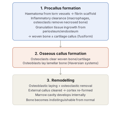
- Procallus formation — (i) a haematoma forms from torn vessels, its fibrin meshwork acting as a scaffold for granulation tissue; (ii) a local inflammatory response exudes fibrin, polymorphs and macrophages, which clear fibrin, red cells, debris and necrosed bone fragments (the latter assisted by osteoclasts); (iii) ingrowth of granulation tissue begins, with neovascularisation and mesenchymal cell proliferation from periosteum and endosteum forming a weak soft-tissue callus joining the fracture ends; (iv) within days, osteogenic cells of the inner periosteal layer lay down collagen and osteoid within the granulation tissue, which calcifies into woven bone callus spanning a wide zone on either side of the fracture in a spindle (fusiform) shape — in poorly immobilised fractures (e.g. rib fractures), cartilage may form instead alongside or as well as woven bone, at this provisional callus (procallus) stage.
- Osseous callus formation — the procallus acts as scaffolding: incoming osteoclasts clear the woven bone, calcified cartilage disintegrates, and new blood vessels and osteoblasts lay down osteoid that calcifies into lamellar bone, organised concentrically around vessels as a Haversian system.
- Remodelling — ongoing osteoblastic deposition and osteoclastic removal reshape the united bone ends until they become indistinguishable from normal bone: external callus is cleared, compact cortical bone replaces the intermediate callus, and the marrow cavity re-forms within the internal callus.
Complications of fracture healing: fibrous union (occasionally with a false joint, pseudo-arthrosis) if immobilisation is inadequate; non-union if soft tissue becomes interposed between the fracture ends; and delayed union from the same general causes that delay any wound healing — infection, poor blood supply, poor nutrition, movement, old age.
Healing in Other Specialised Tissues
Central nervous system: neurons are permanent cells and, once destroyed, are never replaced; CNS axons show no significant regeneration either. Damaged neuroglia may instead proliferate as astrocytes — gliosis — the CNS equivalent of a glial scar. Peripheral nervous system: peripheral nerves, by contrast, show limited but genuine regenerative capacity.
Clinical Relevance
- The labile/stable/permanent classification directly predicts which tissues can genuinely regenerate after injury and which can only be repaired by scar — this is the reason a myocardial infarct or a destroyed neuron leaves permanent fibrous replacement while a superficial abrasion of skin or gut mucosa restores completely.
- Recognising wound contraction as specific to secondary (not primary) union explains why open wounds left to heal by second intention can still end up smaller than expected purely from myofibroblast activity, independent of any new tissue formed.
- Distinguishing hypertrophied scar (confined to the original wound) from keloid (extending beyond it) is a genuine clinical distinction with management and prognostic implications, not just a naming exercise.
- The VEGF/hypoxia link in angiogenesis is the same biology exploited therapeutically in both directions — pro-angiogenic therapy to revascularise ischaemic myocardium, and anti-angiogenic therapy to starve tumours or halt pathological vessel growth in wet macular degeneration — worth keeping in mind as the connective thread between wound healing and these seemingly unrelated clinical contexts.
Diagram 1: Formation of Granulation Tissue
Draw two boxes in a downward column (inflammation, then clearance), then a single label branching into two side-by-side boxes (angiogenesis and fibrogenesis), which both converge into a final box (cicatrisation/mature scar).
- Inflammation — clot forms, plasma/neutrophil/monocyte exudation, within 24 hours.
- Clearance — neutrophil proteases and macrophage phagocytosis remove debris.
- Ingrowth, split into:
- Angiogenesis — endothelial budding, name VEGF as the principal driver, note the hypoxia trigger.
- Fibrogenesis — myofibroblasts, collagen from day 6, fibronectin cross-linking.
- Cicatrisation — rising collagen, falling vessel/fibroblast density, into mature scar.
Labels required
- Angiogenesis and fibrogenesis must be shown as parallel, simultaneous processes both arising from the same “ingrowth” step, not as sequential events — this is the detail most often lost when the sequence is written out as a flat list instead of drawn.
- VEGF must be named specifically as the angiogenesis driver, and the hypoxia → HIF → VEGF trigger noted, since this is the same mechanism tested in ischaemic and tumour angiogenesis contexts elsewhere.
Errors commonly made
- Describing granulation tissue as simply “new blood vessels,” omitting the fibroblast/collagen component that is equally part of the definition.
- Treating cicatrisation as a separate fourth phase rather than as the natural maturation endpoint of the same ingrowth phase.
Diagram 2: Fracture Healing by Secondary Union
Draw a single downward column of three stages.
- Procallus formation — list its four internal steps in order: haematoma, inflammatory clearance, granulation tissue ingrowth, woven bone/cartilage callus (fusiform shape).
- Osseous callus formation — osteoclasts clear the procallus scaffold while osteoblasts lay down lamellar bone in Haversian systems.
- Remodelling — combined osteoblastic/osteoclastic activity restores normal cortical and medullary architecture.
Labels required
- Woven bone (procallus) must be explicitly distinguished from lamellar bone (osseous callus), since mistaking one for the other is a common histology error — woven bone is the temporary scaffold, lamellar bone is the final structural product.
- The fusiform (spindle) shape of the procallus should be noted as a specific, describable morphologic detail, not left as an unlabelled generic swelling.
Errors commonly made
- Describing fracture healing as a single continuous “callus forms then hardens” process, without the two distinct bone-tissue types (woven, then lamellar) that the exam-relevant sequence actually depends on.
- Omitting remodelling as its own stage, leaving the impression that healing ends once bony union (osseous callus) is achieved, when the bone is not yet structurally normal at that point.
Short Essays (1)
Short Answers (1)
Multiple Choice Questions (3)
My Sticky Notes
Personal revision notes, mnemonics and reminders.
