
Vitamin D Deficiency and Rickets
Function
Maintains plasma Ca+phosphate for bone mineralisation, metabolism, neuromuscular transmission. Deficiency → rickets(children) / osteomalacia(adults) / hypocalcaemic tetany (uncommon — ↓Ca usually corrected first by ↑PTH+bone resorption, so skeletal picture dominates).
Synthesis/activation pathway
Skin: 7-dehydrocholesterol+UVB→cholecalciferol(D3), ~90% of supply. Dark skin=↓production (melanin absorbs UV). Diet: minor (fish, plants as ergosterol, fortified milk). Liver: 25-hydroxylase→25(OH)D (major circulating/storage form, clinically measured). Kidney: 1α-hydroxylase→1,25(OH)₂D (ACTIVE hormone). ↑by PTH+hypophosphataemia; ↓by self-feedback.
Actions of 1,25(OH)₂D: Intestine ↑Ca+phosphate absorption | Bone: normocalcaemia→mineralisation; hypocalcaemia→↑RANKL(osteoblast)→osteoclast activation→Ca mobilisation | Parathyroid: ↓PTH gene transcription (feedback) | Kidney: ↑renal Ca resorption.
Causes of deficiency
↓Endogenous synthesis(sunlight) | dietary deficiency | malabsorption (↓bile salts: biliary obstruction/pancreatic insufficiency/malabsorption syndrome) | deranged metabolism (renal dz→↓1α-OHase; liver dz→↓25-OHase; genetic) | end-organ resistance.
Compensatory sequence: ↓Ca→↑PTH→(1)↑renal 1α-hydroxylase activity(↑active vit D+intestinal Ca absorption) (2)bone Ca mobilisation (3)↓renal Ca excretion (4)↑renal phosphate excretion → Ca restored near-normal BUT hypophosphataemia persists = real driver of impaired mineralisation.
Rickets (children, 6mo-2yr, before epiphyseal closure)
2 primary defects: failed mineralisation + deranged endochondral/intramembranous growth.
Endochondral (long bones): Normal = cartilage proliferation→adequate mineralisation→resorption+osteoid replacement→mineralise to bone→normal vascularisation. Rickets = proliferation continues+INADEQUATE mineralisation→cartilage persists/overgrows (osteoid on poor-mineralised cartilage→enlarged costochondral junctions)→deformed bone (no rigidity)→irregular vessel overgrowth in disorganised weak bone.
Intramembranous (flat bones): osteoblasts lay osteoid → normally mineralises; in rickets osteoid FAILS to mineralise → soft weak flat bones.
Skeletal changes: Craniotabes(earliest, unossified skull areas, square skull, resolves by 12mo) | Harrison’s sulcus(rib indrawing on inspiration) | rachitic rosary(costochondral overgrowth) | pigeon chest(sternal protrusion) | bow legs | knock knees | enlarged distal radial epiphyses | lumbar lordosis.
Biochem: ↓25-OH-D+1,25(OH)₂D | Ca normal/slightly↓ | phosphate↓ | ALP↑ (osteoblastic activity).
Vitamin D-dependent rickets = autosomal dominant, distinct entity, responds to direct 1,25(OH)₂D not plain vit D.
Osteomalacia (adults, after epiphyseal closure)
Same lesion as rickets (unmineralised osteoid) but in mature skeleton.
Morphology: widened/thickened osteoid seams(pink,H&E), ↓mineralisation at osteoid-bone border(basophilic); von Kossa stain: unmineralised osteoid unstained vs calcified bone black; ↑osteoclastic activity, marrow fibrosis.
Clinical: muscular weakness, vague bone pain, trivial-trauma fractures, incomplete/greenstick fractures, Looser’s zones (pseudofractures).
Biochem: same pattern as rickets (Ca normal/↓, phosphate↓, ALP↑). Osteoporosis = clinically similar but biochemically NORMAL (distinct disease).
Hypervitaminosis D
Excess → ↑intestinal Ca+phosphate absorption → hypercalcaemia+hyperphosphataemia+↑bone resorption → ↑urinary Ca/phosphate excretion, renal calculi risk, osteoporosis, widespread metastatic calcification (renal tubules, arteries, myocardium, lungs, stomach — most marked sites).
Clinical relevance
Persistent hypophosphataemia (not transient hypocalcaemia) = real mineralisation-defect driver → raised ALP = most consistent biochemical clue in both conditions. Rickets vs osteomalacia = same biochemistry, different skeletal age (before vs after epiphyseal closure). Osteomalacia vs osteoporosis: same clinical/radiographic look in elderly, but biochemistry distinguishes (abnormal vs normal) — treatment-relevant. Vit D-dependent rickets needing 1,25(OH)₂D specifically (not plain vit D) = reminder not all rickets is nutritional.
Function of Vitamin D
Vitamin D’s principal role is maintaining adequate plasma calcium and phosphorus for bone mineralisation, general metabolic function, and neuromuscular transmission. Deficiency, whatever its cause, produces one or more of three clinical pictures: rickets in growing children, osteomalacia in adults, and hypocalcaemic tetany from disturbed neuromuscular excitability — though tetany is uncommon in practice, because a fall in serum calcium is usually corrected first by increased parathyroid hormone secretion and bone resorption, which shifts the dominant clinical picture toward the skeleton instead.
Synthesis and Activation
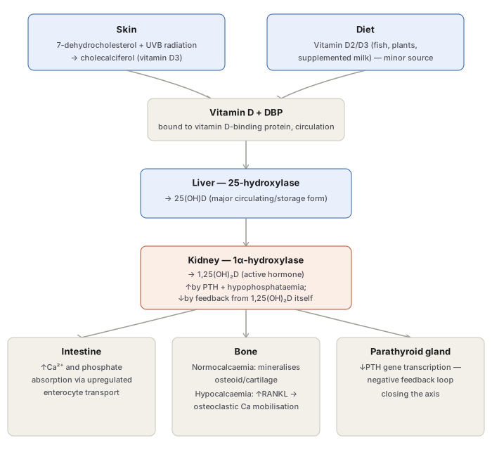
- Skin — UVB radiation converts 7-dehydrocholesterol to cholecalciferol (vitamin D3); under ordinary sun exposure, roughly 90% of the body’s vitamin D is derived this way. Darker skin tones produce less vitamin D per unit sun exposure, since melanin absorbs UV light.
- Diet — a minor contributor (deep-sea fish, plants/grains as ergosterol, fortified milk).
- Liver — 25-hydroxylase converts vitamin D to 25-hydroxyvitamin D (25-OH-D), the major circulating/storage form and the one measured clinically to assess vitamin D status.
- Kidney — 1α-hydroxylase converts 25-OH-D to 1,25-dihydroxyvitamin D [1,25-(OH)₂D], the biologically active hormone. This final activation step is tightly regulated: stimulated by parathyroid hormone (PTH) and by hypophosphataemia, and inhibited by feedback from 1,25-(OH)₂D itself once levels rise sufficiently.
Actions of 1,25-(OH)₂D (acting via a nuclear receptor present in most nucleated cells, inducing target gene transcription):
- Intestine — upregulates enterocyte calcium and phosphate absorption.
- Bone — effect depends on calcium status: in normocalcaemia, supports mineralisation of osteoid matrix and epiphyseal cartilage; in hypocalcaemia, together with PTH, upregulates RANK ligand on osteoblasts, activating osteoclast precursors to mobilise calcium and phosphate from bone to protect serum levels.
- Parathyroid gland — suppresses PTH gene transcription, a negative feedback loop closing the axis.
- Kidney — promotes renal tubular calcium resorption.
Causes of Vitamin D Deficiency
- Reduced endogenous synthesis — inadequate sunlight exposure.
- Dietary deficiency.
- Malabsorption of dietary lipids — from lack of bile salts, as in intrahepatic biliary obstruction, pancreatic insufficiency, or malabsorption syndromes.
- Derangements of vitamin D metabolism — kidney disease (chronic renal failure, nephrotic syndrome, uraemia — impairing 1α-hydroxylation), liver disease (impairing 25-hydroxylation), or genetic disorders.
- End-organ resistance to vitamin D action, despite normal hormone levels.
The compensatory sequence once deficiency develops: falling serum calcium → increased PTH secretion → (1) activation of renal 1α-hydroxylase (raising active vitamin D and intestinal calcium absorption), (2) mobilisation of calcium from bone, (3) reduced renal calcium excretion, (4) increased renal phosphate excretion. Serum calcium is thereby restored toward normal, but hypophosphataemia persists, and it is this persistent hypophosphataemia — not hypocalcaemia — that is chiefly responsible for the impaired bone mineralisation seen in both rickets and osteomalacia.
Rickets (Growing Children)
Two primary defects: failure of bone mineralisation and deranged endochondral and intramembranous bone growth. Rickets classically affects children 6 months to 2 years of age, before epiphyseal closure.
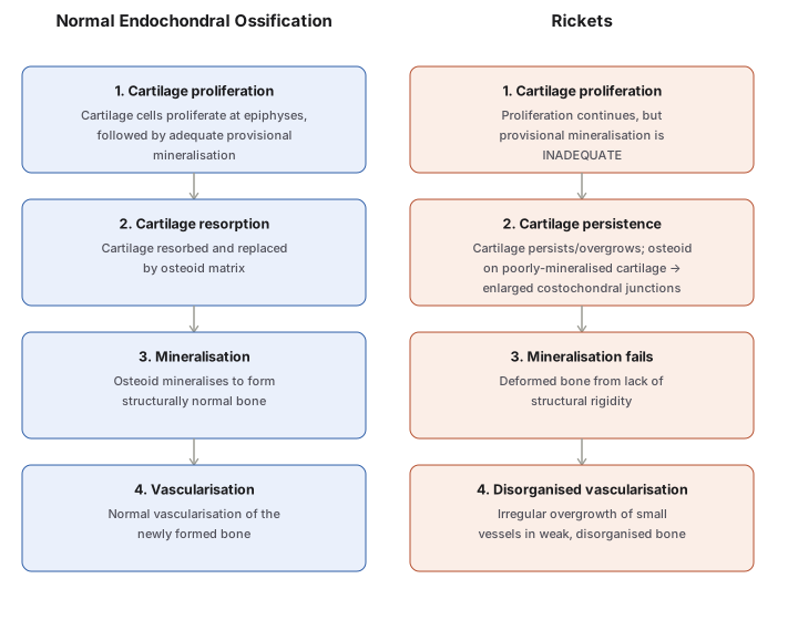
The pathogenesis is best understood by contrasting it with normal bone growth:
- Endochondral ossification (long tubular bones) — normally, cartilage proliferates at the epiphyses with adequate provisional mineralisation, is resorbed and replaced by osteoid, mineralises into bone, and is normally vascularised. In rickets, proliferation continues but mineralisation is inadequate, cartilage persists and overgrows (with osteoid deposited on poorly mineralised cartilage, producing enlarged costochondral junctions), the resulting bone is deformed from lack of structural rigidity, and small vessels overgrow irregularly through disorganised, weak bone.
- Intramembranous ossification (flat bones) — mesenchymal cells normally differentiate into osteoblasts that lay down osteoid which then mineralises; in rickets, osteoid is laid down but fails to mineralise, leaving soft, weak flat bones.
Skeletal changes:
- Craniotabes — the earliest lesion; small round unossified areas of the skull, giving a square, box-like skull; resolves within 12 months of birth.
- Harrison’s sulcus — indrawing of soft ribs on inspiration.
- Rachitic rosary — cartilaginous overgrowth at the costochondral junctions.
- Pigeon-chest deformity — anterior sternal protrusion from respiratory muscle action.
- Bow legs — weak lower-leg bones in ambulatory children.
- Knock knees — enlarged distal femur/proximal tibia-fibula ends.
- Enlarged lower radial epiphyses.
- Lumbar lordosis — from spinal and pelvic involvement.
Biochemical changes: reduced active vitamin D metabolites (25-OH-D and 1,25-(OH)₂D); serum calcium normal or slightly low; serum phosphate low; serum alkaline phosphatase raised (reflecting increased osteoblastic activity).
Vitamin D-dependent rickets is a distinct autosomal dominant disorder of vitamin D metabolism that responds rapidly to direct administration of 1,25-dihydroxyvitamin D, rather than to ordinary vitamin D supplementation.
Osteomalacia (Adults)
The adult counterpart of rickets — failure of mineralisation of osteoid matrix, occurring after epiphyseal closure, from dietary deficiency, poor endogenous synthesis, or conditioned deficiency.
Morphology: widened, thickened osteoid seams (pink on H&E) with decreased mineralisation at the osteoid-bone interface (basophilic); von Kossa’s stain highlights unmineralised osteoid (unstained) against calcified bone (black); increased osteoclastic activity and marrow fibrosis may accompany.
Clinical features: muscular weakness, vague bony pain, fractures after trivial trauma, incomplete/greenstick fractures, and Looser’s zones (pseudofractures) — radiolucent bands at characteristic weak points in bone.
Biochemistry: serum calcium normal or low, phosphate low, alkaline phosphatase raised — essentially the same biochemical signature as rickets, reflecting the shared underlying defect. (Osteoporosis, a distinct age-related skeletal disorder, is clinically similar but biochemically different — normal calcium/phosphate/alkaline phosphatase.)
Hypervitaminosis D
Excess vitamin D increases intestinal calcium and phosphate absorption, producing hypercalcaemia, hyperphosphataemia, and increased bone resorption, with downstream effects: increased urinary calcium/phosphate excretion, predisposition to renal calculi, osteoporosis, and widespread metastatic calcification — most marked in the renal tubules, arteries, myocardium, lungs, and stomach.
Clinical Relevance
- The persistent hypophosphataemia (not the transiently corrected hypocalcaemia) is the real mechanistic driver of impaired mineralisation in both rickets and osteomalacia — this is why raised alkaline phosphatase, not a dramatically low calcium, is the most consistent biochemical clue across both conditions.
- Rickets and osteomalacia share an identical underlying lesion (unmineralised osteoid excess) but differ entirely in clinical presentation because rickets occurs before epiphyseal closure (producing growth-plate deformity) while osteomalacia occurs after (producing weak, pain-prone mature bone) — the same biochemistry, different skeletal age.
- Distinguishing osteomalacia from osteoporosis by biochemistry (abnormal in osteomalacia, normal in osteoporosis) despite similar clinical/radiographic appearance in the elderly is a frequently tested discrimination with direct treatment implications.
- Vitamin D-dependent rickets responding specifically to 1,25-(OH)₂D rather than standard vitamin D supplementation is a useful reminder that not every rachitic presentation reflects simple nutritional deficiency — some reflect a fixed defect in the activation pathway itself.
Diagram 1: Vitamin D Synthesis, Activation, and Downstream Actions
Draw two converging source boxes (skin, diet) feeding into a single circulating vitamin D box, then a linear liver→kidney activation sequence, then three parallel action boxes (intestine, bone, parathyroid) fed from the kidney box.
- Skin (7-dehydrocholesterol + UVB → D3, ~90% of supply) and Diet (D2/D3, minor source) converge.
- Vitamin D + DBP in circulation.
- Liver: 25-hydroxylase → 25(OH)D (storage/measured form).
- Kidney: 1α-hydroxylase → 1,25(OH)₂D (active form); regulated up by PTH/hypophosphataemia, down by self-feedback.
- Three parallel actions: Intestine (↑Ca/phosphate absorption), Bone (mineralise if normocalcaemic / resorb via RANKL if hypocalcaemic), Parathyroid (↓PTH transcription, feedback).
Labels required
- The liver and kidney enzyme names (25-hydroxylase, 1α-hydroxylase) must both appear — this is the detail that separates a student who understands the activation sequence from one who only knows “vitamin D gets activated somewhere.”
- The bone box must show both branches of its calcium-dependent behaviour (mineralise vs resorb) — collapsing this to “vitamin D builds bone” loses the RANKL-mediated resorptive half of the physiology, which is exactly what explains why vitamin D deficiency does not simply cause weak bone directly but works through a compensatory PTH-driven cascade.
- The kidney box’s dual regulation (PTH/hypophosphataemia stimulate; self-product inhibits) must be shown together, since it is a self-contained feedback loop worth recognising as a unit.
Errors commonly made
- Reversing which organ does which hydroxylation — 25-hydroxylation is hepatic, 1α-hydroxylation is renal; swapping them is a common error.
- Treating 25(OH)D as the active hormone rather than the storage/measured form — 1,25(OH)₂D is the biologically active form, a distinction that matters clinically since serum 25(OH)D, not 1,25(OH)₂D, is what is measured to assess vitamin D status.
- Omitting that PTH itself is part of the kidney’s regulatory input, not just a downstream consequence of vitamin D deficiency — the two hormones regulate each other bidirectionally.
Diagram 2: Normal Endochondral Ossification Contrasted With Rickets
Draw two parallel columns (Normal, Rickets), each with four stacked stages matched step-for-step, so the point of divergence at each stage is directly comparable.
- Cartilage proliferation — adequate (normal) vs inadequate (rickets) provisional mineralisation.
- Cartilage resorption/replacement — resorbed and replaced by osteoid (normal) vs persists and overgrows, producing enlarged costochondral junctions (rickets).
- Mineralisation — osteoid mineralises to bone (normal) vs mineralisation fails, deforming the bone (rickets).
- Vascularisation — normal (normal) vs irregular vessel overgrowth in disorganised bone (rickets).
Labels required
- Each rickets-column box must be explicitly tied to the matching normal-column step it diverges from — the value of this diagram is the step-for-step comparison, not the rickets column alone.
- “Enlarged costochondral junctions” (step 2) must be explicitly linked to the clinical finding “rachitic rosary,” since that is the bedside sign this histological step actually explains.
Errors commonly made
- Describing rickets as simply “soft bones” without being able to specify which step of normal ossification fails and why (inadequate provisional mineralisation at the cartilage stage, not a defect in cartilage production itself).
- Conflating the endochondral defect described here with the separate intramembranous ossification defect in flat bones (osteoid laid down normally but failing to mineralise) — the two processes are described separately in
notes.mdand should not be merged into one diagram.
My Sticky Notes
Personal revision notes, mnemonics and reminders.
