
Viral Haemorrhagic Fever
Overview
Viral haemorrhagic fevers = acute viral infections, common features: haemorrhage, shock, sometimes death. Formerly “arbovirus” group (assumed all arthropod-borne — now known incorrect). Classified by transmission route into 4 groups:
- Mosquito-borne — yellow fever, dengue, Rift Valley fever
- Tick-borne — Crimean haemorrhagic fever, Kyasanur Forest disease
- Zoonotic — Korean haemorrhagic fever, Lassa fever
- Unknown route — Marburg, Ebola
Mosquito-borne (Aedes aegypti) = commonest worldwide, esp. developing countries. Two key examples below.
Yellow fever
Oldest known viral haemorrhagic fever, Africa/South America. Monkeys = reservoir (no illness), Aedes aegypti transmits to humans.
Clinical: sudden high fever, chills, myalgia, headache, jaundice, hepatic+renal failure, bleeding, hypotension.
Morphology:
- Liver: midzonal necrosis, Councilman bodies, microvesicular fat
- Kidney: coagulative necrosis of proximal tubules, tubular fat, haemorrhages
Recovery usual, death rate <5% (hepatic/renal failure, petechial brain haemorrhage).
Dengue haemorrhagic fever (DHF)
“Dengue” = African word, “fever with haemorrhages.” Aedes aegypti, peak post-monsoon. First described Philippines 1953; now tropics/subtropics worldwide. Since 1996: seen every year in North India post-monsoon (sourced regional pattern).
4 serotypes, similar syndrome. Two forms:
- Dengue fever (“break-bone fever”) — self-limited, muscle/joint/back pain
- DHF — severe, potentially fatal: cutaneous/intestinal haemorrhage (thrombocytopenia), haemoconcentration, hypovolaemic shock, neuro disturbance. Commonest <15yr.
Pathogenesis: virus infects monocytes/lymphocytes/endothelial cells → complement activation + consumptive coagulopathy (incl. thrombocytopenia). Rapid evolution (hours). Prompt Rx → dramatic recovery. Untreated → dengue shock syndrome → death.
Morphology: focal haemorrhage/congestion, ↑vascular permeability→oedema, coagulopathy+thrombocytopenia, haemoconcentration.
Diagnosis: serology, immunofluorescence/monoclonal Ab, RT-PCR/fluorogenic ELISA.
Lab findings: leucopenia+relative lymphocytosis (± atypical lymphocytes), thrombocytopenia, ↑haematocrit (haemoconcentration), bilateral pleural effusion (CXR), deranged LFTs (↑transaminases, hypoalbuminaemia, reversed A:G), prolonged PT/APTT/TT.
Autopsy: Brain (intracranial haemorrhage, cerebral oedema, encephalitis), Liver (enlarged, hepatocyte/Kupffer necrosis, Reye’s in children), Kidney (petechiae, renal failure), Muscles/joints (perivascular mononuclear infiltrate).
Clinical relevance
Reclassification away from “arbovirus” label = nosology corrected once biology better understood (not all arthropod-borne) — old term persists informally, not taxonomically accurate anymore. Distinguishing break-bone fever (self-limited) from DHF (thrombocytopenia+haemoconcentration+shock risk) = single most important judgment call — same virus, very different monitoring needs. DHF’s lab triad (leucopenia+thrombocytopenia+↑haematocrit) = high-yield pattern — rising haematocrit reflects PLASMA LEAKAGE (capillary permeability), not true polycythaemia, tracked to anticipate shock. Councilman bodies in yellow fever — specific finding but not unique to it (also in other hepatocyte-apoptosis causes) — distinguish by clinical/epidemiologic context, not histology alone.
Overview
Viral haemorrhagic fevers are a group of acute viral infections sharing common features of haemorrhage, shock, and sometimes death. These viruses were formerly grouped as “arthropod-borne (arbo) viruses,” on the assumption that arthropod transmission was universal — this is now known to be incorrect, so the group is classified instead by route of transmission and epidemiologic features into four categories:
- Mosquito-borne — e.g. yellow fever, dengue fever, Rift Valley fever.
- Tick-borne — e.g. Crimean haemorrhagic fever, Kyasanur Forest disease.
- Zoonotic — e.g. Korean haemorrhagic fever, Lassa fever.
- Unknown route — Marburg virus disease and Ebola virus disease.
Mosquito-borne viral haemorrhagic fevers, transmitted by Aedes aegypti, are the most common problem worldwide, especially in developing countries. The two most important examples — yellow fever and dengue haemorrhagic fever — are detailed below.
Yellow Fever
The oldest known viral haemorrhagic fever, restricted to parts of Africa and South America. Monkeys are the natural reservoir, carrying the virus without illness; transmission to humans occurs via Aedes aegypti.
Clinical features: sudden-onset high fever, chills, myalgia, headache, jaundice, hepatic failure, renal failure, bleeding disorders, hypotension.
Morphology — the liver and kidneys bear the major pathologic changes:
- Liver: midzonal necrosis, Councilman bodies (apoptotic hepatocytes), microvesicular fat.
- Kidneys: coagulative necrosis of proximal tubules, fat accumulation in tubular epithelium, haemorrhages.
Most patients recover without sequelae; death rate is under 5%, with fatalities resulting from hepatic or renal failure, or petechial brain haemorrhages.
Dengue Haemorrhagic Fever (DHF)
The name “dengue” derives from an African word meaning “fever with haemorrhages.” Transmitted by Aedes aegypti, with peak transmission during and after the rainy season. First described in the Philippines in 1953; now regularly reported across the tropics and subtropics — South East Asia, Latin America, Pacific Islands — and, since 1996, seen every year in North India during the post-monsoon period, reflecting a genuine, sourced regional pattern rather than an incidental note.
Four dengue virus serotypes exist, all producing a similar clinical syndrome. Dengue presents in two forms:
- Dengue fever (“break-bone fever”) — an uncomplicated, self-limited febrile illness with severe muscle/joint pain and back pain (hence the name).
- Dengue haemorrhagic fever (DHF) — a severe, potentially fatal form with cutaneous and intestinal haemorrhage from thrombocytopenia, haemoconcentration, hypovolaemic shock, and neurologic disturbance. Most common in children under 15.
Pathogenesis
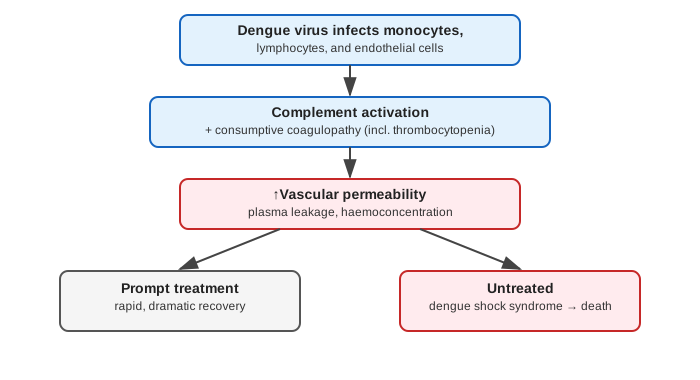
The virus infects blood monocytes, lymphocytes, and endothelial cells, initiating complement activation and a consumptive coagulopathy (including thrombocytopenia). This process can evolve rapidly, over a matter of hours. Prompt treatment at this stage produces rapid, dramatic recovery; untreated cases progress to dengue shock syndrome and death.
Morphology
Organ changes stem from four processes: focal haemorrhages and congestion; increased vascular permeability causing oedema in multiple organs; coagulopathy with thrombocytopenia; and haemoconcentration.
Diagnosis
Serologic antibody testing, direct virus detection (immunofluorescence, monoclonal antibodies), and rapid molecular methods (RT-PCR, fluorogenic ELISA).
Laboratory Findings
- Leucopenia with relative lymphocytosis, sometimes atypical lymphocytes.
- Thrombocytopenia.
- Elevated haematocrit from haemoconcentration.
- Chest X-ray: bilateral pleural effusion.
- Deranged liver function tests — elevated transaminases, hypoalbuminaemia, reversed A:G ratio.
- Prolonged coagulation tests — PT, APTT, TT.
Autopsy Findings
- Brain — intracranial haemorrhage, cerebral oedema, dengue encephalitis.
- Liver — enlarged, with hepatocyte and Kupffer cell necrosis; Reye’s syndrome in children.
- Kidneys — petechial haemorrhages, features of renal failure.
- Muscles and joints — perivascular mononuclear infiltrate.
Clinical Relevance
- The reclassification of viral haemorrhagic fevers away from the “arbovirus” label toward a transmission-route-based scheme (mosquito, tick, zoonotic, unknown) is a useful example of nosology being corrected once the underlying biology (not all these viruses are actually arthropod-borne) was better understood — the old term persists informally but is no longer taxonomically accurate.
- Distinguishing uncomplicated dengue fever (“break-bone fever,” self-limited) from dengue haemorrhagic fever (thrombocytopenia, haemoconcentration, shock risk) is the single most clinically important judgment call in this topic, since the two require entirely different levels of monitoring and intervention despite arising from the same virus.
- DHF’s characteristic laboratory triad — leucopenia, thrombocytopenia, and rising haematocrit (haemoconcentration) — is a compact, high-yield pattern: the rising haematocrit specifically reflects plasma leakage from increased vascular permeability, not true polycythaemia, and is the parameter most closely tracked to anticipate impending shock.
- Councilman bodies in yellow fever are a specific, testable morphologic finding worth distinguishing from other causes of hepatocyte apoptosis (e.g. viral hepatitis) by clinical and epidemiologic context, since the histologic appearance alone (an apoptotic hepatocyte) is not unique to yellow fever.
Diagram 1: DHF Pathogenesis and Outcome Fork
Draw a vertical chain of three boxes forking at the bottom into two outcome boxes.
- Dengue virus infects monocytes, lymphocytes, endothelial cells.
- Complement activation + consumptive coagulopathy (incl. thrombocytopenia).
- ↑Vascular permeability — plasma leakage, haemoconcentration. 4a. Prompt treatment → rapid, dramatic recovery. 4b. Untreated → dengue shock syndrome → death.
Labels required
- The fork must be explicitly labelled by treatment status (prompt vs untreated), not just “good outcome vs bad outcome” — the entire clinical point of this diagram is that the divergence is treatment-dependent and reversible if caught in time.
- Haemoconcentration must be tied explicitly to plasma leakage (↑vascular permeability), not left implying red cell overproduction — this is the same distinction worth making in the lab-findings discussion.
Errors commonly made
- Treating dengue shock syndrome as an inevitable endpoint of DHF rather than a preventable outcome of delayed/absent treatment — the rapid, dramatic recovery branch is equally central to the disease’s actual clinical course.
- Confusing the thrombocytopenia/coagulopathy step with the vascular permeability step as the same event — they are sequential and mechanistically distinct (consumptive coagulopathy first, capillary leak second), even though both contribute to the bleeding-and-shock clinical picture.
My Sticky Notes
Personal revision notes, mnemonics and reminders.
