
Type IV Hypersensitivity
Definition
T-cell mediated, NO antibody. Onset ~24h (tuberculin: induration 8-12h, peak 24-72h), lasts up to 14 days.
Two mechanisms
- CD4+ inflammation (cytokine-mediated) — APC presents Ag in node →
- Th1 → IFN-γ → classically activates macrophages (M1) — microbicidal + tissue damage
- Th17 → IL-17 → neutrophil recruitment
- = classic DTH. Morphology: perivascular mononuclear cuffing (CD4+ T cells + macrophages) → granuloma if Ag persists
- CD8+ CTL cytolysis — direct recognition of Ag on target cell (MHC-I, e.g. virus-infected) → perforin (pores) + granzymes (apoptosis) → direct kill, no cytokine cascade needed
Tuberculin reaction (classic model)
PPD intradermal in sensitised host → reddening/induration 8-12h → peak 24-72h → subsides. Basis of Mantoux test reading time (48-72h, not immediate).
Examples
- Mycobacterial: tuberculin reaction, TB/tuberculoid leprosy granulomas
- Virus-infected cells (CD8+ CTL)
- Tumour cells (CTL surveillance)
- Transplant rejection + GVHD (cellular)
- Contact sensitivity (poison ivy/oak-urushiol, drugs) — Th1(±Th17) → epidermal necrosis, blistering
- Organ-specific autoimmune: Type 1 DM (CTL destroys β-cells, insulin/GAD Ag), MS (Th1/Th17, myelin basic protein, demyelination), RA (Th17/Th1, synovitis), IBD (Th1/Th17), psoriasis (mainly Th17)
Clinical relevance
Mantoux read at 48-72h (not before) = direct consequence of DTH kinetics. Th1/Th17 vs CD8+CTL dominant mechanism → targeted biologics (anti-IL-17 for psoriasis only makes sense if Th17-dominant). Only hypersensitivity type with NO antibody role → serology irrelevant, skin/T-cell testing used instead.
Definition
Type IV (delayed-type, T cell-mediated) hypersensitivity is tissue injury caused by a T-cell-mediated immune response, occurring without antibody formation — the feature that distinguishes it from Types I, II and III. The reaction is slow to develop, beginning around 24 hours after antigen exposure (in the classic tuberculin reaction, reddening and induration appear at 8–12 hours, peak at 24–72 hours) and, unlike the immediate types, the effect is prolonged, sometimes lasting up to 14 days.
Two Mechanisms of T-Cell-Mediated Injury
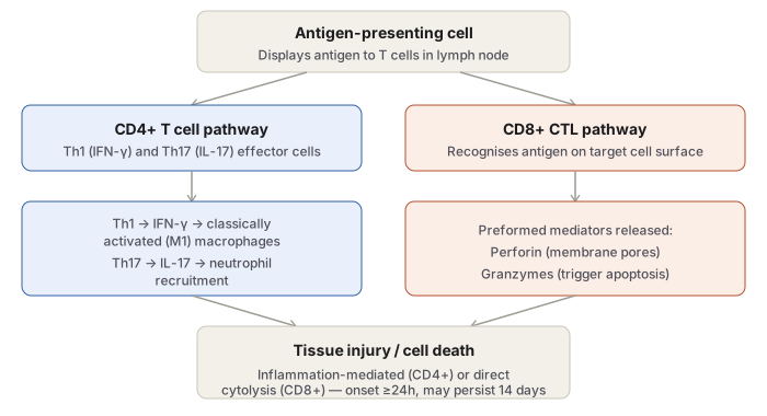
1. CD4+ T-cell-mediated inflammation (cytokine-mediated). Antigen-presenting cells process and display antigen to CD4+ T cells in lymph nodes, generating effector cells of two relevant subsets: Th1 cells, which secrete IFN-γ — the cytokine responsible for classical (M1) macrophage activation, producing microbicidal and tissue-damaging mediators and further pro-inflammatory signalling — and Th17 cells, which secrete IL-17, recruiting neutrophils (and, to a lesser extent, monocytes) into the reaction. This is the prototype mechanism of delayed-type hypersensitivity (DTH), and its classic morphology is a mononuclear (mainly CD4+ T cell and macrophage) infiltrate cuffing venules — “perivascular cuffing” — which, when the antigen cannot be cleared, matures into a granuloma (see Chronic and Granulomatous Inflammation).
2. CD8+ cytotoxic T-lymphocyte (CTL)-mediated cytolysis. Sensitised CD8+ cells directly recognise antigen displayed on the surface of target cells (typically intracellular antigen presented via MHC class I, as in virus-infected cells) and kill them directly, using preformed cytotoxic mediators — perforin (forms membrane pores) and granzymes (proteases that trigger apoptosis once inside the target cell) — without requiring the intervening cytokine/macrophage cascade of the CD4+ pathway.
Both mechanisms can operate together in the same disease process, with the balance between them determining the histologic picture: cytokine-driven inflammation and granuloma formation in one, direct cell killing in the other.
Classic Clinical Model: the Tuberculin Reaction
Intracutaneous injection of purified protein derivative (PPD/tuberculin) in a previously sensitised individual (the PPD or Mantoux skin test) produces reddening and induration at 8–12 hours, peaking at 24–72 hours, then slowly subsiding — the prototype demonstration of DTH kinetics and the clinical basis of tuberculin testing (see Tuberculosis).
Examples of Type IV Reaction
- Reaction against mycobacterial antigen — the tuberculin reaction itself, and the granulomatous tissue response of tuberculosis and tuberculoid leprosy.
- Reaction against virus-infected cells — CD8+ CTL-mediated killing of cells displaying viral antigen.
- Reaction against tumour cells — CTL-mediated tumour immune surveillance.
- Reaction against transplanted tissue — cellular (T-cell-mediated) transplant rejection and graft-versus-host disease (see Transplant Rejection).
- Contact sensitivity — reaction to environmental chemicals (urushiol from poison ivy/oak, certain drugs), producing epidermal necrosis, dermal inflammation, rash and blistering, mediated by Th1 (and possibly Th17) cytokines.
- Organ-specific autoimmune disease driven predominantly by this mechanism: Type 1 diabetes mellitus (T-cell-mediated insulitis and CTL destruction of pancreatic β-cells, targeting insulin and glutamic acid decarboxylase antigens), multiple sclerosis (Th1/Th17-mediated demyelination targeting myelin basic protein, with perivascular CNS inflammation), rheumatoid arthritis (Th17/Th1-driven chronic synovitis and cartilage destruction), inflammatory bowel disease (Th1/Th17-mediated chronic intestinal inflammation), and psoriasis (predominantly Th17-driven skin plaques).
Clinical Relevance
- The tuberculin reaction’s specific timeline (induration detectable only after 8–12 hours, peaking at 24–72 hours) is the pathologic basis for reading a Mantoux test at 48–72 hours rather than immediately — reading it too early would understate a genuinely positive reaction.
- Recognising which arm of Type IV hypersensitivity dominates a given autoimmune disease (Th1/Th17 cytokine-driven inflammation in RA/MS/IBD/psoriasis versus CD8+ CTL-driven direct cytotoxicity in Type 1 diabetes) is increasingly the basis for targeted biologic therapy — anti-IL-17 agents for psoriasis, for instance, only make pharmacological sense once the Th17-dominant mechanism is understood.
- Type IV is the only hypersensitivity type not mediated by antibody at all, which is why serologic tests are irrelevant to diagnosing conditions in this category and skin testing or T-cell-based assays are used instead.
Diagram 1: Two Mechanisms of Type IV Hypersensitivity
Draw a top box (antigen-presenting cell) branching into two parallel columns — CD4+ and CD8+ pathways — that reconverge into a shared “tissue injury” box.
- CD4+ column: Th1 cells (IFN-γ → classically activated macrophages) and Th17 cells (IL-17 → neutrophil recruitment), both feeding into inflammation-mediated injury.
- CD8+ column: direct recognition of target-cell antigen, releasing perforin and granzymes, producing direct cytolysis without an intervening inflammatory step.
Labels required
- Th1 and Th17 must be shown as two distinct sub-branches within the CD4+ column, each with its own cytokine and its own recruited effector cell (macrophages vs neutrophils) — collapsing them into one undifferentiated “CD4+ cytokines” box loses the exam-relevant distinction.
- Perforin and granzyme must both be named on the CD8+ side, since they act at different steps (pore formation vs triggering apoptosis) and are commonly asked as a pair.
Errors commonly made
- Describing Type IV hypersensitivity as a single mechanism (“T cells attack the antigen”) without separating the cytokine-mediated CD4+ route from the direct-killing CD8+ route, which have different kinetics, different morphology, and different diseases as their prototypes.
- Assuming Type IV always involves granuloma formation — granulomas are the outcome only when the CD4+/Th1 route encounters a persistent, poorly degradable antigen (as in tuberculosis), not a universal feature of every Type IV reaction (e.g. contact dermatitis or CD8+-driven viral clearance do not form granulomas).
My Sticky Notes
Personal revision notes, mnemonics and reminders.
