
Type II Hypersensitivity
Definition
IgG/IgM against cell-surface or tissue antigen. Tissue-specific (unlike Type III). Peak 15-30min (myasthenia/thyroiditis slower).
Aetiology
Foreign target antigen (transfusion, HDN) · self-antigen/autoimmune · drug-hapten on cell surface.
3 mechanisms
- Opsonisation + phagocytosis — Fab binds Ag → Fc-complement link → classical pathway → C3b opsonin → phagocyte (spleen) removes cell; ± MAC lysis directly. Examples: AIHA, transfusion reaction, HDN, ITP, drug-induced cytopenia.
- Complement/Fc-mediated inflammation — Ab on FIXED tissue Ag → C3a/C5a recruit neutrophils/monocytes → genuine inflammatory injury. Examples: Goodpasture, glomerulonephritis, vascular graft rejection.
- Cellular dysfunction (no injury/inflammation, often missed) — Ab blocks or stimulates a receptor:
- Block: myasthenia gravis (anti-AChR), pernicious anaemia (anti-intrinsic factor)
- Stimulate: Graves disease (anti-TSH receptor)
Examples list (blood cells)
AIHA (direct Coombs+) · transfusion reaction · erythroblastosis fetalis (maternal IgG crosses placenta) · ITP (anti-GPIIb/IIIa) · autoimmune neutropenia · drug-induced (penicillin/methyldopa/rifampicin hapten).
Examples (tissue)
Graves · myasthenia gravis · pernicious anaemia · Goodpasture (anti-GBM, kidney+lung) · pemphigus vulgaris (anti-desmoglein → bullae) · ANCA vasculitis · rheumatic fever (molecular mimicry: strep Ag → cross-reacts myocardium) · male infertility (antisperm Ab) · Type 1 DM (islet cell Ab) · hyperacute transplant rejection.
Clinical relevance
Mechanism sorting → treatment logic (splenectomy helps phagocytic cytopenias, not receptor-blockade disease). Rheumatic fever = molecular mimicry teaching example. Type II (tissue-specific, fixed Ag) vs Type III (circulating, non-specific deposition) = key distinction.
Definition
Type II (antibody-mediated, cytotoxic) hypersensitivity is tissue injury caused by humoral antibodies — IgG or IgM — directed against antigens present on the surface of specific target cells or in tissue matrix, generally appearing within 15–30 minutes of antigen exposure (though myasthenia gravis and thyroiditis may take longer to manifest clinically). Unlike Type III, Type II is tissue-specific: the antibody targets a defined antigen on a defined cell or tissue rather than forming complexes that deposit indiscriminately wherever blood is filtered.
Aetiology
Type II reactions occur when antibody is raised against a genuinely foreign target-cell antigen (as in transfusion reaction or haemolytic disease of the newborn), against a self-antigen through failure of tolerance (autoimmune disease), or against a normal cell-surface protein modified by an adsorbed drug metabolite acting as a hapten (drug-induced cytotoxicity). Participating effectors include the complement system, tissue macrophages, platelets, NK cells, neutrophils and eosinophils, with IgG and IgM as the principal antibody classes.
Pathogenesis
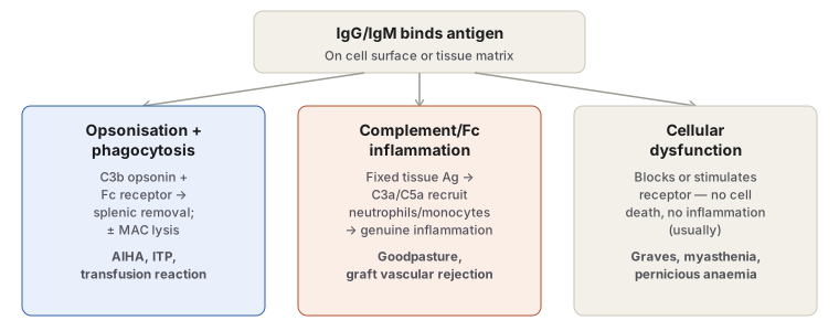
Antibody binding to a target antigen produces injury through three genuinely distinct mechanisms, and distinguishing which one is operating in a given disease is the central organising idea of this topic:
1. Opsonisation and phagocytosis. The antigen on the target cell surface binds the Fab portion of IgG or IgM, forming an antigen–antibody complex. The unattached Fc fragment links to complement, activating the classical pathway (C1 cleaves C4 and C2 to generate C3b). Activated C3b bound to the cell acts as an opsonin, and phagocytes — bearing receptors for both antibody Fc tails and C3b — bind and ingest the opsonised cell, typically in the spleen (which is why splenectomy benefits some antibody-mediated cytopenias). In parallel, full complement activation can proceed to the membrane attack complex (MAC), directly lysing the target cell.
2. Complement- and Fc-receptor-mediated inflammation. Rather than opsonising and removing a single cell, antibody bound to a fixed tissue antigen (e.g. basement membrane) activates complement in situ; the resulting C3a/C5a recruit and activate neutrophils and monocytes via their Fc and complement receptors, producing genuine inflammatory tissue injury — the mechanism underlying immune-complex-independent forms of glomerulonephritis and vascular rejection.
3. Antibody-mediated cellular dysfunction. Antibody against an essential surface receptor or protein can derange cell function without causing direct injury, complement activation, or inflammation at all — either by blocking normal receptor engagement (anti-acetylcholine-receptor antibody in myasthenia gravis impairs neuromuscular transmission; anti-intrinsic-factor antibody in pernicious anaemia blocks vitamin B12 absorption) or by stimulating the receptor inappropriately (anti-TSH-receptor antibody in Graves disease drives excess thyroid hormone secretion). This third mechanism is the one most often missed, since it produces disease without the cell death or inflammation that “cytotoxic hypersensitivity” implies by name.
Examples
Cytotoxic antibodies to blood cells (the classical, most commonly tested group):
- Autoimmune haemolytic anaemia — autoantibody against red cell membrane antigen; detected by the direct Coombs (antiglobulin) test.
- Transfusion reactions — incompatible/mismatched donor blood.
- Haemolytic disease of the newborn (erythroblastosis fetalis) — maternal IgG anti-red-cell isoantibody crosses the placenta and destroys foetal red cells.
- Idiopathic thrombocytopenic purpura (ITP) — autoantibody against platelet surface components (GPIIb/IIIa).
- Autoimmune neutropenia/agranulocytosis — autoantibody against leucocytes.
- Drug-induced cytotoxicity — a drug or its metabolite (penicillin, methyldopa, rifampicin) acts as a hapten on the blood cell surface, and antibody against the drug–protein complex destroys the cell.
Cytotoxic/dysfunctional antibodies against tissue components:
- Graves disease — anti-TSH-receptor antibody stimulates thyroid epithelium.
- Myasthenia gravis — anti-acetylcholine-receptor antibody blocks neuromuscular transmission (with emerging evidence that complement-mediated injury to the motor end-plate also contributes, not purely functional blockade).
- Pernicious anaemia — anti-intrinsic-factor antibody impairs vitamin B12 absorption.
- Goodpasture syndrome — antibody against basement membrane antigen shared by kidney glomeruli and lung alveoli, producing complement/Fc-mediated inflammation (nephritis, pulmonary haemorrhage).
- Pemphigus vulgaris — antibody against desmoglein (epidermal intercellular junction protein) activates proteases and disrupts intercellular adhesion, producing skin bullae.
- ANCA-associated vasculitis — antibody against neutrophil granule proteins drives neutrophil degranulation and vessel wall inflammation.
- Acute rheumatic fever — antibody raised against streptococcal cell wall antigen cross-reacts with myocardial antigen (molecular mimicry), producing myocarditis and arthritis.
- Male infertility — antisperm antibody impairs sperm motility and causes cellular injury.
- Type 1 diabetes mellitus — islet cell autoantibodies react against pancreatic islet tissue.
- Hyperacute transplant rejection — pre-formed antibody against donor antigen, discussed further under Transplant Rejection.
Clinical Relevance
- Sorting a Type II example into one of the three mechanisms (phagocytic removal, inflammatory injury, or functional dysfunction) predicts both the histologic picture and the treatment logic — a phagocytosis-driven cytopenia may respond to splenectomy, while a pure receptor-blockade disease like myasthenia gravis will not, since there is no cell to remove and no inflammation to suppress in the same way.
- Acute rheumatic fever is the clearest teaching example of molecular mimicry in this category: the antibody is genuinely directed at a foreign (streptococcal) antigen, but cross-reacts with self myocardial tissue, which is why the pathology appears autoimmune despite the trigger being an actual infection.
- Recognising that Type II and Type III both involve antigen–antibody complex formation, but differ in whether the antigen is tissue-specific and fixed (Type II) versus circulating and non-specifically deposited (Type III), is the single distinction examiners use most often to test whether the two have been properly separated in a student’s understanding.
Diagram 1: Three Mechanisms of Type II Hypersensitivity
Draw a single top box (“antibody binds antigen”) branching into three side-by-side boxes, each carrying its mechanism name, a one-line description, and two or three named example diseases.
- Opsonisation and phagocytosis — C3b opsonin + Fc receptor → splenic removal, ± direct complement (MAC) lysis. Examples: AIHA, ITP, transfusion reaction.
- Complement/Fc-mediated inflammation — antibody on a fixed tissue antigen recruits neutrophils/monocytes via C3a/C5a → genuine inflammatory injury. Examples: Goodpasture syndrome, vascular graft rejection.
- Cellular dysfunction — antibody blocks or stimulates a receptor without cell death or inflammation. Examples: Graves disease, myasthenia gravis, pernicious anaemia.
Labels required
- Each branch must carry its own named example diseases directly underneath it — the exam-relevant skill is sorting a given disease into the correct one of the three mechanisms, not just knowing the three mechanisms exist.
- The third branch must explicitly note the absence of cell death or inflammation, since this is what most distinguishes it from the other two and is the branch most often forgotten when this topic is recalled as “cytotoxic hypersensitivity.”
Errors commonly made
- Sorting every Type II example into the phagocytosis/lysis branch by default, because “cytotoxic reaction” is the topic’s other name, and missing that Graves disease and myasthenia gravis involve no cell destruction at all.
- Confusing branch 2 (inflammation from antibody on a fixed tissue antigen) with Type III immune-complex disease — the distinguishing feature is that the antigen here is tissue-specific and already in place, not a circulating complex depositing wherever blood is filtered.
Short Essays (1)
Multiple Choice Questions (1)
My Sticky Notes
Personal revision notes, mnemonics and reminders.
