
Turner Syndrome
Overview
Primary hypogonadism in phenotypic females. Partial/complete monosomy of X short arm. Incidence ~1/3000 female births. Physical features VARIABLE — hypogonadism = only consistent finding.
Karyotypic subtypes
- ~57% — 45,X (entire X missing) = “classic”
- ~14% — structural X abnormalities → partial monosomy. Order of frequency: isochromosome long arm [46,X,i(X)(q10)] > ring chromosome [46,X,r(X)] > deletion [46,X,del(Xp)/del(Xq)]
- ~29% — mosaics (45,X + other line): 45,X/46,XX | 45,X/46,XY | 45,X/47,XXX | 45,X/46,X,i(X)(q10)
5-10% of mosaics carry Y sequences (complete Y or translocated fragments) → ↑gonadoblastoma risk — karyotype-directed screening important.
Karyotypic heterogeneity → phenotypic heterogeneity: classic 45,X = fullest phenotype; mosaics/deletion variants = may show ONLY primary amenorrhoea.
Pathogenesis — 2 independent mechanisms
1. Streak ovary formation: both X’s normally active in oogenesis, essential for ovarian development. Normal: 7 million oocytes (fetal)→400,000(menarche)→<10,000(menopause). Turner: ovaries form NORMALLY early in embryogenesis, but 2nd-X absence → ACCELERATED oocyte loss, complete by ~age 2. “Menopause before menarche.” → streak ovaries (fibrous, no follicles/ova) → 1° amenorrhoea, infertility, failed 2° sex characteristics.
2. SHOX haploinsufficiency (short stature): SHOX gene (Xp22.33) escapes X-inactivation, has active Y homolog too → normal males+females both have 2 active copies. Turner: 1 copy lost → SHOX haploinsufficiency → growth deficit. (SHOX deletions found in 2-5% of otherwise-normal short children — confirms independent causal role.) SHOX loss alone does NOT explain cardiac/endocrine features — other X genes involved (less characterised).
Clinical features
Growth/skeletal: short stature (<3rd percentile), cubitus valgus, shield chest+widely-spaced nipples, high-arched palate
Neck/lymphatic: nuchal swelling in infancy (distended lymphatics)→webbed neck (older children), low posterior hairline, hand/foot lymphoedema
Congenital malformations: horseshoe kidney, bicuspid aortic valve, coarctation of aorta — cardiovascular = MOST COMMON cause of childhood death
Reproductive/endocrine: failed 2° sex characteristics (infantile genitalia, minimal breast development, sparse pubic hair), 1° amenorrhoea (most patients), streak ovaries. Hypothyroidism (autoantibody) up to 50%, esp. with isochromosome Xp.
Intellect: usually normal; subtle visuospatial processing deficits.
Diagnosis: adult + short stature + 1° amenorrhoea → strong suspicion. Confirm by karyotyping.
Clinical relevance
Short stature + 1° amenorrhoea = single most actionable diagnostic pattern → trigger for karyotyping. SHOX escape-from-X-inactivation biology explains why short stature is SO consistent even with mild karyotype abnormality (single-gene dosage effect vs broader less-understood gene set for cardiac/endocrine). “Menopause before menarche” phrase = captures ovaries form normally, defect = accelerated postnatal attrition (not failed formation). Y-material mosaics = real actionable gonadoblastoma screening implication (45,X/46,XY specifically). CV malformations = leading childhood death cause → cardiac eval (coarctation, bicuspid valve) = genuine diagnosis-time priority.
Overview
Turner syndrome is characterised by primary hypogonadism in phenotypic females, resulting from partial or complete monosomy of the short arm of the X chromosome. Incidence approximately 1 in 3000 female live births. Physical features are notably variable across affected individuals — hypogonadism is the only consistently present finding.
Karyotypic Subtypes
Three categories of karyotypic abnormality are recognised on routine cytogenetics:
- ~57% — missing an entire X chromosome: 45,X karyotype (the “classic” form).
- ~14% (one-third of the remaining 43%) — structural abnormalities of the X chromosome, producing partial monosomy. In order of frequency: isochromosome of the long arm [46,X,i(X)(q10)], with loss of the short arm; ring chromosome [46,X,r(X)], from deletion of portions of both arms; and deletion of part of the short or long arm [46,X,del(Xp) or del(Xq)].
- ~29% (two-thirds of the remaining 43%) — mosaics, with a 45,X cell population alongside one or more other cell lines — examples: 45,X/46,XX; 45,X/46,XY; 45,X/47,XXX; 45,X/46,X,i(X)(q10).
5–10% of mosaic Turner patients carry Y-chromosome sequences (a complete Y as in 45,X/46,XY, or Y fragments translocated elsewhere) — these patients carry a higher risk of gonadal tumour (gonadoblastoma), making karyotype-directed screening clinically important.
Karyotypic heterogeneity drives phenotypic heterogeneity: classic monosomy-X patients show the fullest phenotype, while mosaics or structural-deletion variants may have minimal findings and present with primary amenorrhoea alone.
Pathogenesis
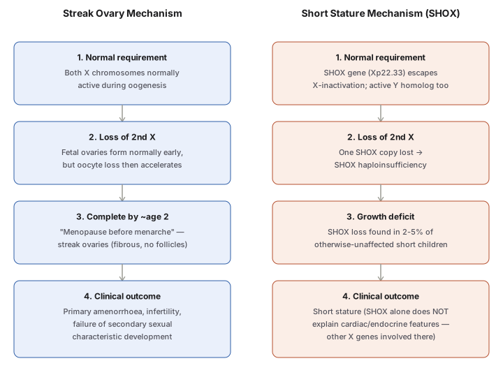
Turner syndrome’s phenotype arises from (at least) two distinct, independent gene-loss mechanisms:
1. Streak ovary formation: both X chromosomes are normally active during oogenesis and are essential for normal ovarian development — during normal fetal development, ovaries contain as many as 7 million oocytes, dwindling to ~400,000 by menarche and <10,000 by menopause. In Turner syndrome, fetal ovaries develop normally early in embryogenesis, but absence of the second X chromosome causes accelerated oocyte loss, complete by around age 2 years — in effect, “menopause occurs before menarche.” The ovaries are reduced to atrophic fibrous strands devoid of ova and follicles (streak ovaries), producing primary amenorrhoea, infertility, and failure of secondary sexual characteristic development.
2. SHOX haploinsufficiency and short stature: the short stature homeobox (SHOX) gene at Xp22.33 is one of the genes that escapes X-inactivation and is unique in having an active homolog on the Y chromosome — so unaffected males and females both normally carry two active copies. Loss of one copy (from the missing/structurally abnormal X in Turner syndrome) causes SHOX haploinsufficiency, directly producing the growth deficit responsible for short stature. (SHOX deletions are found in 2–5% of otherwise-unaffected short-statured children, confirming the gene’s independent causal role in growth.) However, SHOX loss alone cannot explain the cardiac and endocrine abnormalities of Turner syndrome — these depend on other, less well-characterised genes on the X chromosome.
Clinical Features
Growth and skeletal: growth retardation → short stature (below the 3rd percentile); cubitus valgus (increased carrying angle of the arms); shield-like chest with widely spaced nipples; high-arched palate.
Neck/lymphatic: swelling of the nape of the neck in infancy (distended lymphatic channels), manifesting as webbing of the neck in older children; low posterior hairline; lymphoedema of the hands and feet.
Congenital malformations: horseshoe kidney, bicuspid aortic valve, coarctation of the aorta — cardiovascular abnormalities are the most common cause of death in childhood.
Reproductive/endocrine: failure to develop secondary sexual characteristics in adolescence — infantile genitalia, minimal breast development, sparse pubic hair; primary amenorrhoea in most patients; streak ovaries on histology (as above). Hypothyroidism from autoantibodies occurs frequently — up to 50% of patients, particularly those with an isochromosome Xp.
Intellect: usually within normal limits, though subtle visuospatial information processing defects have been noted.
Diagnosis: in an adult, the combination of short stature + primary amenorrhoea should prompt strong suspicion; diagnosis is established by karyotyping.
Clinical Relevance
- The combination of short stature and primary amenorrhoea is the single most clinically actionable diagnostic pattern in this topic — it is the trigger that should prompt karyotyping for Turner syndrome in an adult patient.
- Recognising the SHOX gene’s independent, escape-from-X-inactivation biology explains why short stature is such a consistent Turner finding even in patients with relatively mild karyotypic abnormalities — it is a dosage effect on a single well-characterised gene, distinct from the broader, less-understood gene set responsible for cardiac/endocrine features.
- “Menopause before menarche” is a genuinely useful phrase for retaining the streak-ovary mechanism — it captures that ovarian development is initially normal and the defect is one of accelerated postnatal oocyte attrition, not a failure of ovarian formation itself.
- Y-chromosome material in a subset of mosaic Turner patients carrying real gonadoblastoma risk is a direct, actionable screening implication of the karyotype — distinguishing 45,X/46,XY mosaicism from other mosaic forms is not just an academic classification detail.
- Cardiovascular malformations as the leading cause of childhood death in Turner syndrome means cardiac evaluation (specifically screening for coarctation and bicuspid aortic valve) is a genuine clinical priority at diagnosis, not an incidental finding to note in passing.
Diagram 1: Two Independent Gene-Loss Mechanisms Producing the Turner Phenotype
Draw two parallel, independent columns (Streak Ovary Mechanism, Short Stature/SHOX Mechanism) — not a normal-vs-disease contrast but two separate consequences of the same underlying X-chromosome loss, each with four stacked stages.
- Normal requirement — both X’s active in oogenesis (ovary) vs SHOX escapes X-inactivation with active Y homolog (stature).
- Loss of 2nd X — initially normal ovarian formation (ovary) vs one SHOX copy lost, haploinsufficiency (stature).
- Time course/evidence — accelerated oocyte loss complete by age 2 (ovary) vs SHOX deletions found in 2-5% of otherwise-unaffected short children (stature).
- Clinical outcome — primary amenorrhoea/infertility/failed secondary sexual characteristics (ovary) vs short stature, with the explicit caveat that SHOX alone doesn’t explain cardiac/endocrine features (stature).
Labels required
- The phrase “menopause before menarche” must appear in the ovary column — it is the single most memorable and most tested description of Turner ovarian pathogenesis.
- The stature column must explicitly state that SHOX loss explains short stature ONLY, not the syndrome’s cardiac or endocrine features — this caveat prevents over-extending a single-gene mechanism to explain the whole phenotype.
- Both columns should be visually presented as parallel/independent, not sequential — a common error is assuming one mechanism causes the other, when in fact both are independent downstream consequences of the same chromosomal loss.
Errors commonly made
- Describing Turner ovaries as failing to form in the first place, rather than forming normally and then undergoing accelerated postnatal oocyte loss — the distinction matters because it explains why ovarian tissue is present (as fibrous streaks) rather than absent.
- Assuming SHOX haploinsufficiency explains the entire Turner phenotype rather than specifically the growth deficit — a frequent overreach that this diagram’s explicit caveat is designed to prevent.
- Treating the two mechanisms shown here as sequential or causally linked, rather than recognising them as parallel, independent consequences of losing the same chromosome.
Multiple Choice Questions (1)
My Sticky Notes
Personal revision notes, mnemonics and reminders.
