
Transplant Rejection
Graft types
Autograft (self) · isograft (identical twin) · allograft (same species, different genotype — most transplants) · xenograft (different species, always rejected).
Recognition
HLA = major antigen recognised. Identical HLA essentially only in twins (~25% siblings share haplotype by chance).
- Direct pathway — donor APC presents donor HLA directly to recipient T cell → drives CTL-mediated ACUTE rejection
- Indirect pathway — recipient APC processes/presents donor Ag → drives CHRONIC rejection + alloantibody production (donor HLA picked up by host B cells)
Both → CD8+ CTL + CD4+ (mainly Th1) effector cells. Allograft T-cell frequency >> anti-microbial T-cell frequency → unusually strong rejection response.
Mechanisms
Cell-mediated (dominant) — CD8+ direct kill + CD4+ hypersensitivity-type inflammation. Humoral — preformed Ab (transfusion/pregnancy/prior transplant sensitisation) OR complement-dependent cytotoxicity/ADCC/immune complexes in non-sensitised.
3 types of rejection
- Hyperacute (min-hours) — PREFORMED Ab vs graft endothelium → complement+clotting → fibrinoid necrosis, thrombosis, cortical infarction. Arthus-like histology. Prevented by cross-matching (now uncommon).
- Acute (days-months; principal cause of EARLY failure; can flare late if immunosuppression stopped):
- Cellular (more common) — CD8+CTL + CD4+ cytokines → tubulointerstitial (tubulitis) OR vascular (endotheliitis/intimal arteritis) pattern. Responds well to immunosuppression.
- Antibody-mediated/humoral — Ab + complement on endothelium → glomerular/peritubular capillary injury, focal thrombosis. B-cell-predominant infiltrate, LESS marked than cellular. Poor response to standard immunosuppression.
- Chronic (months-years; leading cause of LATE failure) — immunologic or ischaemic. Kidney: intimal fibrosis, graft arteriosclerosis, interstitial fibrosis, tubular atrophy, glomerulopathy (BM duplication), sparse infiltrate. Refractory to treatment.
Graft survival
HLA matching (best for living-related kidney; less relevant for heart/lung/liver/islet given modern immunosuppression + urgency). Drugs: cyclosporine/tacrolimus (block IL-2 transcription), rapamycin (blocks IL-2 response). Cost: opportunistic infection, CMV/polyomavirus reactivation, EBV lymphoma, HPV SCC.
GVHD
Immunocompetent donor cells → immunodeficient/non-rejecting host (classic: BM transplant). Fever, weight loss, anaemia, dermatitis, diarrhoea, malabsorption, pneumonia, hepatosplenomegaly. Severity ∝ HLA disparity.
Clinical relevance
Rejection type dictates management (hyperacute=graft lost; acute cellular=treatable; acute humoral=refractory to standard tx; chronic=refractory). Cross-matching = diagnostic test that transformed outcomes (eliminated most hyperacute rejection). Immunosuppression trade-off = same secondary-immunodeficiency logic as elsewhere.
Types of Grafts
Classified by the genetic relationship between donor and recipient:
- Autograft — donor and recipient are the same individual.
- Isograft — donor and recipient are genetically identical (e.g. identical twins).
- Allograft — donor and recipient are the same species but genetically distinct — the overwhelming majority of clinical transplants (skin, kidney, bone marrow, heart, lung, liver, pancreas, cornea).
- Xenograft — donor and recipient are different species; invariably rejected due to the degree of genetic disparity.
Only allografts (and, to a lesser extent, xenografts) provoke a clinically significant rejection response; autografts and isografts do not, since there is no genetic disparity for the recipient immune system to recognise.
Recognition of Graft Alloantigens
The major antigens the recipient recognises as foreign are HLA molecules, encoded by the highly polymorphic MHC locus (see Chemical Mediators of Inflammation and related HLA discussion) — genuinely identical HLA type between unrelated individuals is essentially confined to identical twins (with roughly 25% of ordinary siblings sharing the same HLA haplotype by chance). Recipient T cells recognise donor (allogeneic) HLA antigens by two distinct pathways:
- Direct pathway — donor antigen-presenting cells within the graft present donor HLA directly to recipient T cells. This pathway is thought to dominate CTL-mediated acute rejection.
- Indirect pathway — donor antigens are instead picked up by the recipient’s own APCs, processed like any other foreign antigen, and presented to recipient T cells (and, when donor HLA is presented to helper T cells by recipient B cells, this route also generates anti-HLA alloantibodies). This pathway is thought to play the larger role in chronic rejection.
Both pathways activate CD8+ T cells (which differentiate into cytotoxic T lymphocytes) and CD4+ T cells (which become cytokine-producing, mainly Th1, effector cells). Because the frequency of T cells capable of recognising foreign HLA is far higher than the frequency specific for any single microbe, allograft immune responses are unusually strong — which is why transplant rejection, left unchecked, can destroy a graft rapidly and why powerful immunosuppression is required.
Mechanisms of Rejection
Rejection involves both cell-mediated immunity (the dominant mechanism — CD8+ cytotoxic T cells directly destroying graft cells, and CD4+ T cells driving a hypersensitivity-type inflammatory reaction against the graft) and humoral immunity, via preformed circulating antibody (from prior sensitisation — blood transfusion, pregnancy, or previous transplantation) or via complement-dependent cytotoxicity, antibody-dependent cell-mediated cytotoxicity (ADCC), and immune-complex formation in previously unsensitised recipients.
Types of Rejection
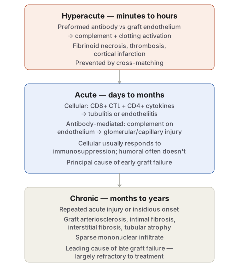
1. Hyperacute rejection — appears within minutes to hours of graft implantation, mediated entirely by preformed antibody against donor antigen (natural anti-blood-group IgM, or anti-HLA antibody from prior transfusion/pregnancy/transplantation). Once blood flow is restored, antibody binds graft endothelium and activates complement and clotting, producing endothelial injury, thrombosis and ischaemic necrosis. Grossly the organ becomes swollen, oedematous, cyanotic and haemorrhagic rather than gaining the expected pink perfused colour. Histologically the picture resembles an Arthus reaction: fibrinoid necrosis of arteriolar/arterial walls, luminal thrombotic occlusion, neutrophil accumulation in arterioles/glomeruli/peritubular capillaries, progressing to cortical infarction. Pre-transplant cross-matching (testing recipient serum against donor lymphocytes) has made hyperacute rejection uncommon in practice.
2. Acute rejection — becomes evident within days to months of transplantation (the principal cause of early graft failure), or can appear suddenly much later if immunosuppression is reduced or stopped. Two overlapping patterns occur, usually together in the same graft:
- Acute cellular rejection (more common) — extensive interstitial infiltration by lymphocytes (mainly T cells), plasma cells, monocytes and some polymorphs, with tubular injury (tubulitis) in a tubulointerstitial pattern, or — in a vascular pattern — inflamed vessels with swollen endothelium and lymphocytes wedged between endothelium and vessel wall (endotheliitis/intimal arteritis). Recognising pure cellular rejection matters clinically because it usually responds well to immunosuppressive therapy.
- Acute antibody-mediated (humoral) rejection — antibody binds vascular endothelium and activates complement via the classical pathway, producing glomerular and peritubular capillary inflammation with complement deposition and focal small-vessel thrombosis; the mononuclear infiltrate is less prominent than in cellular rejection and is predominantly B-lymphocytic. This form responds poorly to standard immunosuppression.
3. Chronic rejection — an indolent process developing over months to years, either following repeated acute rejection episodes or arising insidiously, and now the leading cause of long-term graft failure since treatment of acute rejection has improved. Mechanisms may be immunologic (T cells and alloantibody driving fibroblast/smooth muscle proliferation) or ischaemic. In the kidney: progressive renal impairment (rising serum creatinine) with intimal fibrosis, graft arteriosclerosis, interstitial fibrosis and tubular atrophy; glomerulopathy with basement membrane duplication and peritubular capillary basement membrane multilayering reflect chronic endothelial injury; the mononuclear infiltrate is typically sparse (unlike acute rejection), and renal allografts may develop glomerulonephritis either transmitted from the original donor kidney or arising de novo.
Increasing Graft Survival
HLA matching improves survival, most valuably for living-related kidney transplants; for heart, lung, liver and islet transplantation, urgency and anatomic compatibility (size) usually outweigh HLA matching given modern immunosuppression. All allografts (except between identical twins) require ongoing immunosuppression: cyclosporine and tacrolimus suppress T-cell-mediated immunity by inhibiting cytokine gene transcription (chiefly IL-2), and rapamycin blocks T-cell proliferative response to IL-2. Immunosuppression carries its own cost — increased susceptibility to opportunistic fungal, viral and other infection; reactivation of latent viruses (CMV, polyomavirus); and increased risk of virus-driven malignancy (EBV-associated lymphoma, HPV-induced squamous cell carcinoma) — the therapeutic trade-off inherent to all transplant immunosuppression.
Graft-versus-Host (GVH) Disease
GVHD is the mirror-image problem, arising when immunocompetent cells are transplanted into an immunodeficient (or otherwise unable-to-reject) recipient — the classic setting being bone marrow/haematopoietic stem cell transplantation, where donor-derived immunocompetent T cells instead recognise the recipient’s tissues as foreign and attack them. Clinical features include fever, weight loss, anaemia, dermatitis, diarrhoea, intestinal malabsorption, pneumonia and hepatosplenomegaly; severity correlates with the degree of genetic (HLA) disparity between donor and recipient.
Clinical Relevance
- Correctly assigning a rejection episode to hyperacute, acute-cellular, acute-humoral or chronic determines management directly: hyperacute rejection is essentially untreatable and the graft is usually lost; acute cellular rejection typically responds well to immunosuppression; acute humoral rejection responds poorly to standard regimens and needs antibody-directed therapy; chronic rejection is largely refractory to available treatment.
- The near-elimination of hyperacute rejection through routine pre-transplant cross-matching is one of transplant medicine’s clearest examples of a purely diagnostic test (rather than a new drug) transforming clinical outcomes.
- Immunosuppression’s trade-off — reduced rejection risk against increased opportunistic infection and virus-driven malignancy — is the same balance that underlies the broader concept of secondary immunodeficiency, and is directly relevant to why transplant recipients are screened and monitored for CMV, EBV-associated lymphoma and HPV-related lesions long after the transplant itself.
Diagram 1: Types of Transplant Rejection by Timing and Mechanism
Draw a single downward column of three stages, each carrying its timeframe, mechanism, and characteristic histology.
- Hyperacute (minutes–hours) — preformed antibody, complement/clotting activation, fibrinoid necrosis and thrombosis; note that cross-matching largely prevents this today.
- Acute (days–months) — split cellular (tubulitis/endotheliitis, responds to immunosuppression) from antibody-mediated (complement on endothelium, responds poorly); label as the principal cause of early graft failure.
- Chronic (months–years) — vascular/interstitial fibrosis, sparse infiltrate, refractory to treatment; label as the leading cause of late graft failure.
Labels required
- Each stage’s timeframe must be explicit and distinct, since the three-way timing distinction (minutes-hours / days-months / months-years) is itself the most frequently tested fact in this topic.
- The acute stage must show both its cellular and antibody-mediated sub-patterns with their differing treatment responsiveness, since conflating them loses the clinically important point that only one of the two responds reliably to standard immunosuppression.
Errors commonly made
- Describing all three types of rejection as points on a single severity spectrum rather than as mechanistically distinct entities — hyperacute rejection does not “become” acute rejection if untreated; it is a separate, antibody-driven process that acts within hours and is essentially unrelated in mechanism to acute cellular rejection.
- Assuming chronic rejection is simply cumulative damage from repeated acute rejection episodes, when it can also arise insidiously without any preceding acute episode, and its mechanism (fibrosis-driving cytokines, ischaemic injury) differs from the cell-killing/complement mechanisms of acute and hyperacute rejection.
My Sticky Notes
Personal revision notes, mnemonics and reminders.
