
Thalassaemia
Overview
Hereditary disorder — reduced globin chain SYNTHESIS (quantitative defect; contrast haemoglobinopathies like sickle cell = structural defect). First described in Mediterranean region. Also seen Middle East, Indian subcontinent, SE Asia, Africa.
India: seen nationwide, ~35 million carry β-thalassaemia. High incidence: Punjabis, Bengalis, Gujaratis, Parsis. India’s most significant inherited Hb disorder.
Normal adult Hb: HbA (α2β2) 95-98%, HbA2 (α2δ2) 1.5-3.5%, HbF (α2γ2) <1%.
2 α-genes from each parent (4 total, chr16). 2 β-genes total, 1 from each parent (chr11) — this gene-number difference explains why α and β thalassaemia genetics look so different.
α-thalassaemia (gene deletion, severity = number deleted)
- 4 genes deleted → Hb Bart’s hydrops fetalis — Hb Bart’s (γ4), high O2 affinity, poor O2 release → severe hypoxia → fatal in utero/soon after birth. Electrophoresis: 80-90% Hb Bart’s, NO HbA/A2/F.
- 3 genes deleted → HbH disease — HbH (β4), precipitates as Heinz bodies. Well-compensated haemolytic anaemia, worse in pregnancy/infection. Splenomegaly, cholelithiasis common.
- 2 genes deleted → α-thal trait — asymptomatic, microcytic no anaemia. HbA2 normal/low (opposite of β-thal trait).
- 1 gene deleted → silent carrier
β-thalassaemia (mostly point mutations, NOT deletion)
β⁰ = no β-chain made. β⁺ = reduced β-chain made.
Pathogenesis of anaemia: ↓β-chain → excess unpaired α-chains → some pair with γ→HbF; rest precipitate as Heinz bodies → membrane damage → splenic pitting → RE phagocytosis (spleen/liver) → chronic haemolysis + ineffective erythropoiesis + hepatosplenomegaly + iron overload.
3 clinical types:
- β-thal major (Cooley’s anaemia) — homozygous (β0/β0 or β+/β+), severe, transfusion-dependent. 25% recurrence risk if both parents carry trait.
- β-thal intermedia — heterozygous combos, moderate, no regular transfusion needed.
- β-thal minor (trait) — heterozygous, mild/asymptomatic.
Rare variants: δβ-thal minor (both β+δ absent, ↑HbF, normal/↓HbA2), Hb Lepore (β-δ gene fusion).
β-thal major clinical features: anaemia from 4-6 months age (γ→β switch time) · marked hepatosplenomegaly · bone expansion → thalassaemic facies + jaw malocclusion · iron overload (transfusion) → growth failure, delayed puberty, diabetes, liver/heart damage.
β-thal major labs: severe anaemia, microcytic hypochromic, anisopoikilocytosis, basophilic stippling, target cells, tear-drop cells, normoblasts. Electrophoresis: HbA 0-50%, HbF 50-98% (markedly ↑).
Summary table
| Type | Hb | Electrophoresis | Genotype |
|---|---|---|---|
| Hb Bart’s | 3-10 | Bart’s 100% | 4-gene del |
| HbH disease | 2-12 | HbF10%,HbH2-4% | 3-gene del |
| α-thal trait | 10-14 | near normal | 2-gene del |
| β-thal major | <5 | HbA0-50%,HbF50-98% | β/β thal |
| β-thal intermedia | 5-10 | variable | mixed |
| β-thal minor | 10-12 | HbA2 4-9%,HbF1-5% | βA/β thal |
Clinical relevance
HbA2: ↑ in β-thal trait, normal/↓ in α-thal trait — key discriminator when both look identical (microcytic, no anaemia) on film. 25% recurrence risk (both parents trait carriers) = basis for premarital/antenatal carrier screening in high-prevalence Indian communities. Iron overload in β-thal major comes from BOTH disease process (↑gut absorption from ineffective erythropoiesis) AND transfusion — chelation therapy needed from start of transfusion programme, not just after overload appears.
Overview
Thalassaemias are hereditary disorders of reduced synthesis of one or more globin polypeptide chains — a quantitative defect, in contrast to haemoglobinopathies (such as sickle cell disease), which produce structurally abnormal but normally-synthesised globin. First described around the Mediterranean (the name derives from Greek thalassa, “the sea”), thalassaemia is now recognised across the Middle East, Indian subcontinent, South-East Asia, and in people of African descent.
In India, thalassaemia occurs nationwide, and an estimated 35 million people carry β-thalassaemia; incidence is particularly high among Punjabis (including those who migrated from Pakistan after Partition), Bengalis, Gujaratis, and Parsis. Given this scale, thalassaemia represents the single most significant inherited haemoglobin disorder in the country.
Normal adult haemoglobin distribution: HbA (α₂β₂) 95–98%, HbA2 (α₂δ₂) 1.5–3.5%, HbF (α₂γ₂) <1% (higher in infants under 6 months). Two α-globin genes are inherited from each parent (four total, chromosome 16); two β-globin genes are inherited, one from each parent (chromosome 11) — this gene dosage difference underlies the very different genetics of α- and β-thalassaemia.
α-Thalassaemia
Caused by deletion of one or more of the four α-globin genes; severity depends directly on the number of genes deleted.
| Genes deleted | Syndrome | Haemoglobin | Genotype effect |
|---|---|---|---|
| 4 (homozygous) | Hb Bart’s hydrops fetalis | 3–10 g/dL | Total suppression of α-chain synthesis |
| 3 | HbH disease | 2–12 g/dL | Haemolytic anaemia |
| 2 | α-thalassaemia trait | 10–14 g/dL | Microcytic/hypochromic, no anaemia |
| 1 | Silent carrier | Normal | Asymptomatic |
- Hb Bart’s hydrops fetalis — complete absence of α-chain synthesis; the resulting haemoglobin, Hb Bart’s (γ4), has very high oxygen affinity and releases oxygen poorly, causing severe tissue hypoxia. Incompatible with life — fatal in utero or shortly after birth; if live-born, the picture resembles severe Rh haemolytic disease. Electrophoresis shows 80–90% Hb Bart’s with some Hb-H and Hb-Portland, but no HbA, HbA2, or HbF.
- HbH disease — three-gene deletion, producing HbH (β4), which precipitates as Heinz bodies. Presents as a well-compensated haemolytic anaemia, severity intermediate between β-thalassaemia minor and major, worsening in pregnancy or infection; splenomegaly and cholelithiasis are common. HbH inclusions are demonstrated with brilliant cresyl blue stain.
- α-thalassaemia trait — two-gene deletion, asymptomatic; suspected in refractory microcytic hypochromic anaemia once iron deficiency and β-thalassaemia minor are excluded. Small amounts of Hb Bart’s may be seen at birth, disappearing by adulthood; HbA2 is normal or slightly low, the opposite of β-thalassaemia trait.
β-Thalassaemia
Molecular pathogenesis is more complex than in α-thalassaemia — gene deletion is rare (seen only in hereditary persistence of fetal haemoglobin); instead, most cases arise from point mutations affecting the β-globin gene’s promoter, splice junctions, or coding sequence. β⁰ denotes complete absence of β-chain synthesis; β⁺ denotes partial, reduced synthesis.
Pathogenesis of Anaemia
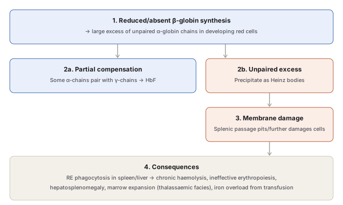
Deficient β-chain synthesis leaves a large excess of unpaired α-chains in developing red cells. Some are removed by pairing with γ-chains to form HbF; the remainder precipitate rapidly as Heinz bodies, damaging the red cell membrane. During passage through splenic sinusoids, these damaged cells are further “pitted” (stripped of the precipitated aggregates) and are then phagocytosed by reticuloendothelial cells of the spleen and liver — producing anaemia, hepatosplenomegaly, and excess tissue iron. Contributing factors also include shortened red cell lifespan, genuinely ineffective erythropoiesis, and haemodilution from expanded plasma volume. β-thalassaemia minor, by contrast, has only mild ineffective erythropoiesis, haemolysis, and lifespan shortening.
Clinical Classification
- β-thalassaemia major (Cooley’s/Mediterranean anaemia) — homozygous state (β⁰/β⁰ or β⁺/β⁺), the most common severe congenital haemolytic anaemia, requiring regular transfusion. Statistically, 25% of offspring of two β-thalassaemia trait carriers will inherit the homozygous (major) state.
- β-thalassaemia intermedia — heterozygous combinations (β⁰/β or β⁺/β) of intermediate severity, not requiring regular transfusion.
- β-thalassaemia minor (trait) — heterozygous, mild, usually asymptomatic.
Less common variants: δβ-thalassaemia minor (both β and δ chain synthesis absent, elevated HbF but normal/reduced HbA2, unlike ordinary β-thalassaemia minor), and Hb Lepore syndrome (non-homologous fusion of β- and δ-genes producing an abnormal fusion haemoglobin, with total absence of normal β-chain synthesis).
Clinical Features of β-Thalassaemia Major
- Anaemia appearing within the first 4–6 months of life, as the γ-to-β chain production switch occurs.
- Marked hepatosplenomegaly — from excess red cell destruction, extramedullary haematopoiesis, and iron overload.
- Bone expansion — marked erythroid hyperplasia produces thalassaemic facies and jaw malocclusion.
- Iron overload — from repeated transfusion, damaging endocrine organs (growth failure, delayed puberty, diabetes mellitus), liver, and heart.
Laboratory Findings (β-Thalassaemia Major)
Severe anaemia; blood film shows severe microcytic hypochromic morphology, marked anisopoikilocytosis, basophilic stippling, numerous target cells, tear-drop cells, and normoblasts; haemoglobin electrophoresis shows HbA reduced or absent (0–50%) with HbF markedly elevated (50–98%).
Summary Table
| Type | Hb level | Electrophoresis | Genotype | Syndrome |
|---|---|---|---|---|
| Hb Bart’s hydrops fetalis | 3–10 g/dL | Hb Bart’s (γ4) 100% | 4-gene deletion | Fatal in utero/early infancy |
| HbH disease | 2–12 g/dL | HbF 10%, HbH 2–4% | 3-gene deletion | Haemolytic anaemia |
| α-thalassaemia trait | 10–14 g/dL | Near-normal | 2-gene deletion | Microcytic, no anaemia |
| β-thalassaemia major | <5 g/dL | HbA 0–50%, HbF 50–98% | β-thal/β-thal | Severe, transfusion-dependent |
| β-thalassaemia intermedia | 5–10 g/dL | Variable | Multiple mechanisms | Severe, transfusion not routinely required |
| β-thalassaemia minor | 10–12 g/dL | HbA2 4–9%, HbF 1–5% | βA/β-thal | Usually asymptomatic |
Clinical Relevance
- The reciprocal HbA2 pattern is a key discriminator worth holding in memory as a pair: β-thalassaemia trait raises HbA2, while α-thalassaemia trait leaves HbA2 normal or low — this single value helps separate the two traits when both present with an identical microcytic, non-anaemic blood picture.
- The 25% recurrence risk when both parents carry β-thalassaemia trait is the concrete basis for premarital/antenatal carrier screening programmes in high-prevalence Indian populations (Punjabi, Bengali, Gujarati, Parsi communities specifically named in the epidemiology above), where identifying trait carriers before conception is the only way to prevent transfusion-dependent β-thalassaemia major in the next generation.
- Recognising that iron overload in β-thalassaemia major arises from the disease process itself (increased intestinal absorption from ineffective erythropoiesis) as well as from transfusion means iron chelation therapy is a necessary part of management from the start of a regular transfusion programme, not an afterthought once overload is already clinically apparent.
Diagram 1: Pathogenesis of Anaemia in β-Thalassaemia Major
Draw a top box (reduced β-globin synthesis) branching into two outcomes for the resulting excess α-chains, converging back into a shared consequences box.
- Reduced/absent β-globin synthesis → excess unpaired α-chains. 2a. Partial compensation — some α-chains pair with γ-chains → HbF. 2b. Unpaired excess — precipitate as Heinz bodies.
- Membrane damage — splenic passage pits/further damages the cell (following only from branch 2b).
- Consequences — RE phagocytosis → chronic haemolysis, ineffective erythropoiesis, hepatosplenomegaly, marrow expansion (thalassaemic facies), iron overload.
Labels required
- Both fates of the excess α-chains (paired into HbF vs precipitated as Heinz bodies) must be shown as parallel branches from the same starting point — the disease is not simply “too few β-chains,” it is specifically the unpaired excess α-chains that drive the pathology.
- The spleen’s dual role must be clear: it both damages passing cells further (“pitting”) and is the site of final phagocytic destruction — not merely a passive filter.
Errors commonly made
- Describing β-thalassaemia anaemia as if it were simply “too little haemoglobin,” missing that the actual membrane-damaging, haemolysis-driving lesion is the toxic excess of unpaired α-chains, not the β-chain deficit itself.
- Confusing this pathogenesis with α-thalassaemia’s mechanism — in α-thalassaemia the excess/toxic chains are β- or γ-chains (forming HbH or Hb Bart’s), the mirror image of β-thalassaemia’s excess α-chains; the two must not be described with the same diagram or the same chain labels.
Long Essays (1)
Short Essays (1)
My Sticky Notes
Personal revision notes, mnemonics and reminders.
