
Syphilis
Definition and organism
Venereal disease, Treponema pallidum (spirochaete). Related: yaws, pinta, bejel.
T. pallidum: coiled, motile, 10µm. Not stainable conventionally → DGI (dark ground illumination), fluorescent Ab, silver impregnation, PCR. Not culturable in vitro (rabbit/chimp models). Killed by cold/heat/antiseptics.
Transmission
Sexual intercourse (main) · intimate contact (lips/tongue/fingers) · transfusion · transplacental (congenital).
Immunology and serology
No toxin — damage = host immune response (same principle as TB/leprosy).
- Treponemal tests (specific, sensitive) — FTA-ABS, MHA-TP, TPHA, Serodia TP-PA
- Non-treponemal/reaginic tests (cardiolipin-lecithin-cholesterol Ag, screening) — RPCF (rapid dx), VDRL/RPR (Wassermann-derived, = STS)
Stages of acquired syphilis
- Primary (2-4wk) — chancre: painless papule→ulcer, indurated, regional lymphadenitis, heals without scar. Histo: plasma cells, periarteritis/endarteritis, endothelial proliferation. Ab+ only 1-3wk after chancre. Infectivity: high.
- Secondary (2-3mo) — mucous patches, skin eruptions, condyloma lata, painless lymphadenopathy. Ab always +. Most infective stage.
- Latent period (2-3yr)
- Tertiary — least infective, most destructive.
- Gumma: solitary rubbery lesion, central coagulative necrosis (less destructive than caseation, outlines faintly visible) + palisaded macrophages + plasma cells + giant cells + fibroblasts. Sites: liver (hepar lobatum), testis, bone, brain.
- Diffuse: CVS (thoracic aortitis → aneurysm, AR, coronary ostial narrowing) + neurosyphilis (meningovascular / tabes dorsalis / general paresis)
Congenital syphilis
16wk gestation exposure. Saddle nose · Hutchinson’s teeth (peg-shaped, widely spaced) · mucocutaneous 2° lesions · epiphysitis/periostitis · interstitial keratitis · hepatic + pulmonary interstitial fibrosis. Stillborn: premature, macerated skin, hepatosplenomegaly, epiphysitis.
Unifying histology
Perivascular plasma-cell infiltrate + endothelial proliferation — present at every stage/site; diagnostic clue even without genital-lesion history.
Clinical relevance
Infectivity ↓ but destruction ↑ across stages (secondary = most infective, tertiary = most destructive) — public health contact-tracing logic. Reactive VDRL/RPR needs treponemal confirmation (reaginic tests can false-positive). Plasma-cell/endarteritis pattern = diagnostic clue on unexpected-site biopsy (liver, aorta, CNS).
Definition
Syphilis is a venereal (sexually transmitted) disease caused by the spirochaete Treponema pallidum — one of a group of related treponemal diseases that also includes yaws, pinta and bejel. Like tuberculosis and leprosy, it is a classical example of chronic granulomatous inflammation with a plasma-cell-rich histologic signature running through all its stages.
Causative Organism
T. pallidum is a coiled, actively motile spiral filament roughly 10 µm long. It cannot be visualised by conventional staining and instead requires dark ground illumination (DGI) of fresh material, fluorescent antibody technique, silver impregnation, or PCR-based nucleic acid amplification. The organism has never been cultivated in artificial media — experimental infection is instead produced in rabbits and chimpanzees — and is rapidly killed by cold, heat and antiseptics, which is relevant to how specimens must be handled for DGI to succeed.
Incidence and Transmission
Incidence fell sharply after penicillin became available in 1943 but the disease remains globally present, most heavily in Sub-Saharan Africa, South America and South-East Asia; men who have sex with men are at higher risk, and some African countries carry a high burden of congenital syphilis with an associated high stillbirth rate. Transmission occurs by sexual intercourse (the dominant route, producing lesions on glans penis, vulva, vagina or cervix), by intimate contact with lesions on lips, tongue or fingers, by transfusion of infected blood, and by transplacental (materno-foetal) spread, producing congenital syphilis.
Immunology and Serology
T. pallidum produces no endotoxin or exotoxin; as with tuberculosis and leprosy, the tissue damage of syphilis is a consequence of the host’s own immune response to the organism, not direct bacterial toxicity.
Diagnosis relies on two categories of serologic test, and distinguishing them by what they actually detect is a frequently tested point:
- Treponemal tests measure antibody directed specifically against T. pallidum antigen and are the more sensitive and specific group: fluorescent treponemal antibody-absorbed (FTA-ABS), agglutination assays (microhaemagglutination assay for T. pallidum/MHA-TP, and the more sensitive Serodia TP-PA), and T. pallidum passive haemagglutination (TPHA).
- Non-treponemal tests measure non-specific reaginic IgM/IgG antibodies directed against a cardiolipin–lecithin–cholesterol complex rather than against the organism itself, and are more widely used for screening: the Reiter protein complement fixation (RPCF) test (preferred for rapid diagnosis), and VDRL/RPR, descended from Wassermann’s original complement-fixation test against an antigen derived from syphilitic tissue — together constituting the traditional Standard Test for Syphilis (STS).
Stages of Acquired Syphilis
Acquired syphilis progresses through three defined stages, distinguished by the timing and character of the lesion, with infectivity and organism burden falling as the disease progresses even as tissue destruction rises:
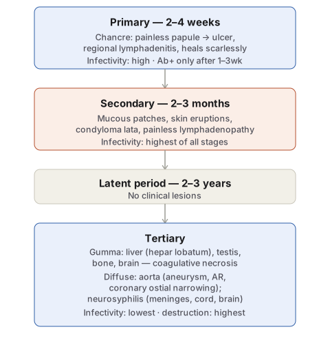
Primary syphilis. The chancre appears 2–4 weeks after exposure, on genitals or at extragenital sites: initially a painless papule that ulcerates centrally, maturing into an indurated, centrally ulcerated lesion with regional lymphadenitis. It heals without scarring even untreated. Histologically the chancre shows a dense plasma-cell-rich infiltrate, perivascular mononuclear (chiefly plasma cell) cuffing with periarteritis and endarteritis, and vascular endothelial proliferation. Antibody tests turn positive only 1–3 weeks after the chancre appears; spirochaetes are demonstrable in the exudate by DGI.
Secondary syphilis. In inadequately treated primary disease, mucocutaneous lesions and painless lymphadenopathy appear 2–3 months after exposure — mucous patches of the mouth, pharynx and vagina, generalised skin eruptions, and condyloma lata in the anogenital region. Antibody tests are always positive by this stage, and this is the most infective stage of the disease, with spirochaetes readily demonstrated in the mucocutaneous lesions.
Tertiary syphilis. After a latent period of roughly 2–3 years from first exposure, tertiary lesions appear; this stage is the least infective, with spirochaetes demonstrable only with difficulty, but the most tissue-destructive. Two lesion types occur:
- Syphilitic gumma — a solitary, localised, rubbery lesion with central necrosis, occurring in liver, testis, bone or brain; in the liver it produces the scarred, lobulated hepar lobatum. Histologically, gumma shows central coagulative necrosis resembling caseation but less destructive (necrosed cell outlines remain faintly visible), surrounded by a zone of palisaded macrophages, abundant plasma cells, some lymphocytes, giant cells and fibroblasts.
- Diffuse lesions, from widespread spirochaetal dissemination, chiefly affecting the cardiovascular and nervous systems. Cardiovascular syphilis targets the thoracic aorta: syphilitic aortitis weakens and dilates the aortic wall, producing aortic aneurysm, aortic valve incompetence, and narrowing of the coronary ostia. Neurosyphilis presents as meningovascular syphilis (chiefly meninges), tabes dorsalis (spinal cord), or general paresis (brain).
Congenital Syphilis
Congenital syphilis may develop in a foetus beyond 16 weeks’ gestation exposed to maternal spirochaetaemia. Major morphologic features include: saddle-shaped nose deformity (destruction of the nasal bridge); Hutchinson’s teeth — small, widely spaced, peg-shaped permanent teeth; mucocutaneous lesions of acquired secondary syphilis; bony lesions (epiphysitis, periostitis); interstitial keratitis with corneal opacity; diffuse hepatic fibrosis; and interstitial pulmonary fibrosis. A stillborn foetus with congenital syphilis is typically premature, with macerated skin, hepatosplenomegaly and syphilitic epiphysitis.
Across every stage and organ, the underlying histology of syphilis is consistent: a perivascular plasma-cell-rich inflammatory infiltrate with endothelial cell proliferation, and spirochaetes demonstrable within the affected tissue — this shared histologic signature is what unifies chancre, gumma, and diffuse tertiary lesions as manifestations of the same disease process at different stages.
Clinical Relevance
- The inverse relationship between infectivity and tissue destruction across the three stages — most infective at secondary, most destructive at tertiary — is a recurring exam point and also the clinical logic behind why secondary syphilis, not tertiary, is the public-health-relevant stage for contact tracing.
- Treponemal versus non-treponemal serology is tested less as trivia and more because their false-positive/negative profiles differ in practice: non-treponemal (reaginic) tests can be falsely positive in unrelated conditions with anticardiolipin antibody production, which is why a reactive VDRL/RPR is confirmed with a treponemal-specific test before a diagnosis is made.
- Recognising the shared “perivascular plasma cells plus endarteritis” histology across chancre, gumma and diffuse lesions is what allows a pathologist to suspect syphilis on a biopsy from an unexpected site (liver, aorta, CNS) even without a preceding history of a genital lesion.
Diagram 1: Stages of Acquired Syphilis
Draw a single downward timeline of four stages.
- Primary (2–4 weeks) — chancre, regional lymphadenitis, heals without scarring; note that antibody tests lag the lesion by 1–3 weeks.
- Secondary (2–3 months) — mucocutaneous lesions and painless lymphadenopathy; label this stage explicitly as the most infective of the three.
- Latent period (2–3 years) — no clinical lesions, included to make clear that tertiary disease is not a direct continuation of secondary disease but follows a genuine quiescent interval.
- Tertiary — split into gumma (liver, testis, bone, brain) and diffuse lesions (aorta, nervous system); label this stage as the least infective but most tissue-destructive.
Labels required
- Infectivity must be marked at each stage, and must visibly fall from primary/secondary to tertiary, since the inverse infectivity-versus-destruction relationship across the three stages is the single most examined concept in this topic.
- The latent period must appear as its own stage, not be compressed into the arrow between secondary and tertiary, since its existence is what explains why tertiary disease can appear years after all symptoms have resolved.
Errors commonly made
- Assuming infectivity rises as the disease progresses (reasoning that a “worse” stage should be more contagious), when secondary — not tertiary — is actually the most infective stage.
- Describing gumma and diffuse tertiary lesions as separate diseases rather than as two expressions of the same tertiary-stage process, one localised and one from widespread dissemination.
My Sticky Notes
Personal revision notes, mnemonics and reminders.
