
Sickle Cell Anaemia
Overview
Haemoglobinopathy from sickle haemoglobin (HbS) — red cells sickle at low oxygen tension. Highest frequency in black Africans/Central Africa (falciparum malaria endemic — HbS carriers protected). In India: seen in tribal populations of central India (MP, Chhattisgarh, Odisha, Maharashtra); overall India’s main haemoglobin disorders are thalassaemia and HbD.
3 forms: heterozygous (sickle trait, AS) · homozygous (sickle cell anaemia, SS) · double heterozygous (sickle-β-thal, SC, SD).
Sickle cell trait (AS)
Benign carrier state, 1 abnormal gene. No symptoms unless severe hypoxia (→ crisis). No anaemia, normal-looking cells. Dx: sickling test (sodium metabisulfite) + electrophoresis shows 35-40% HbS.
Sickle cell anaemia (SS)
2 abnormal genes (1 from each parent). Severe, shortened life expectancy.
Pathogenesis:
- Point mutation — valine replaces glutamic acid at β-globin residue 6 → HbS
- Deoxygenation → HbS polymerises into rod-like fibres → distorts cell into sickle shape
- Reversible sickling (early) → repeated cycles damage membrane → irreversible sickling (stays sickled even with normal O2)
- Factors ↑sickling: ↓HbF proportion (HbF resists sickling, protects), ↑HbS concentration, ↑total Hb, ↑deoxygenation, acidosis, dehydration, ↑2,3-BPG
Clinical features (symptoms start after 6 months, once HbF replaced by HbS):
- Anaemia — severe, chronic, extravascular haemolysis + aplastic crises; often mild symptoms relative to Hb level (HbS releases O2 easily)
- Vaso-occlusion: microinfarcts (abdomen/chest/back/joints → painful crises); macroinfarcts (spleen→autosplenectomy, bone→aseptic necrosis/osteomyelitis, lung, kidney→cortical necrosis, CNS→stroke, retina, skin ulcers)
- Constitutional: impaired growth, ↑infection risk (poor splenic function)
Lab: Hb 6-9 g/dl. Film: sickle cells, target cells, Howell-Jolly bodies (splenic atrophy). Sickling test +ve. Electrophoresis: NO HbA, mostly HbS, HbF 2-20%.
Double heterozygous states
Sickle-β-thal, sickle-C (SC), sickle-D (SD) — all milder than SS. Dx by electrophoresis.
Other haemoglobinopathies (regional)
- HbD (Gln for Glu at β121) — NW India/Pakistan/Iran, ~3% Punjabi Sikhs
- HbE (Lys for Glu at β26) — SE Asia, India, Burma, Sri Lanka
- HbC (Lys for Glu at β6) — West Africa/American blacks
- Unstable Hbs (Hb-Köln etc) — precipitate as Heinz bodies → congenital Heinz body haemolytic anaemia, AD or spontaneous
Clinical relevance
Trait vs disease distinction matters clinically — trait carriers can sickle under severe hypoxia (high altitude, anaesthesia, severe illness) → relevant for pre-op/pre-travel screening. HbF protects against sickling → basis for hydroxyurea therapy (raises HbF). Mild symptoms despite low Hb (HbS unloads O2 easily) → vaso-occlusive crises, not anaemia itself, drive most morbidity/presentations.
Overview
Sickle cell syndromes are the most important and widely prevalent group of haemoglobinopathies, produced by sickle haemoglobin (HbS), which causes red cells to deform (“sickle”) under low oxygen tension. Sickle syndromes reach their highest frequency in the black African population and in Central Africa, where falciparum malaria is endemic — HbS carriers are relatively protected against severe falciparum malaria, the classic example of heterozygote advantage. In India, HbS is found particularly among tribal populations of central India (e.g. parts of Madhya Pradesh, Chhattisgarh, Odisha, and Maharashtra), though the country’s dominant haemoglobinopathies overall are thalassaemia and HbD.
Sickle syndromes occur in three forms:
- Heterozygous — sickle cell trait (AS).
- Homozygous — sickle cell anaemia (SS).
- Double heterozygous — sickle-β-thalassaemia, sickle-C disease (SC), sickle-D disease (SD).
Sickle Cell Trait (AS)
A benign heterozygous carrier state — only one abnormal gene inherited. Clinically silent except under severe hypoxia, which can precipitate a sickle crisis. No anaemia; red cells appear normal at baseline. Diagnosis: sickling demonstrated under reduced oxygen tension using sodium metabisulfite; haemoglobin electrophoresis shows 35–40% HbS.
Sickle Cell Anaemia (SS)
The homozygous state — an abnormal β-globin gene inherited from each parent — producing severe disease with protean manifestations and reduced life expectancy.
Pathogenesis
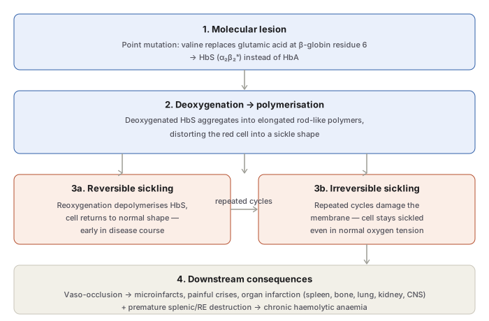
- Molecular lesion — a single point mutation substitutes valine for glutamic acid at residue 6 of the β-globin chain, producing HbS (α₂βˢ₂) instead of HbA.
- Polymerisation — on deoxygenation, HbS molecules aggregate into elongated rod-like polymers, distorting the biconcave disc into the characteristic sickle/crescent shape. This occurs both within intact cells and in free solution.
- Reversible → irreversible sickling — early sickling reverses on reoxygenation, but repeated cycles progressively damage the membrane until cells remain sickled (“irreversibly sickled cells”) even at normal oxygen tension.
- Factors accelerating sickling: proportion of non-HbS haemoglobin (HbF-containing cells resist sickling and protect against co-polymerisation; HbA-containing cells readily co-polymerise with HbS), intracellular HbS concentration, total haemoglobin concentration, degree of deoxygenation, acidosis and dehydration, and raised intracellular 2,3-BPG.
Clinical Features
Symptoms typically begin after the 6th month of life, once HbF is largely replaced by HbS; infection and folate deficiency worsen the clinical picture.
- Anaemia — severe, chronic, predominantly extravascular haemolysis, punctuated by aplastic crises. Symptoms are often mild relative to the haemoglobin level, since HbS releases oxygen to tissues more readily than HbA.
- Vaso-occlusive phenomena — recurrent obstruction of capillary flow by sickled cells:
- Microinfarcts — abdomen, chest, back, joints — the cause of recurrent painful crises.
- Macroinfarcts — spleen (splenic sequestration, eventual autosplenectomy), bone marrow (bone pain), bone (aseptic necrosis, osteomyelitis), lung (pulmonary infection/infarction), kidney (renal cortical necrosis), CNS (stroke), retina, and skin (ulcers).
- Constitutional features — impaired growth and development, and markedly increased infection susceptibility from progressive loss of splenic function.
Laboratory Findings
- Moderate-to-severe anaemia (Hb 6–9 g/dL).
- Blood film: sickle cells, target cells, and features of splenic atrophy (Howell-Jolly bodies).
- Positive sickling test (sodium metabisulfite).
- Haemoglobin electrophoresis: no HbA, predominance of HbS, HbF 2–20%.
Double Heterozygous States
Combinations of HbS with another haemoglobinopathy — sickle-β-thalassaemia, sickle-C disease (SC), sickle-D disease (SD) — all behave as milder forms of sickle cell disease; diagnosis rests on haemoglobin electrophoresis to separate the individual haemoglobins.
Other Structural Haemoglobinopathies (Relevant Regional Context)
- HbD (HbD-Punjab/Los Angeles) — glutamine for glutamic acid at β-121; found in North-West India, Pakistan, and Iran, affecting about 3% of Punjabi Sikhs.
- HbE — lysine for glutamic acid at β-26; predominant in South-East Asia, India, Burma, and Sri Lanka; occurs as trait, homozygous disease (compensated haemolysis), or double heterozygous states with thalassaemia (HbE-β-thalassaemia, HbE-α-thalassaemia).
- HbC — lysine for glutamic acid at β-6; West Africa and American blacks.
- Unstable haemoglobins (e.g. Hb-Köln, Hb-Hammersmith) — denature and precipitate as Heinz bodies, causing congenital non-spherocytic (Heinz body) haemolytic anaemia; autosomal dominant or spontaneous mutation.
Clinical Relevance
- The sickle trait/sickle cell anaemia distinction is a genuine bedside safety issue, not just an academic classification: trait carriers are asymptomatic at normal oxygen tension but can sickle under severe hypoxia (high-altitude flight, anaesthesia, severe illness) — a fact directly relevant to pre-operative and pre-travel screening in populations where HbS is prevalent.
- HbF’s protective effect against sickling is the biological basis for hydroxyurea therapy in sickle cell anaemia, which works chiefly by raising HbF levels and thereby reducing the proportion of sickling-prone cells.
- Recognising that anaemia symptoms in SS can be disproportionately mild relative to the haemoglobin level (because HbS unloads oxygen more readily) helps explain why some patients tolerate a haemoglobin of 6–9 g/dL comparatively well, while vaso-occlusive crises — not the anaemia itself — dominate morbidity and drive most acute presentations.
Diagram 1: Mechanism of Sickling
Draw a single downward sequence: molecular lesion → polymerisation → a fork into reversible/irreversible sickling (with a “repeated cycles” arrow from reversible to irreversible) → converging into downstream consequences.
- Molecular lesion — Val for Glu at β6 → HbS.
- Deoxygenation → polymerisation — rod-like HbS fibres distort cell shape. 3a. Reversible sickling — resolves on reoxygenation (early disease). 3b. Irreversible sickling — membrane damage from repeated cycles, persists despite normal oxygenation.
- Downstream consequences — vaso-occlusion (microinfarcts, crises, organ infarction) and premature splenic/RE destruction (chronic haemolysis).
Labels required
- The exact mutation (valine for glutamic acid, β-globin position 6) must be stated precisely — this exact substitution is one of the most frequently tested single facts in this topic.
- The reversible → irreversible transition must be explicitly linked to “repeated cycles” of sickling/unsickling as the cause of the membrane damage, not presented as an unexplained jump.
- Both downstream consequences (vaso-occlusion and haemolysis) must be shown as parallel outcomes of the same sickled cell, not sequential steps of each other.
Errors commonly made
- Naming the wrong amino acid substitution or the wrong chain (it is the β-chain, not α-chain, and valine replaces glutamic acid, not the reverse).
- Treating all sickling as irreversible from the outset, missing that reversibility is present early in the disease and is lost only after repeated cycles of membrane damage.
- Attributing anaemia in sickle cell disease solely to vaso-occlusion, rather than recognising chronic extravascular haemolysis (premature splenic/RE destruction of sickled cells) as the separate, parallel mechanism.
My Sticky Notes
Personal revision notes, mnemonics and reminders.
