
Radiation Carcinogenesis
Classification
Physical carcinogens = Radiation (UV + ionising, most important) + Non-radiation physical agents.
UV Light
Sources: sunlight, UV lamps, welder’s arc. Limited to epidermis; melanin-dependent (fair skin/albino/Australia-NZ/outdoor workers ↑risk).
Causes: SCC, BCC (↑ with cumulative dose), Malignant melanoma (↑ with intense intermittent exposure — sunbathing/tanning beds).
Mechanism: pyrimidine dimer formation → repaired by NER (nucleotide excision repair) → unrepaired (overwhelmed OR congenital defect) → RAS/p53 mutation.
DNA-repair-defect diseases (proof of mechanism):
- Xeroderma pigmentosum — NER defect → skin Ca <20yo
- Ataxia telangiectasia — DS-break repair defect → leukaemia, ↑radiosensitivity too
- Bloom syndrome — genomic instability → all cancer types
- Fanconi anaemia — cross-link repair defect → ↑overall cancer risk
Ionising Radiation
Sources: X-ray, α/β/γ, radionuclides, protons, neutrons.
Most frequent: all leukaemias except CLL. Also thyroid (papillary), skin, breast, ovary, uterus, lung, myeloma, salivary gland.
Risk ↑ with dose + high-LET (neutron, α > X-ray, γ).
Mechanism: direct DNA bond breakage OR indirect free-radical (water radiolysis) → double-strand breaks = most mutagenic. → chromosome breakage, translocation/inversion, point mutation.
Evidence: radiation dermatitis (early X-ray workers) · osteosarcoma (radium-dial girls) · 10x lung Ca (radioactive-element miners) · Hiroshima/Nagasaki (leukaemia after ~7yr latency; thyroid/breast/colon/lung Ca) · Chernobyl (1985) · therapeutic irradiation (head/neck→thyroid Ca, ankylosing spondylitis, thymus in children, in-utero) · Thorotrast (2x malignancy, withdrawn) · cellphone/EMF (WHO 2011 caution re: glioma/acoustic neuroma, unresolved).
Non-Radiation Physical Carcinogenesis
Gallbladder/urinary stones → organ Ca · burn/trauma scars → skin Ca (Marjolin’s ulcer) · asbestos → lung Ca + mesothelioma · hardwood dust → paranasal sinus adenoCa · surgical implants/foreign bodies → occasional sarcoma.
Clinical relevance
Repair-defect diseases test mechanism understanding, not just association. Cumulative vs intermittent-intense UV pattern → different counselling for SCC/BCC vs melanoma. 7yr leukaemia latency + longer solid-tumour latency → surveillance planning post-exposure. AT’s double-strand-break defect links radiosensitivity + cancer risk in one condition.
Overview
Radiation, whatever its source — sunlight, radiographs, nuclear fission, or radionuclides — is carcinogenic. It is conventionally divided into ultraviolet (UV) light and ionising radiation, the two most important physical carcinogens, with a smaller group of non-radiation physical agents completing the physical carcinogenesis category.
Ultraviolet (UV) Light Carcinogenesis
Sources: sunlight, UV lamps, welder’s arcs. Carcinogenicity is confined to the epidermis (UV does not penetrate deeply) and depends on the degree of skin pigmentation — melanin absorbs and scatters UV, so darker skin is relatively protected.
Epidemiological evidence: highest incidence in fair-skinned people of European descent, in albinos (who lack protective melanin entirely), in inhabitants of sun-drenched regions such as Australia and New Zealand, and in outdoor workers with chronic occupational sun exposure. UV light causes squamous cell carcinoma, basal cell carcinoma, and malignant melanoma of the skin. The pattern of exposure differs by tumour type: non-melanoma skin cancers (SCC, BCC) correlate with total cumulative UV dose, whereas melanoma correlates instead with intense, intermittent exposure — the pattern typical of sunbathing or tanning-bed use rather than chronic outdoor work.
Mechanism: UV light’s key carcinogenic effect is DNA damage in the form of pyrimidine dimer formation — adjacent pyrimidine bases become covalently cross-linked. This damage is normally corrected by the nucleotide excision repair (NER) pathway. Cancer results when repair fails, either because:
- Exposure is excessive enough to overwhelm an otherwise-normal repair system, or
- The repair system itself is congenitally defective, as demonstrated by a group of inherited DNA-repair-defect diseases that prove the mechanism:
- Xeroderma pigmentosum — defective nucleotide excision repair specifically; presents with skin cancer developing before age 20.
- Ataxia telangiectasia — defective repair of double-strand breaks (also increases sensitivity to ionising radiation); predisposes to leukaemia.
- Bloom syndrome — genomic instability disorder; predisposes broadly to all cancer types.
- Fanconi anaemia — defective DNA cross-link repair; increased overall cancer risk.
Unrepaired pyrimidine dimers, once fixed by cell division, lead to mutation of key regulatory genes, particularly RAS and p53.
Ionising Radiation Carcinogenesis
Sources: all forms of ionising radiation — X-rays, α-, β-, and γ-rays, radioactive isotopes, protons, and neutrons — whether from occupational, therapeutic, accidental, or environmental exposure.
Tumours most associated: all forms of leukaemia except chronic lymphocytic leukaemia (CLL) are the most frequent consequence; also thyroid carcinoma (typically papillary), skin cancer, breast, ovarian, uterine, and lung carcinoma, multiple myeloma, and salivary gland tumours.
Dose and LET dependence: carcinogenic risk rises with total dose and is greater for radiation of high linear energy transfer (LET) — neutrons and α-particles are more oncogenic per unit dose than X-rays or γ-rays.
Mechanism: ionising radiation damages DNA either directly, by breaking chemical bonds in the DNA strand, or indirectly, through generation of free radicals from the radiolysis of water. Double-stranded DNA breaks are the most mutagenic form of damage produced. The resulting genetic injury manifests as chromosome breakage, chromosomal rearrangement (translocation, inversion), and, less frequently, point mutation. The oncogenic outcome is further modulated by radiation type, total dose, dose-rate, frequency of exposure, and host factors (age, tissue radiosensitivity, DNA-repair capacity).
Historical and epidemiological evidence:
- Early radiation dermatitis and subsequent skin cancer in the first generation of X-ray workers and radiotherapists, before protective measures were understood.
- Osteosarcoma in the “radium-dial-painting girls” who ingested radium while hand-painting luminous watch dials.
- Increased lung cancer (roughly 10-fold) in unprotected miners of radioactive elements.
- Hiroshima and Nagasaki survivors — markedly increased leukaemia incidence after an average latent period of about 7 years, together with increased mortality from thyroid, breast, colon, and lung carcinoma.
- The Chernobyl nuclear accident (1985) — continuing excess cancer incidence in the surrounding population.
- Therapeutic irradiation — of the head and neck in children (papillary thyroid cancer years later), of the spine in ankylosing spondylitis, and of an enlarged thymus in infancy; also carcinogenic risk from in-utero irradiation exposure.
- Thorotrast, a thorium-containing radiographic contrast medium used mid-20th century, was withdrawn after being found to roughly double malignancy risk in those who received it.
- The debate over electromagnetic fields/cellphone radiation remains unresolved — the WHO (2011) issued a cautionary classification regarding possible glioma and acoustic neuroma risk with long-term heavy use, but no conclusive causal link has been established for other outcomes.
Non-Radiation Physical Carcinogenesis
A smaller group of physical agents cause cancer through chronic irritation, inflammation, or foreign-body reaction rather than radiation:
- Gallbladder and urinary tract stones — chronic irritation predisposing to organ carcinoma (gallbladder, bladder).
- Burn scars and trauma scars — chronic wound sites predisposing to skin carcinoma (Marjolin’s ulcer).
- Asbestos fibres — bronchogenic carcinoma and malignant mesothelioma of pleura/peritoneum.
- Hardwood dust (cutting, engraving occupations) — adenocarcinoma of the paranasal sinuses.
- Inert surgical implants and embedded foreign bodies — occasional focus for sarcoma formation.
Clinical Relevance
- The UV-repair-defect diseases (xeroderma pigmentosum, ataxia telangiectasia, Bloom syndrome, Fanconi anaemia) are a favourite way examiners test whether the mechanism of UV carcinogenesis, not just the association, has been understood — each disease “proves” the pyrimidine-dimer/NER pathway by showing what happens when repair fails congenitally rather than from overexposure.
- The cumulative-dose (SCC/BCC) versus intense-intermittent (melanoma) distinction in UV exposure pattern has direct counselling relevance — chronic sun protection reduces SCC/BCC risk, while advice against intermittent intense exposure (sunburn episodes, tanning beds) specifically targets melanoma risk.
- The 7-year latency for leukaemia after atomic bomb exposure, and the decades-long latency for solid tumours and therapeutic-irradiation-associated thyroid cancer, are clinically relevant to long-term surveillance planning after any significant radiation exposure (occupational, therapeutic, or accidental).
- Recognising double-strand DNA breaks as the most mutagenic lesion from ionising radiation links directly back to ataxia telangiectasia (defective double-strand break repair), which explains that condition’s combined radiosensitivity and cancer predisposition.
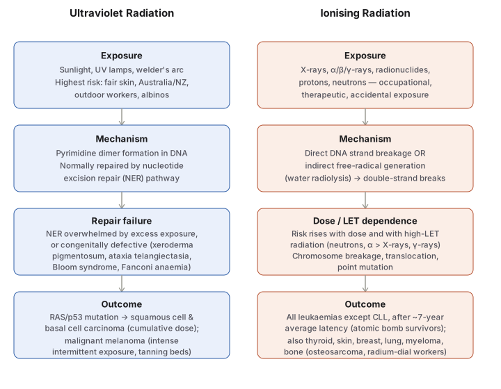
Diagram 1: UV versus Ionising Radiation — Mechanism Compared
Draw two parallel columns (UV Radiation, Ionising Radiation), each with four stacked stages: Exposure → Mechanism → Modulating factor → Outcome. Do not connect the two columns — they are compared side by side, not sequential.
UV column:
- Exposure — sunlight/UV lamps/welder’s arc; fair skin, Australia/NZ, outdoor workers at greatest risk.
- Mechanism — pyrimidine dimer formation, normally repaired by nucleotide excision repair (NER).
- Repair failure — NER overwhelmed by excess exposure, or congenitally absent (four named diseases).
- Outcome — SCC/BCC (cumulative dose) vs melanoma (intense intermittent exposure).
Ionising column:
- Exposure — X-ray/particulate/radionuclide, occupational/therapeutic/accidental.
- Mechanism — direct strand breakage or indirect free-radical damage, double-strand breaks most mutagenic.
- Dose/LET dependence — risk ∝ dose and LET (neutron, α > X-ray, γ).
- Outcome — leukaemia (~7yr latency, dominant) plus thyroid/skin/breast/lung/bone.
Labels required
- The four DNA-repair-defect diseases (xeroderma pigmentosum, ataxia telangiectasia, Bloom syndrome, Fanconi anaemia) must all appear in the UV “repair failure” box by name — each is independently examinable and each targets a slightly different repair defect, not interchangeable synonyms for “DNA repair disease.”
- The cumulative-dose vs intense-intermittent distinction between non-melanoma skin cancer and melanoma must be explicit in the UV outcome box — this is the single most commonly tested nuance of UV carcinogenesis.
- “Double-strand breaks = most mutagenic lesion” must be called out explicitly in the ionising mechanism box, since it is the fact that mechanistically links back to ataxia telangiectasia’s combined radiosensitivity/cancer phenotype.
Errors commonly made
- Treating “UV causes skin cancer” and “ionising radiation causes leukaemia” as the whole answer, without being able to state the actual DNA lesion (pyrimidine dimer vs double-strand break) or the repair pathway (NER) involved.
- Reversing the UV exposure-pattern rule — attributing melanoma to cumulative dose and SCC/BCC to intermittent intense exposure, rather than the correct (and counter-intuitive) opposite assignment.
- Omitting that ataxia telangiectasia patients are hypersensitive to ionising radiation as well as prone to cancer — the two facts are mechanistically the same defect (double-strand break repair), not two unrelated features of the syndrome.
My Sticky Notes
Personal revision notes, mnemonics and reminders.
