
Primary Myelofibrosis
Overview
Primary myelofibrosis (chronic idiopathic myelofibrosis/agnogenic myeloid metaplasia/myelosclerosis) = clonal myeloproliferative disorder, neoplastic stem cells proliferate OUTSIDE marrow (extramedullary haematopoiesis — liver, spleen), no identifiable cause. Same family as CML, polycythaemia vera, ET.
Secondary myelofibrosis = different entity — associated with other marrow disorders or toxic/irradiation injury.
Pathogenesis
Unknown cause. Various chromosomal abnormalities but no specific marker (unlike CML’s BCR-ABL). Three growth-factor processes:
- Marrow fibrosis — TGF-β overproduction
- Osteosclerosis — osteonectin
- Marrow angiogenesis — ↑VEGF
Clinical features
Late middle life, gradual onset, both sexes equal. Anaemia+constitutional symptoms (fatigue, weakness, anorexia). MASSIVE splenomegaly — often dominant feature (discomfort, pain, dyspnoea). Hepatomegaly ~half. Bleeding (petechial) ~20%. Less common: lymphadenopathy, jaundice, ascites, bone pain, hyperuricaemia.
Lab findings
Anaemia — usually mild (unless PV features coexist). Leucocytosis at presentation → leucopenia later. Thrombocytosis initially → thrombocytopenia advanced (tracks proliferative→burnt-out progression).
Smear (highly characteristic): bizarre RBC shapes, TEARDROP poikilocytes (dacrocytes), basophilic stippling, nucleated RBCs, immature leucocytes (= leucoerythroblastic reaction), basophilia, giant platelets.
Marrow aspiration → “DRY TAP” (fibrotic, can’t aspirate) → need TREPHINE BIOPSY: focal hypercellularity, ↑reticulin, variable collagen, well-preserved megakaryocyte clusters. Extramedullary haematopoiesis confirmed by liver biopsy/splenic aspiration.
Treatment/prognosis
No specific cure. Anaemia/ineffective erythropoiesis don’t respond to erythropoietin/androgens. Splenectomy for symptomatic relief in some. WORSE prognosis than PV or ET.
Clinical relevance
Teardrop poikilocyte = one of the most specific morphologic clues in haematology — with leucoerythroblastic picture, immediately suggests marrow infiltration/fibrosis (myelofibrosis or metastatic marrow replacement), not a nonspecific finding. “Dry tap” is diagnostically meaningful itself (reflects fibrotic marrow), not a failed procedure — should prompt trephine biopsy. Platelet trajectory (thrombocytosis→thrombocytopenia) = single continuous process (hyperproliferative→progressive failure), not two disconnected findings. Massive splenomegaly = direct consequence of extramedullary haematopoiesis (spleen actively producing blood cells), not passive congestion/infiltration.
Overview
Primary myelofibrosis — also called chronic idiopathic myelofibrosis, agnogenic myeloid metaplasia, or myelosclerosis — is a clonal myeloproliferative disorder characterised by proliferation of neoplastic stem cells at sites outside the bone marrow (extramedullary haematopoiesis), especially in the liver and spleen, without an identifiable underlying cause. It belongs to the same family of chronic myeloproliferative neoplasms as Chronic Myeloid Leukaemia, Polycythaemia, and Essential Thrombocythaemia.
Secondary myelofibrosis, by contrast, develops in association with other well-defined marrow disorders, or as a result of toxic chemical/irradiation injury — it is not the same entity as primary myelofibrosis, though the marrow fibrosis can look morphologically similar.
Pathogenesis
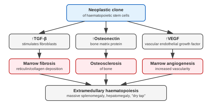
Exact aetiology is unknown; various chromosomal abnormalities have been reported, but no single specific cytogenetic marker defines the disease (unlike CML’s BCR-ABL). Three distinct growth-factor-driven processes underlie the disease’s characteristic findings:
- Marrow fibrosis — from overproduction of transforming growth factor-β (TGF-β).
- Osteosclerosis of bone — related to osteonectin.
- Marrow angiogenesis — driven by increased vascular endothelial growth factor (VEGF).
Clinical Features
Onset in late middle life, gradual, affecting both sexes equally:
- Anaemia with constitutional symptoms — fatigue, weakness, anorexia.
- Massive splenomegaly — often the dominant clinical feature, producing abdominal discomfort, pain, and dyspnoea.
- Hepatomegaly — present in about half of cases.
- Bleeding problems (petechial and other) — in about 20% of cases.
- Less common: lymphadenopathy, jaundice, ascites, bone pain, hyperuricaemia.
Laboratory Findings
- Anaemia — usually mild, unless features of coexistent polycythaemia vera are present.
- Leucocytosis at presentation, but leucopenia may develop later as the disease progresses.
- Thrombocytosis initially, but advanced disease shows thrombocytopenia — the platelet count trajectory tracks the overall progression from proliferative to “burnt-out,” fibrotic marrow failure.
- Peripheral blood smear — a distinctive and highly characteristic picture: bizarre red cell shapes, teardrop poikilocytes (dacrocytes), basophilic stippling, nucleated red cells, immature leucocytes (together constituting a leucoerythroblastic reaction), basophilia, and giant platelet forms.
- Bone marrow aspiration typically yields a “dry tap” — the fibrotic marrow cannot be aspirated. Trephine biopsy is therefore required, showing focal hypercellularity, increased reticulin network, and variable collagen, with well-preserved clusters of megakaryocytes.
- Extramedullary haematopoiesis can be confirmed by liver biopsy or splenic aspiration.
Treatment and Prognosis
No specific curative therapy exists for primary myelofibrosis itself. The anaemia and ineffective erythropoiesis are difficult to treat and generally do not respond to erythropoietin or androgens. Splenectomy may be necessary in some cases for symptomatic relief of massive splenomegaly. Overall, primary myelofibrosis carries a poorer prognosis than either polycythaemia vera or essential thrombocythaemia.
Clinical Relevance
- The teardrop poikilocyte (dacrocyte) on peripheral smear is one of the most specific morphologic clues in haematology — its presence, especially alongside a leucoerythroblastic picture, should immediately raise suspicion for marrow infiltration/fibrosis (myelofibrosis, or marrow replacement by metastatic tumour), rather than being read as a nonspecific red cell shape change.
- The “dry tap” on bone marrow aspiration is itself a diagnostically meaningful finding, not simply a failed procedure — it reflects the fibrotic marrow’s physical inability to yield aspirate, and should prompt trephine biopsy rather than being written off as technical failure.
- The platelet count’s evolution from thrombocytosis early to thrombocytopenia late captures the disease’s overall arc — from a hyperproliferative marrow state to progressive marrow failure as fibrosis advances — a pattern worth understanding as a single continuous process rather than two disconnected findings.
- Massive splenomegaly in myelofibrosis is not incidental but is the direct anatomic consequence of the disease’s defining feature (extramedullary haematopoiesis) — the spleen becomes a site of active blood cell production, not merely an organ passively enlarged by congestion or infiltration.
Diagram 1: Growth-Factor-Mediated Mechanism
Draw a top box (neoplastic clone) forking into three parallel columns, each three boxes deep, converging into a single bottom box.
- Top — neoplastic clone of haematopoietic stem cells. 2a/2b/2c — ↑TGF-β / ↑Osteonectin / ↑VEGF (three parallel growth factors). 3a/3b/3c — Marrow fibrosis / Osteosclerosis / Marrow angiogenesis (each factor’s direct effect).
- Bottom, convergent — Extramedullary haematopoiesis → massive splenomegaly, hepatomegaly, “dry tap.”
Labels required
- Each growth factor must be paired with its own specific downstream effect (TGF-β→fibrosis, osteonectin→osteosclerosis, VEGF→angiogenesis) — these are three separate, factor-specific processes, not one generic “growth factor excess” arrow.
- The convergence box must name all three downstream clinical consequences (splenomegaly, hepatomegaly, dry tap) so the diagram closes the loop back to the disease’s actual clinical presentation.
Errors commonly made
- Treating marrow fibrosis as the primary event that causes the neoplastic proliferation, rather than as a secondary consequence of the neoplastic clone’s growth factor output — the fibrosis is reactive, not the initiating lesion.
- Missing that the “dry tap” on aspiration is a direct, predictable consequence of the marrow fibrosis shown in this diagram, rather than an unrelated technical detail to memorise separately.
My Sticky Notes
Personal revision notes, mnemonics and reminders.
