
Pneumoconiosis
Overview
Originally = mineral dust lung disease (now broadened to organic particulates ± chemical fume/vapour). Classic 3: coal, silica, asbestos (occupational; asbestos uniquely → family risk via clothing residue).
Determinants: particle size/shape, solubility, retained amount, co-irritants (esp smoking), host factors. Size: >5-10μm trapped before distal airways | <0.5μm move in/out, minimal deposition | 1-5μm = MOST dangerous (lodge at distal airway bifurcations). Coal=inert(needs large burden); silica/asbestos/beryllium=strong response at LOW doses.
Alveolar macrophage = key cell. Phagocytosis→inflammasome activation→IL-1→fibroblast proliferation+collagen. Lymphatic spread→immune amplification. Smoking worsens ALL mineral dusts, most with asbestos.
Coal Workers’ Pneumoconiosis (context)
Commonest pneumoconiosis. 20-30yr bituminous coal exposure. Spectrum: anthracosis(benign, no disease, also in smokers/urban dwellers) → simple CWP(macules/nodules, minimal dysfunction) → PMF(<10% of simple CWP progress; confluent >2cm black scars). PMF risk↑: older age, ↑dust burden, ↑duration, concurrent TB, +silica. Caplan’s syndrome = RA+pneumoconiosis(coal/silica/asbestos), necrotic/cavitating/calcifying nodules, immunologic (RF+ANA+). CWP = NO increased lung cancer risk (key distinguishing feature).
Silicosis
Most prevalent chronic occupational disease worldwide. Crystalline SiO2, quartz=most fibrogenic. Occupations: mining(granite/sandstone/slate/coal/gold/tin/copper), quarrying, tunnelling, sandblasting, grinding, ceramics, foundry; India: pencil/slate/agate-grinding (~3 million at-risk workers, ICMR).
Pathogenesis: silica(~0.5-5μm) phagocytosed → CYTOTOXIC to macrophages → kills them → repetitive phagocytosis-necrosis cycle. Inflammasome activation→IL-1/TNF/lipid mediators/free radicals/fibrogenic cytokines. + T&B lymphocyte activation→↑IgG/IgM, ANA, RF, immune complexes.
Morphology: discrete hard silicotic nodules(1-5mm), upper zones initially, hyalinised sparse-cellularity centre, WHORLED/concentric collagen laminations, birefringent silica (polarised light, central). May coalesce→complicated silicosis/PMF, calcify, cavitate(ischaemic necrosis/TB/Caplan’s). Pleural+hilar node involvement.
Clinical: slow/insidious, dyspnoea main symptom, obstructive or restrictive pattern. Egg-shell calcification of hilar nodes = characteristic sign. Complications: TB (silica IMPAIRS macrophage mycobacterial killing — specific mechanism, not vague immunosuppression; silicotuberculosis nodules show central caseation), RA(Caplan’s), cor pulmonale. Modestly ↑lung cancer risk (unlike CWP).
Asbestosis/Asbestos-Related Disease
Fibrous crystalline hydrated silicate, fire-resistant, insulation use. 2 forms: Serpentine (curly/flexible = chrysotile/“white asbestos,” >90% commercial use) vs Amphibole (straight/rigid = crocidolite/“blue,” amosite/“brown,” tremolite, anthophyllite, actinolite — LESS common but MORE linked to malignant pleural tumours, esp. crocidolite). Aphorism: “coal=lot of dust+little fibrosis; asbestos=little dust+lot of fibrosis.”
Pathogenesis: fibres phagocytosed→interstitium→pleura/nodes via lymphatics. Coated glycoprotein+haemosiderin→beaded/dumbbell asbestos bodies (Prussian-blue+). Inflammasome+phagolysosomal damage+free radicals+fibrogenic cytokines(IL-1)→DIFFUSE interstitial fibrosis (NOT nodular, unlike silica). Carcinogenic: tumour initiator+promoter — free radicals near mesothelium + adsorbed toxic chemicals(incl. tobacco carcinogens) on fibres → basis of smoking synergy.
3 disease categories (>1 decade exposure usually):
- Asbestosis (parenchymal) — small firm lungs, pleural thickening, subpleural/basal fibrosis, non-specific interstitial fibrosis + asbestos bodies (defining feature); ± emphysema; hilar nodes LESS involved than silicosis
- Pleural disease — pleural effusion(~5% workers, serious, +subpleural asbestosis) | visceral pleural fibrosis(dense, encasing) | pleural plaques(MOST COMMON asbestos lesion; circumscribed/firm/bilateral/hyalinised/often calcified/visible on CXR; usually NO asbestos bodies within — distinct from asbestosis!)
- Tumours — bronchogenic carcinoma (most common malignancy in workers; 5x non-smoker-worker vs non-smoker-general; 10x smoker-worker vs other smokers = SYNERGISTIC not additive) | malignant mesothelioma (uncommon overall but 30-80% of mesothelioma cases have asbestos history; occurs even with LIGHT exposure incl. household contacts) | also oesophagus/stomach/colon/kidney/larynx/lymphoid Ca
Clinical: slow/insidious, may be asymptomatic years despite radiographic changes; interstitial fibrosis→dyspnoea+cough; advanced→Caplan’s, cor pulmonale, tumours.
Clinical relevance
3-way contrast = highest-yield structure: coal=mild/nodular/no Ca risk | silica=cytotoxic/nodular/modest Ca risk+TB susceptibility | asbestos=diffuse(not nodular)/strong Ca risk(smoking-synergistic)+mesothelioma — each traces to distinct particle-macrophage interaction. Silicosis-TB link = SPECIFIC mechanism (macrophage mycobacterial-killing impairment), not vague immunosuppression. Asbestos-smoking synergy(5x→10x) = classic true-synergy (not additive) teaching example. Pleural plaques usually asbestos-body-FREE ≠ asbestosis (parenchymal, asbestos-body-DEFINING) — related but distinct entities, common conflation to avoid.
Overview
Pneumoconiosis originally described lung disease from inhaled mineral dust, though the term has broadened to include organic particulates and, by some definitions, chemical fume/vapour-induced lung disease. The three classic, most common mineral-dust pneumoconioses are caused by coal dust, silica, and asbestos — occupational exposures in each case, though asbestos uniquely extends cancer risk to family members of exposed workers (via residual fibres on clothing).
Determinants of lung damage from inhaled dust: particle size and shape, solubility/physicochemical composition, amount retained in the lung, co-exposure to other irritants (especially tobacco smoke), and host factors (clearance efficiency, immune status). Particle size is critical: particles >5–10 μm are largely trapped before reaching distal airways; particles <0.5 μm tend to move in and out of alveoli without significant deposition; particles in the 1–5 μm range lodge most efficiently at the bifurcations of distal airways and cause the most disease. Coal dust is relatively inert (large amounts must accumulate before disease appears); silica, asbestos, and beryllium provoke a much stronger immune/fibrotic response at lower doses.
The pulmonary alveolar macrophage is the key cell type initiating and perpetuating the inflammatory/fibrotic response: after phagocytosing particles, many trigger inflammasome activation and IL-1 release, initiating fibroblast proliferation and collagen deposition; particles reaching the lymphatics (directly or within migrating macrophages) can also trigger an immune response, amplifying the local reaction. Tobacco smoking worsens the effect of every inhaled mineral dust, most markedly with asbestos.
Coal Workers’ Pneumoconiosis (Context/Comparison)
Commonest pneumoconiosis, from prolonged (20–30 year) inhalation of coal dust, chiefly in bituminous coal miners. Spectrum: anthracosis (benign, asymptomatic carbon pigment deposition, common in urban dwellers/smokers too, no true disease) → simple CWP (coal macules/nodules, dust-laden macrophages, minimal reticulin/collagen increase, little dysfunction) → progressive massive fibrosis (PMF), developing in a minority (<10%) of simple CWP, with confluent >2 cm dense collagenous black scars. PMF risk rises with older age, higher coal dust burden, longer exposure, concomitant TB, and additional silica exposure. Caplan’s syndrome (rheumatoid pneumoconiosis) — rheumatoid arthritis developing alongside CWP (or silicosis/asbestosis), with rounded necrotic/cavitating/calcifying nodules on an immunologic basis (rheumatoid factor, antinuclear antibodies detectable). CWP notably does not increase bronchogenic carcinoma risk (once smoking is accounted for) — a key distinguishing feature from silica and asbestos exposure.
Silicosis
Currently the most prevalent chronic occupational disease worldwide, from prolonged inhalation of crystalline silicon dioxide (silica), the most fibrogenic form being quartz. At-risk occupations: mining (granite, sandstone, slate, coal, gold, tin, copper), quarrying, tunnelling, sandblasting, grinding, ceramics, foundry work, abrasive manufacture; in India specifically, pencil/slate/agate-grinding industries carry high risk (an estimated 3 million Indian workers at high potential risk per ICMR).
Pathogenesis: silica particles (roughly 0.5–5 μm) are phagocytosed by alveolar macrophages, but silica is directly cytotoxic to the macrophages that engulf it, killing them and setting up a repetitive cycle of phagocytosis and necrosis as new macrophages take up the released silica. Silica-laden macrophages/dust reach the interstitium and, via lymphatics, the regional lymph nodes. Silica activates the inflammasome, releasing IL-1, TNF, lipid mediators, free radicals, and fibrogenic cytokines; concurrently, T and B lymphocyte activation raises serum immunoglobulins (IgG, IgM), antinuclear antibodies, rheumatoid factor, and circulating immune complexes.
Morphology: discrete, well-circumscribed, hard silicotic nodules (1–5 mm), initially in the upper lung zones, each with a hyalinised, sparsely cellular centre in whorled/concentric laminations of collagen, surrounded by dust-laden macrophages and a rim of lymphocytes/plasma cells; birefringent silica particles are visible under polarised light, mainly centrally. Nodules may coalesce into complicated silicosis/PMF, calcify, or cavitate (ischaemic necrosis, TB, or Caplan’s syndrome). Pleural and hilar lymph node involvement occurs.
Clinical features: slow, insidious onset; dyspnoea is the main complaint; may progress to obstructive or restrictive patterns; egg-shell calcification of hilar nodes is a characteristic radiographic sign. Complications: TB (silica impairs macrophage killing of mycobacteria, hence increased susceptibility — silicotuberculosis nodules show central caseation), rheumatoid arthritis (Caplan’s), cor pulmonale. Silicosis carries a modestly increased lung cancer risk (unlike CWP).
Asbestosis and Asbestos-Related Disease
Asbestos is a family of fibrous, fire-resistant crystalline hydrated silicates, historically valued for thermal/electrical insulation. Two major geometric forms: serpentine (curly, flexible fibres — chrysotile/“white asbestos,” >90% of commercial use) and amphibole (straight, rigid fibres — crocidolite/“blue asbestos,” amosite/“brown asbestos,” tremolite, anthophyllite, actinolite); amphiboles, though less commonly used, are more strongly linked to malignant pleural tumours, particularly crocidolite. The aphorism captures the contrast with coal: “coal is a lot of dust and little fibrosis; asbestos is little dust and a lot of fibrosis.”
Pathogenesis: inhaled fibres are phagocytosed by alveolar macrophages, reaching the interstitium and, via lymphatics, the pleura and regional nodes. Fibres coated with glycoprotein and endogenous haemosiderin form the characteristic beaded/dumbbell-shaped asbestos bodies (Prussian-blue positive). All asbestos types are fibrogenic — inflammasome activation, phagolysosomal membrane damage, free radical release, and macrophage-derived fibrogenic cytokines (e.g. IL-1) drive diffuse interstitial fibrosis (unlike silica’s discrete nodules). Asbestos is also carcinogenic, acting as both tumour initiator and promoter, partly via free radicals localising near the mesothelium and partly via toxic chemicals (including tobacco carcinogens) adsorbed onto fibre surfaces — the likely basis of the strong smoking-asbestos synergy for bronchogenic carcinoma.
Three categories of disease result from prolonged (usually >1 decade) exposure:
- Asbestosis (parenchymal interstitial fibrosis) — small, firm lungs with pleural thickening; subpleural/basal fibrosis; histologically non-specific interstitial fibrosis plus characteristic asbestos bodies; emphysematous change may coexist; hilar node involvement is less prominent than in silicosis.
- Pleural disease — three patterns: pleural effusion (~5% of asbestos workers, usually serious, with subpleural asbestosis); visceral pleural fibrosis (dense thickening encasing the lung); pleural plaques (the most common asbestos-related lesion — circumscribed, firm, bilateral, hyalinised collagenous nodules, often calcified and radiographically visible, typically without asbestos bodies within them).
- Tumours — bronchogenic carcinoma (the most common malignancy in asbestos workers; incidence 5× higher in non-smoking asbestos workers vs non-smoking general population, and 10× higher in smoking asbestos workers vs other smokers — a markedly synergistic, not merely additive, interaction) and malignant mesothelioma (uncommon overall, but asbestos exposure history is present in 30–80% of mesothelioma cases; can occur even with light exposure, including in asbestos workers’ household contacts). Other associated cancers: oesophagus, stomach, colon, kidney, larynx, and lymphoid malignancies.
Clinical features: slow, insidious; may be asymptomatic for years despite radiographic pleural plaques/parenchymal change; interstitial fibrosis eventually brings dyspnoea with dry or productive cough; advanced cases may develop Caplan’s syndrome, cor pulmonale, or the associated tumours described above.
Clinical Relevance
- The coal/silica/asbestos three-way contrast is the highest-yield organising structure in this topic: coal = mild/nodular/no cancer risk; silica = cytotoxic/nodular/modest cancer risk + TB susceptibility; asbestos = diffuse (not nodular)/strong cancer risk (synergistic with smoking) + mesothelioma — each difference traces back to a distinct particle-macrophage interaction.
- Silicosis’s link to tuberculosis via impaired macrophage mycobacterial killing (rather than a vague “immunosuppression”) is a specific, testable mechanism worth distinguishing from the general principle that any chronic lung disease predisposes to infection.
- The asbestos-smoking synergy for bronchogenic carcinoma (roughly multiplicative, not additive — 5× non-smoker risk becomes 10× in smokers relative to non-exposed smokers) is one of medicine’s clearest teaching examples of true synergistic (rather than simply additive) carcinogen interaction, with direct occupational-counselling relevance.
- Recognising pleural plaques as usually asbestos-body-free (unlike the parenchymal lesion, asbestosis, which is defined by the presence of asbestos bodies) prevents a common conflation of “any asbestos-related pleural or lung finding” with “asbestosis” specifically — the two are related but pathologically distinct entities requiring different histologic criteria.
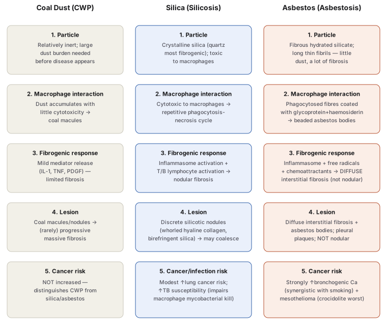
Diagram 1: Three Mechanisms of Pneumoconiosis Compared
Draw three parallel columns (Coal Dust/CWP, Silica/Silicosis, Asbestos/Asbestosis), each with five stacked, step-matched rows: particle behaviour → macrophage interaction → fibrogenic response → lesion produced → cancer/infection risk.
- Particle — inert, needs large burden (coal) vs cytotoxic to macrophages (silica) vs fibrous, little dust causes much fibrosis (asbestos).
- Macrophage interaction — accumulates without cytotoxicity (coal) vs kills macrophages, repetitive phagocytosis-necrosis cycle (silica) vs coated fibres form beaded asbestos bodies (asbestos).
- Fibrogenic response — mild mediator release, limited fibrosis (coal) vs inflammasome + lymphocyte activation, nodular fibrosis (silica) vs inflammasome + free radicals, diffuse fibrosis (asbestos).
- Lesion — coal macules/nodules, rare PMF (coal) vs discrete whorled silicotic nodules (silica) vs diffuse interstitial fibrosis + pleural plaques (asbestos).
- Cancer/infection risk — none (coal) vs modest lung cancer risk + TB susceptibility (silica) vs strong, smoking-synergistic bronchogenic carcinoma + mesothelioma (asbestos).
Labels required
- Row 4 must explicitly contrast “nodular” (silica) against “diffuse” (asbestos) — this single word distinction is the most direct way to separate the two conditions’ histology on sight, and dropping it collapses the diagram’s main teaching value.
- Row 5 must carry the specific mechanism for silica’s TB link (impaired macrophage mycobacterial killing) rather than a generic “weakened immunity” label — the mechanism itself is what is tested.
- The coal column’s “no increased cancer risk” (row 5) must be shown explicitly, not left blank — it is the key negative finding that distinguishes coal from the other two dusts, and a blank cell reads as “unknown” rather than “specifically absent.”
Errors commonly made
- Describing all three pneumoconioses as producing the same kind of fibrosis — the nodular-vs-diffuse distinction (silica vs asbestos) is a frequently tested, easily blurred detail.
- Attributing lung cancer risk to coal dust exposure by over-generalising from silica/asbestos — CWP is specifically the pneumoconiosis without increased cancer risk once smoking is accounted for.
- Treating asbestos bodies as present in every asbestos-related lesion — they define asbestosis (the parenchymal lesion) but are typically absent from pleural plaques, a distinct lesion type covered in
notes.md.
Multiple Choice Questions (2)
My Sticky Notes
Personal revision notes, mnemonics and reminders.
