
Phagocytosis
Definition
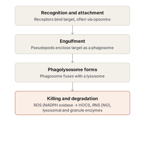
Recognition, internalisation, and digestion of microbes/necrotic debris by neutrophils and macrophages. Three steps: recognition and attachment → engulfment → killing/degradation.
Recognition and attachment
Receptors: TLRs (microbial products) · G protein-coupled (N-formyl methionine) · cytokine receptors (IFN-γ) · mannose receptors · scavenger receptors · opsonin receptors.
Opsonins and their receptors:
| Opsonin | Receptor |
|---|---|
| IgG | FcγRI |
| C3b | CR1, CR3 |
| Collectins (mannose-binding lectin) | C1q |
Bruton disease (defective B-cell maturation) → no IgG → defective opsonisation.
Engulfment
Actin polymerisation → pseudopods → phagosome → fuses with lysosome → phagolysosome. Granule contents may leak extracellularly → bystander tissue damage.
Chediak-Higashi syndrome: autosomal recessive, LYST gene defect → failed phagosome-lysosome fusion → giant granules, neutropenia, delayed killing, recurrent infection + albinism + nerve defects + bleeding tendency.
Killing and degradation (4 mechanisms)
- ROS: NADPH oxidase → superoxide (respiratory burst) → H₂O₂ (dismutation) → myeloperoxidase + Cl⁻ → HOCl (bleach’s active agent, most powerful O₂-dependent mechanism) → also → hydroxyl radical.
- RNS: iNOS (cytokine-activated macrophages) → NO from L-arginine → + superoxide → peroxynitrite.
- Lysosomal enzymes: acid hydrolases, elastase.
- Granule enzymes/antimicrobial proteins: azurophil (primary) + specific (secondary) granules. α1-antitrypsin restrains neutrophil elastase — deficiency → tissue damage (emphysema).
NETs — non-phagocytic killing
Fibrillar chromatin mesh (histones + DNA) + granule proteins, released with neutrophil nucleus loss (NETosis). Traps/concentrates antimicrobials. Nuclear antigen source implicated in lupus.
Inherited defects — map to the 3 steps
| Step | Disease | Defect |
|---|---|---|
| Recognition (opsonisation) | Bruton disease | No IgG |
| Engulfment | Chediak-Higashi syndrome | Failed phagolysosome fusion (LYST) |
| Killing | Chronic granulomatous disease | NADPH oxidase defect (X-linked or AR) → granulomas form to compensate |
Clinical relevance
Three steps = three distinct immunodeficiency categories, each hitting a different step. HOCl/MPO = most powerful O2-dependent killing mechanism. Unchecked granule enzyme release = tissue injury as an extension of a normally protective process (esp. with antiprotease deficiency).
Definition
Phagocytosis is the process by which neutrophils and macrophages recognise, internalise, and digest microorganisms, necrotic cell debris, and other foreign particulate material. It is central to both innate host defence and the clearance of dead tissue at a site of injury, and it proceeds through three sequential steps: recognition and attachment, engulfment, and killing or degradation of the ingested material.
Recognition and Attachment
Phagocytic cells identify their targets through a range of surface receptors that recognise either the target directly or a coating applied to it.
- Toll-like receptors (TLRs) — recognise microbial products directly.
- G protein-coupled receptors — recognise bacterial N-formyl methionine residues.
- Cytokine receptors — respond to interferon-gamma and other activating cytokines.
- Mannose receptors — bind terminal mannose and fucose residues on microbial glycoproteins and glycolipids.
- Scavenger receptors — bind low-density lipoprotein particles as well as a variety of microbes.
- Opsonin receptors — recognise particles that have been coated by opsonins, proteins that mark a target for more efficient phagocytosis.
Opsonisation substantially enhances phagocytic efficiency. The major opsonins and their corresponding leukocyte receptors are IgG antibodies (recognised by the Fc receptor, FcγRI), the complement breakdown product C3b (recognised by complement receptors CR1 and CR3), and collectins such as mannose-binding lectin (recognised via C1q). Because antibody opsonisation depends on functioning B cells, a defect in B-cell maturation — as in Bruton disease — produces defective opsonisation and consequently impaired phagocytosis.
Engulfment
Once a particle is recognised and bound, the phagocyte extends pseudopods of cytoplasm around it, a process dependent on polymerisation of actin filaments. These pseudopods close around the particle to form a membrane-bound vacuole, the phagosome. The phagosome then fuses with a lysosome to form a phagolysosome, into which lysosomal granules discharge their contents. During this process, some granule contents may also be released into the extracellular space, where they are capable of damaging nearby normal (“innocent bystander”) tissue.
Chediak-Higashi syndrome, an autosomal recessive disorder caused by a defect in the gene encoding the lysosomal trafficking regulator protein LYST, illustrates the clinical consequence of failed phagosome-lysosome fusion: affected leukocytes show giant granules on peripheral smear, delayed microbial killing, and neutropenia, producing increased susceptibility to infection, alongside associated albinism, peripheral nerve defects, and a bleeding tendency from platelet dysfunction.
Killing and Degradation
The ingested material is destroyed within the phagolysosome by four categories of microbicidal agent.
Reactive oxygen species. Activation of NADPH oxidase (phagocyte oxidase) in the phagosomal membrane reduces oxygen to superoxide anion — the respiratory burst. Superoxide is converted to hydrogen peroxide by spontaneous dismutation. Hydrogen peroxide alone is insufficient to kill most microbes, but myeloperoxidase, present in neutrophil azurophilic granules, converts it in the presence of a halide such as chloride into hypochlorous acid (HOCl) — the same active agent found in household bleach — a potent antimicrobial oxidant that destroys microbes by halogenation and by oxidative degradation of lipids and proteins. Hydrogen peroxide can also be converted to the highly destructive hydroxyl radical.
Reactive nitrogen species. Nitric oxide, generated from L-arginine by inducible nitric oxide synthase (iNOS) when macrophages are activated by cytokines such as interferon-gamma, kills microbes by a mechanism similar to reactive oxygen species. Within the macrophage, nitric oxide combines with superoxide to form peroxynitrite, a highly reactive radical that damages the lipids, proteins, and nucleic acids of both microbe and host cell.
Lysosomal enzymes. Acid hydrolases degrade dead microorganisms, and elastase within lysosomal granules has direct bactericidal activity.
Granule enzymes and antimicrobial proteins. Neutrophils carry two principal granule populations — smaller specific (secondary) granules and larger azurophil (primary) granules — whose actively secreted contents can degrade microbes and dead tissue, but which may also damage host tissue if released unchecked. The serum antiprotease α1-antitrypsin normally restrains this potential for collateral damage by inhibiting neutrophil elastase; its deficiency permits sustained proteolytic activity that can damage tissue, as seen in the emphysema associated with α1-antitrypsin deficiency.
Neutrophil Extracellular Traps: A Non-phagocytic Killing Mechanism
Neutrophils also possess a killing mechanism that does not require phagocytosis. In response to infectious pathogens and inflammatory mediators, neutrophils can release neutrophil extracellular traps (NETs) — a fibrillar meshwork of nuclear chromatin, including histones and associated DNA, that binds and concentrates antimicrobial granule proteins and enzymes. Formation of a NET is accompanied by loss of the neutrophil’s nucleus and a distinctive form of neutrophil death termed NETosis. NETs trap and concentrate antimicrobial substances at the site of infection, limiting microbial spread. Because NET formation releases nuclear chromatin into the extracellular space, it has been implicated as a source of the nuclear antigens targeted in systemic autoimmune diseases such as lupus.
Inherited Defects in Microbicidal Activity
Chronic granulomatous disease is a group of inherited disorders caused by defects in the genes encoding components of phagocyte oxidase (NADPH oxidase), producing a decreased respiratory burst — an X-linked form affects the membrane component of the enzyme, and an autosomal recessive form affects a cytoplasmic component. The resulting defect in oxidative killing leaves patients susceptible to recurrent bacterial infection. Because the initial neutrophil-mediated defence is inadequate, a chronic, macrophage-rich inflammatory reaction develops in an attempt to contain the infection, with activated macrophages aggregating into granulomas — the feature that gives the disease its name.
Clinical Relevance
- The three-step structure of phagocytosis — recognition, engulfment, killing — maps directly onto three distinct categories of inherited immunodeficiency, each defective at a different step: impaired opsonisation (Bruton disease) affects recognition, Chediak-Higashi syndrome affects engulfment through failed phagolysosome fusion, and chronic granulomatous disease affects killing through defective oxidative burst.
- Hypochlorous acid generation via myeloperoxidase is the single most powerful oxygen-dependent microbicidal mechanism available to the neutrophil, which is why myeloperoxidase deficiency, though usually clinically mild because alternative killing pathways compensate, is still relevant when evaluating a patient with recurrent infection.
- Recognising that phagocyte granule enzymes can damage host tissue as readily as microbial tissue explains why conditions of unchecked or prolonged neutrophil activation — and deficiency of protective antiproteases such as α1-antitrypsin — produce tissue injury as a direct extension of an otherwise protective process.
Diagram 1: The Three Steps of Phagocytosis
Draw a single neutrophil in cross-section engulfing a bacterium, shown as a sequence of four small panels within the same cell outline.
- The bacterium outside the cell, coated with small Y-shaped opsonin symbols, approaching a cluster of receptors on the cell membrane.
- Pseudopods extending from the cell membrane around the bacterium, beginning to enclose it.
- The bacterium fully enclosed within a membrane-bound vacuole inside the cytoplasm — the phagosome — with a nearby lysosome (a smaller, denser granule) approaching it.
- The phagosome and lysosome fused into a single vacuole — the phagolysosome — with the bacterium shown fragmenting inside it, and small radiating marks indicating the release of reactive oxygen species and enzymes.
Labels required
- Each of the three named steps (recognition and attachment, engulfment, killing and degradation) must be labelled at the correct panel, with the phagosome and phagolysosome specifically named as separate, sequential structures.
- At least two named opsonins (IgG, C3b) should be labelled on the bacterium in panel 1.
- Panel 4 must name at least two killing mechanisms specifically — reactive oxygen species (via NADPH oxidase and myeloperoxidase) and lysosomal enzymes — not a generic “digestion” label.
Errors commonly made
- Merging the phagosome and phagolysosome into a single step, when they are two distinct, separately named structures before and after lysosomal fusion.
- Omitting opsonins from the recognition step, which is frequently asked about as its own separate short question.
- Labelling the killing step generically instead of naming specific microbicidal mechanisms.
Diagram 2: Where the Three Inherited Defects Interrupt the Pathway
Draw the same three-step sequence as a simple horizontal strip of three boxes (recognition → engulfment → killing), with one named disease positioned beneath the step it disrupts.
- Beneath “Recognition”: Bruton disease — no IgG, defective opsonisation.
- Beneath “Engulfment”: Chediak-Higashi syndrome — failed phagosome-lysosome fusion.
- Beneath “Killing”: Chronic granulomatous disease — defective NADPH oxidase, decreased respiratory burst.
Labels required
- Each disease must be positioned directly beneath the specific step it interrupts, not listed separately beside the diagram.
- A one-line consequence should accompany each — recurrent infection is common to all three, but the mechanism differs and should be stated.
Errors commonly made
- Placing chronic granulomatous disease under “engulfment” instead of “killing” — a common mix-up since both involve leukocyte dysfunction, but CGD specifically spares engulfment and fails only at the oxidative killing step.
- Listing the three diseases without visually anchoring each to its corresponding step, which defeats the purpose of a diagram over a plain list.
My Sticky Notes
Personal revision notes, mnemonics and reminders.
