
Pathological Calcification
Definition
Pathological or heterotopic calcification is deposition of calcium salts in tissue other than bone or tooth enamel, often accompanied by small amounts of iron, magnesium, and other minerals. Two forms are recognised, distinguished by where the deposit occurs and why.
| Dystrophic calcification | Metastatic calcification | |
|---|---|---|
| Site | Dying or dead tissue | Normal tissue |
| Serum calcium | Normal | Raised |
| Calcium metabolism | Normal | Deranged |
| Reversibility | Generally irreversible | Reversible once the metabolic disorder is corrected |
Morphology and staining
Gross: fine white, gritty granules or clumps. Microscopic: basophilic, amorphous, granular deposits, intracellular or extracellular; heterotopic bone may form. Von Kossa stain gives a black reaction; alizarin red S stains red; iron often accompanies the deposit, giving a positive Perl’s Prussian blue reaction.
Dystrophic calcification
Causes in necrotic tissue: caseous necrosis of tuberculosis (most common site), liquefactive necrosis of a chronic abscess, fat necrosis (enzymatic and traumatic, through calcium soap formation), dead parasites (hydatid cyst, schistosome eggs, cysticercosis), infarcts, thrombi (phleboliths), haematomas near bone, Gamna-Gandy bodies of the congested spleen, breast carcinoma microcalcification, congenital toxoplasmosis of the brain.
Causes in degenerated tissue: ageing or damaged heart valves, atheromas, Mönckeberg’s medial calcific sclerosis (pipestem rigidity of the artery without luminal narrowing, no inflammation), thyroid goitre, tumour stroma (fibroids, breast carcinoma, thyroid adenoma), psammoma bodies (papillary thyroid carcinoma, papillary serous tumours of the ovary, meningioma, papillary renal cell carcinoma), longstanding epidermal or pilar cysts, calcinosis cutis, senile cartilage and pineal gland calcification, old hyalinised scars.
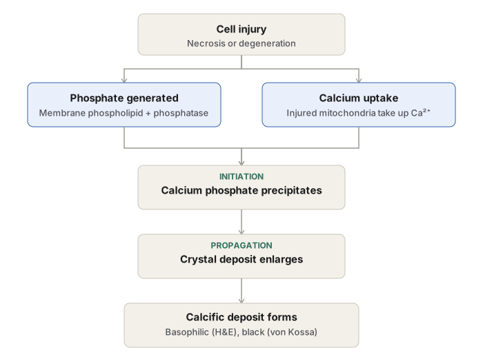
Pathogenesis: initiation — membrane damage releases phospholipid, associated phosphatases generate phosphate ions, injured mitochondria take up excess calcium, calcium and phosphate precipitate together; propagation — the crystal deposit enlarges through further structural rearrangement.
Metastatic calcification
Causes: hyperparathyroidism (primary, from adenoma or hyperplasia; or secondary, from chronic renal failure), bone destruction (multiple myeloma, leukaemia, metastatic tumour), vitamin D-related disorders (intoxication, sarcoidosis), renal failure with phosphate retention, prolonged immobilisation, milk-alkali syndrome.
Sites, in order of preference: lungs (alveolar septa, the most common site), kidneys (tubular basement membrane, causing nephrocalcinosis), blood vessels (internal elastic lamina), stomach (gastric mucosal interstitium).
Pathogenesis: raised serum calcium binds inorganic phosphate, precipitating preferentially at sites where local acid secretion or pH shift favours it; reversible once the systemic disorder is treated.
Clinical relevance
Calcification of an ageing or damaged heart valve can severely compromise its motion. Mammographic microcalcification is an early diagnostic clue to breast carcinoma. Dystrophic calcification with a normal serum calcium should prompt evaluation for local tissue injury; metastatic calcification should always prompt investigation for the cause of the underlying hypercalcaemia.
Definition
Pathological, or heterotopic, calcification is the abnormal deposition of calcium salts in tissues other than bone and tooth enamel, where such mineral deposition is normal. Small amounts of iron, magnesium, and other minerals often accompany the calcium. Two mechanistically distinct forms are recognised, and distinguishing them is the central task in this topic: dystrophic calcification, occurring in dying or dead tissue while calcium metabolism remains normal, and metastatic calcification, occurring in apparently normal tissue as a consequence of deranged calcium metabolism and a raised serum calcium level.
Morphology of Calcification
- Gross: fine white granules or clumps, often gritty or sand-like to touch.
- Microscopic: basophilic, amorphous, granular, clumped deposits, which may be intracellular, extracellular, or both. Heterotopic bone formation (ossification) occasionally occurs at the site.
- Special stains: the von Kossa silver impregnation method produces a black reaction; alizarin red S stains the deposits red. Calcification is often accompanied by iron deposition, giving a positive Prussian blue reaction on Perl’s stain.
Both forms of pathological calcification are morphologically similar to one another and resemble the normal mineral of bone, even though their causes and clinical implications differ.
Dystrophic Calcification
Definition
Deposition of calcium salts in dying or dead tissue. Calcium metabolism and serum calcium level remain normal — the abnormality is confined to the damaged tissue itself.
Causes
In necrotic tissue
- Caseous necrosis of tuberculosis, the most common site — living tubercle bacilli may persist even within a calcified lymph node or lung lesion.
- Liquefactive necrosis in a chronic abscess.
- Enzymatic fat necrosis, as in acute pancreatitis, and traumatic fat necrosis of the breast, where liberated free fatty acids form calcium soaps.
- Dead parasites — hydatid cysts, schistosome eggs, cysticercosis.
- Infarcts, which may calcify over time.
- Thrombi, particularly venous, producing phleboliths.
- Haematomas adjacent to bone.
- Gamna-Gandy bodies — calcific deposits admixed with haemosiderin on fibrous tissue in the chronically congested spleen.
- Microcalcification in breast carcinoma, detected by mammography.
- Congenital toxoplasmosis, visualised as calcification in the infant brain.
In degenerated tissue
- Ageing or damaged heart valves.
- Atheromas of the aorta and coronary arteries.
- Mönckeberg’s medial calcific sclerosis — calcification in the degenerated tunica media of large and medium-sized muscular arteries in elderly patients, particularly of the extremities and the genital tract (including the uterine myometrium). Grossly this produces pipestem-like rigidity of the artery without significant narrowing of the lumen; microscopically the media is replaced by acellular hyalinised fibrous tissue with basophilic calcific granules, and inflammation is conspicuously absent.
- Goitre of the thyroid and the stroma of certain tumours, including uterine fibroids, breast carcinoma, and thyroid adenoma.
- Psammoma bodies — laminated calcific spherules formed by progressive deposition of mineral over a single necrotic cell, seen in papillary thyroid carcinoma, papillary serous cystadenoma and cystadenocarcinoma of the ovary, meningioma, and papillary renal cell carcinoma.
- Longstanding cysts, such as epidermal and pilar cysts, whose walls may calcify.
- Calcinosis cutis, an idiopathic condition producing irregular nodular calcium deposits in skin and subcutaneous tissue.
- Senile degenerative change in costal, tracheal, or bronchial cartilage, and in the pineal gland.
- Old, densely hyalinised scars.
Pathogenesis
Dystrophic calcification mirrors the way normal bone mineral forms, through binding of phosphate to calcium ions, and proceeds in two phases.
- Initiation. Cell injury, whether degeneration or necrosis, damages membranes and releases membrane phospholipids. Phosphatases associated with these phospholipids generate phosphate ions, while injured mitochondria in the same dying cells take up excess calcium. The calcium and phosphate generated by these two processes precipitate together as calcium phosphate.
- Propagation. Structural rearrangement of the calcium and phosphate groups propagates the deposit further, building mineral crystals.
Metastatic Calcification
Definition
Deposition of calcium salts in normal tissue, secondary to hypercalcaemia arising from a systemic derangement of calcium metabolism.
Causes
- Hyperparathyroidism, whether primary — from a parathyroid adenoma or hyperplasia — or secondary, from chronic renal failure.
- Destruction of bone tissue, from primary bone marrow tumours such as multiple myeloma and leukaemia, or from metastatic tumour deposits in bone.
- Vitamin D-related disorders, including vitamin D intoxication and sarcoidosis, in which macrophages activate a vitamin D precursor.
- Renal failure, in which phosphate retention leads to secondary hyperparathyroidism.
- Prolonged immobilisation, causing disuse atrophy of bone and consequent hypercalcaemia.
- Milk-alkali syndrome, from excessive oral intake of calcium and calcium carbonate.
Sites
Metastatic calcification can occur widely, but shows a preference for particular tissues.
- Lungs — the most common site, at the alveolar septa.
- Kidneys — the basement membrane of the renal tubules, producing nephrocalcinosis.
- Blood vessels — the internal elastic lamina of systemic arteries and pulmonary veins.
- Stomach — the interstitial tissue of the gastric mucosa, related to its acid-secreting environment.
Pathogenesis
Elevated serum calcium drives excessive binding of calcium to inorganic phosphate, forming calcium phosphate precipitates preferentially at sites where local acid secretion or rapid shifts in pH favour precipitation, which accounts for the tissue preferences listed above. Because the driving abnormality is systemic rather than structural, metastatic calcification is reversible once the underlying disorder of calcium metabolism is corrected.
Dystrophic Calcification Compared with Metastatic Calcification
| Feature | Dystrophic calcification | Metastatic calcification |
|---|---|---|
| Site of deposition | Dying or dead tissue | Normal tissue |
| Calcium metabolism | Normal | Deranged |
| Serum calcium level | Normal | Raised |
| Reversibility | Generally irreversible | Reversible on correction of the metabolic disorder |
| Typical causes | Caseous necrosis, fat necrosis, infarcts, thrombi, ageing or damaged heart valves, atheromas, Mönckeberg’s sclerosis, certain tumours, cysts | Hyperparathyroidism, bone-destructive lesions, renal failure, vitamin D-related disorders, immobilisation |
Clinical Relevance
- Dystrophic calcification is not always a passive marker of past injury; calcification of an ageing or already damaged heart valve can severely compromise its motion and produce clinically significant valvular dysfunction.
- Microcalcification detected on mammography is a key early radiological clue to breast carcinoma, an instance where dystrophic calcification assists diagnosis rather than simply following it.
- Because dystrophic calcification occurs with normal serum calcium, its presence should prompt a search for local tissue injury rather than a work-up for a systemic calcium disorder; the reverse applies to metastatic calcification, which should always prompt investigation for the cause of hypercalcaemia.
- Extensive metastatic calcification of the lungs may be visible radiographically, and nephrocalcinosis can progress to renal damage.
Diagram 1: Dystrophic and Metastatic Calcification Compared
Draw two simplified human body outlines side by side, left labelled “dystrophic calcification,” right labelled “metastatic calcification.”
Left body: mark discrete, scattered symbols only at sites of local damage — a lung apex labelled “caseous TB focus,” a patch on the aorta wall labelled “atheroma,” a nodule at the breast labelled “fat necrosis / microcalcification,” a nodule on a limb artery labelled “Mönckeberg’s sclerosis,” and a heart valve labelled “ageing/damaged valve.” Beside the body, tag a small blood vessel “serum calcium: normal.”
Right body: mark diffuse, symmetric symbols at the tissues with a preference for metastatic deposition — both lungs (labelled “alveolar septa”), both kidneys (labelled “nephrocalcinosis”), the stomach fundus (labelled “acid-secreting mucosa”), and a blood vessel wall diffusely (labelled “internal elastic lamina”). Beside the body, tag a small blood vessel “serum calcium: raised,” and draw a parathyroid gland icon with an arrow feeding into the vessel to indicate the systemic driver.
Labels required
- Serum calcium status explicitly tagged on both bodies — this is the single most important discriminator between the two diagrams.
- Every marked site individually named, not left as an unlabelled symbol.
- The parathyroid or systemic driver icon present on the metastatic side, showing that the deposition originates from a body-wide cause rather than a local one.
Errors commonly made
- Marking scattered, localised lesions on the metastatic-side body instead of diffuse, symmetric, organ-preferential deposits, which reverses the actual distribution.
- Omitting the serum calcium tag on either diagram.
- Leaving out any of the four preferred metastatic sites — lung, kidney, stomach, blood vessels.
- Using identical symbols on both bodies without a legend distinguishing a local-damage marker from a systemic-deposit marker.
Diagram 2: Pathogenesis of Dystrophic Calcification
Draw a vertical flowchart of five boxes.
- Cell injury — necrosis or degeneration.
- Two boxes side by side at this level: membrane damage releases phospholipid, and associated phosphatase activity generates phosphate ions; injured mitochondria take up excess calcium.
- Both arrows converge into: calcium and phosphate precipitate together as calcium phosphate — labelled “initiation.”
- Structural rearrangement of the crystal groups — labelled “propagation.”
- Growing calcific deposit — basophilic on haematoxylin and eosin, black with von Kossa stain, red with alizarin red S.
Labels required
- “Initiation” and “propagation” must be explicitly labelled as the two named phases.
- Both parallel contributors — phosphate generation and mitochondrial calcium uptake — shown as separate boxes converging into one, not merged into a single step.
- The staining results attached as a note on the final box, linking the mechanism to how the deposit is identified.
Errors commonly made
- Showing only one contributor, commonly phosphate release alone, and omitting mitochondrial calcium uptake.
- Failing to separate initiation from propagation as two distinct labelled phases.
- Leaving the staining methods disconnected from the mechanism diagram.
Long Essays (1)
Multiple Choice Questions (3)
My Sticky Notes
Personal revision notes, mnemonics and reminders.
