
Oncogenic Viruses
Overview
~20% of human cancers = virally induced. First observed: Sanarelli 1889 (rabbit myxomatosis/poxvirus); human wart contagiousness 1907 = first human evidence.
Transmission: horizontal (most) / parenteral / vertical. 2 groups: DNA oncogenic viruses, RNA oncogenic viruses (retroviruses). Both have gag(antigen)/pol(polymerase)/env(envelope) genes.
Evidence pillars: epidemiology + viral DNA in tumour genome + in-vitro transformation + in-vivo transforming gene expression + in-vitro gene product effects. Persistence alone ≠ sufficient — one step in multistep carcinogenesis.
General mechanism
DNA virus: infected cell → Replication (lysis, no transformation) OR Integration (mutation → transformation). Needs T-antigen expression.
RNA virus (retrovirus): reverse transcriptase → viral RNA→single-strand DNA→double-strand DNA (provirus) → integrates into host genome → mutation/transformation → (if permissive) replication/budding.
DNA oncogenic viruses
HPV — first virus linked to any human tumour.
- Low-risk 1,2,4,7 → skin warts. Low-risk 6,11 → genital warts (minimal malignancy).
- High-risk 16,18(31,33,45) → cervical Ca+precursors (75-100% DNA+), anal/vaginal/vulval/penile/oral SCC.
- 5,8 → epidermodysplasia verruciformis →1/3 SCC.
- Mechanism: integration→↑E6+E7. E6: degrades p53 + ↑TERT(telomerase)→immortalise. E7: binds RB→releases E2F→G1→S + inactivates p21. High-risk E6/E7 = higher affinity than low-risk (RK detail). Integrated (vs episomal) genome = more genomic instability.
EBV — B lymphocytes+epithelium. → Burkitt lymphoma, NPC, PTLD, CNS lymphoma(AIDS), Hodgkin, infectious mononucleosis.
- Burkitt: >90% EBV+, 100% ↑Ab titres. Endemic(African)>AIDS>sporadic association → immunosuppression cofactor (malaria endemic overlap).
- NPC: SE Asia/Chinese/Eskimo. 100% EBV DNA+.
- Mechanism: LMP1 (mimics CD40)→NF-kB+JAK/STAT→B-cell proliferation; ↑BCL2(blocks apoptosis). EBNA-2→↑cyclin D. Immunocompetent: LMP1 controlled, need extra hit = t(8;14) MYC translocation (RK confirms).
HHV-8/KSHV → Kaposi sarcoma (AIDS=multicentric+aggressive), pleural effusion lymphoma, Castleman’s. Overexpress v-cyclin, v-IRF, LANA.
HBV/HCV → 70-80% HCC worldwide. HBV region = 200x HCC risk. Multifactorial: chronic injury-regen cycling→DNA damage; defective immune clearance; regen growth factors; NF-kB blocks apoptosis; HBx gene. No dedicated viral oncoprotein (unlike HPV/EBV/HHV8). HBV+aflatoxin synergy.
RNA oncogenic viruses (retroviruses)
Only HTLV causes human cancer (acute-transforming/slow-transforming groups = animal tumours only: Rous sarcoma, MMTV).
HTLV-1 → ATLL. Japan/West Indies endemic. Latency 20-30yr(long). CD4+ tropism (like HIV). Immunosuppression cofactor. HTLV-2 → hairy cell leukaemia (T-cell variant).
Mechanism: no v-onc, no fixed insertion site (unlike other 2 groups). pX region → TAX + REX. TAX: ↑T-cell replication genes + NF-kB→cytokine autocrine loop; ↑cyclins, inactivates p16/p53. Polyclonal→monoclonal (further TAX mutations).
Non-viral biologic agents
Parasites: Schistosoma haematobium→bladder SCC (Egypt); Clonorchis sinensis→cholangiocarcinoma. Fungus: Aspergillus flavus→aflatoxin→HCC(+HBV synergy). Bacteria: H. pylori→gastric lymphoma+carcinoma.
Summary table
HPV16/18→cervical Ca | HPV5/8→skin Ca(EV) | EBV→Burkitt+NPC+PTLD+Hodgkin | HHV8→Kaposi+PEL | HBV/HCV→HCC | HTLV1→ATLL | HTLV2→hairy cell leukaemia variant
Clinical relevance
HBV & HPV vaccines = rare cancer-preventing vaccines. High/low-risk HPV affinity difference explains genotyping significance. Immunosuppression cofactor (Burkitt/NPC/ATLL) → higher risk in transplant/HIV patients, underlies PTLD surveillance. HBV/HCV mechanism (no oncoprotein) contrasts with HPV/EBV/HHV8 (oncoprotein-driven) — viral carcinogenesis is multiple distinct strategies, not one mechanism.
Overview
Roughly 20% of all human cancers worldwide are attributable to persistent infection with a transmissible biologic agent — chiefly viruses, but also select parasites, a fungus, and a bacterium. The association was first observed by Sanarelli (1889, myxomatosis of rabbits caused by a poxvirus), and the contagious nature of the common human wart was established in 1907 — the first indication of a viral cause for any human neoplasm.
Oncogenic viruses transmit by three routes — horizontal (contact, ingestion, inhalation, the majority of infections), parenteral (inoculation, inter-human or animal/insect-to-human spread), and vertical (parent to offspring) — and fall into two broad groups by nucleic acid content: DNA oncogenic viruses and RNA oncogenic viruses (retroviruses). Both types typically carry three core genes: gag (group antigen), pol (polymerase), and env (envelope protein).
Evidence supporting a causal viral role in a given human cancer rests on five pillars: epidemiological data, presence of viral DNA in the tumour cell genome, in-vitro transformation of human cells by the virus, in-vivo expression of transforming viral genes in premalignant/malignant tissue, and in-vitro demonstration that specific viral gene products affect proliferation/survival. Persistent viral infection alone is not sufficient for oncogenesis — it is one step in the multistep process of carcinogenesis, generally acting either by activating growth-promoting pathways or by inhibiting tumour-suppressor products in the infected cell.
General Mechanisms
DNA Oncogenic Viruses
An infected host cell has one of two fates:
- Replication — the virus replicates, lyses the host cell, and releases virions; this kills the cell but does not transform it.
- Integration — viral DNA integrates into host cell DNA, inducing mutation and neoplastic transformation.
Transformation requires expression of virus-specific T-(transforming) antigen immediately after infection.
RNA Oncogenic Viruses (Retroviruses)
Retroviruses carry the enzyme reverse transcriptase, an RNA-dependent DNA synthetase:
- Reverse transcriptase synthesises a single strand of viral DNA from the viral RNA template (the reverse of normal transcription).
- A DNA-dependent DNA synthetase copies this into double-stranded viral DNA, the provirus.
- The provirus integrates into host genomic DNA, inducing mutation and transformation.
- In permissive host cells, integration is followed by transcription of proviral genes into the gag, pol, and env components, which assemble at the plasma membrane and are released by budding (viral replication).
DNA Oncogenic Viruses in Human Cancer
Human Papillomavirus (HPV)
Papillomaviruses were the first viruses implicated in any human neoplasm. Different HPV types produce distinct lesions:
- Low-risk types 1, 2, 4, 7 — common skin warts (verruca vulgaris).
- Low-risk types 6, 11 — genital warts (condyloma acuminata); minimal malignant potential.
- High-risk types 16, 18 (also 31, 33, 45) — viral DNA found in 75–100% of invasive cervical cancers and their precursors; also implicated in squamous cell carcinoma of the anus, perianal region, vagina, vulva, penis, and oral cavity/oropharynx.
- Types 5, 8 — epidermodysplasia verruciformis, a genetic cell-mediated-immunity defect with multiple skin warts, roughly a third progressing to squamous cell carcinoma in sun-exposed lesions.
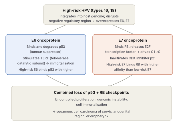
Mechanism: high-risk HPV integrates into the host genome, disrupting a negative regulatory region and driving overexpression of viral oncoproteins E6 and E7:
- E6 binds and degrades p53 and stimulates TERT (telomerase catalytic subunit), contributing to immortalisation.
- E7 binds RB, releasing sequestered E2F transcription factor to drive G1→S progression, and inactivates the CDK inhibitor p21.
- High-risk E6/E7 bind their respective targets (p53, RB) with higher affinity than the corresponding low-risk-HPV proteins — the molecular basis of the risk stratification. Cells with integrated (rather than episomal) HPV genome also show significantly greater genomic instability.
Epstein-Barr Virus (EBV)
EBV infects B lymphocytes and epithelial cells, and is implicated in Burkitt’s lymphoma, anaplastic nasopharyngeal carcinoma, post-transplant lymphoproliferative disease, primary CNS lymphoma in AIDS, Hodgkin’s lymphoma, and infectious mononucleosis (a self-limiting primary infection).
Burkitt’s lymphoma (African endemic and sporadic forms): >90% of cases are EBV-positive; 100% show elevated EBV antibody titres; EBV immortalises infected B cells in culture. Linkage to EBV is strongest in the African endemic form and in AIDS-associated cases, weakest in sporadic disease — pointing to immunosuppression as a necessary cofactor, consistent with malaria’s immunosuppressive effect being endemic in the same regions where endemic Burkitt’s lymphoma is common.
Nasopharyngeal carcinoma: prevalent in South-East Asia (especially Chinese populations) and Eskimos; 100% of cases carry EBV DNA in tumour nuclei with high antibody titres, though genetic susceptibility likely contributes to the geographic distribution.
Mechanism: latently infected cells express viral oncoprotein LMP1 (latent membrane protein), the most crucial step — it mimics the CD40 receptor, activating B-cell proliferation via NF-κB and JAK/STAT pathways, and upregulates BCL2 to block apoptosis. EBNA-2 (EBV nuclear antigen) activates cyclin D. In immunocompetent hosts, LMP1 is normally controlled by the immune system, so lymphoma emerges only after an additional characteristic mutation — the t(8;14) translocation activating the MYC oncogene.
Human Herpesvirus 8 (HHV-8 / KSHV)
Also called Kaposi’s sarcoma-associated herpesvirus. Causes Kaposi’s sarcoma (AIDS-associated disease is multicentric and more aggressive than sporadic KS), pleural effusion lymphoma, and multicentric Castleman’s disease. Viral DNA is present in all KS tumour cell nuclei. Latently infected cells overexpress v-cyclin, v-interferon regulatory factor (v-IRF), and LANA (latency-associated nuclear antigen), driving increased proliferation and survival.
Hepatitis B and C Viruses (HBV, HCV)
HBV (a DNA hepadnavirus) and HCV (an RNA virus) together account for 70–80% of hepatocellular carcinoma worldwide. HBV-infected regions show roughly 200-fold higher HCC risk than uninfected populations in the same area. Mechanism is multifactorial rather than a single fixed molecular event:
- Chronic infection drives repetitive cycles of hepatocyte inflammation, death, and regeneration, causing cumulative DNA damage.
- Defective host immune response to persistent infection promotes tumour development.
- Regenerative hepatocyte proliferation is stimulated by growth factors/cytokines from activated immune cells (angiogenesis, cell survival factors).
- Activated immune cells produce NF-κB, inhibiting apoptosis.
- The HBV genome’s HBx gene activates growth-signalling pathways.
- Neither HBV nor HCV encodes a dedicated viral oncoprotein — unlike HPV, EBV, or HHV-8.
- HBV-infected hepatocytes chronically injured are also rendered more vulnerable to co-carcinogens such as aflatoxin.
RNA Oncogenic Viruses (Retroviruses) in Human Cancer
Of the three retrovirus subgroups (acute transforming, slow transforming, HTLV), only human T-cell lymphotropic virus (HTLV) causes human cancer — the acute and slow transforming groups are implicated in animal tumours only (Rous sarcoma virus, leukaemia-sarcoma viruses, mouse mammary tumour virus).
HTLV-1 causes adult T-cell leukaemia/lymphoma (ATLL), endemic in parts of Japan and the West Indies, with an unusually long 20–30 year latent period. Transmitted sexually, by blood, or via breastfeeding. As with Burkitt’s lymphoma, HTLV-1 has tropism for a specific lymphocyte subset (CD4+ T cells, as does HIV) and immunosuppression plays a supportive role. HTLV-2 causes the T-cell variant of hairy cell leukaemia.
Mechanism differs from both other retroviral groups — HTLV-1 has no v-onc and no fixed insertion site for insertional mutagenesis. Instead, a unique genomic region, pX, encodes viral oncoproteins TAX and REX. TAX upregulates cellular genes controlling T-cell replication and interacts with NF-κB, stimulating cytokine/interleukin gene expression and autocrine T-cell proliferation; it also activates cyclins and inactivates tumour suppressors CDKN2A/p16 and p53. Proliferation is initially polyclonal, becoming monoclonal (leukaemic) once further TAX-driven mutations accumulate.
Non-Viral Biologic Carcinogens
- Parasites — Schistosoma haematobium (urinary bladder infection, endemic in Egypt) → squamous cell carcinoma of the bladder; Clonorchis sinensis (liver fluke, hepatic ducts) → cholangiocarcinoma.
- Fungus — Aspergillus flavus (stored grain) → aflatoxin → hepatocellular carcinoma, especially with concurrent HBV infection.
- Bacteria — Helicobacter pylori (chronic gastric mucosal colonisation, causing chronic gastritis/peptic ulcer) → gastric lymphoma and gastric carcinoma with prolonged infection.
Summary of Virus–Cancer Associations
| Virus | Human cancer |
|---|---|
| HPV (types 16, 18) | Cervical cancer & precursors; SCC of anus, vagina, vulva, penis, oral cavity |
| HPV (types 5, 8) | Skin cancer in epidermodysplasia verruciformis |
| EBV | Burkitt’s lymphoma, nasopharyngeal carcinoma, post-transplant lymphoproliferative disease, Hodgkin’s lymphoma |
| HHV-8 (KSHV) | Kaposi’s sarcoma, pleural effusion lymphoma |
| HBV, HCV | Hepatocellular carcinoma |
| HTLV-1 | Adult T-cell leukaemia/lymphoma |
| HTLV-2 | T-cell variant of hairy cell leukaemia |
Clinical Relevance
- Understanding viral oncogenesis has translated directly into prevention: hepatitis B vaccination lowers HBV-related HCC incidence in high-risk populations, and HPV vaccination protects against HPV-associated precancerous cervical lesions — a rare case in oncology where a vaccine functionally prevents cancer.
- The affinity difference between high-risk and low-risk HPV E6/E7 proteins for p53 and RB respectively is the molecular explanation for why only certain HPV types are oncogenic despite widespread infection with the low-risk types — directly relevant to interpreting HPV genotyping results.
- The immunosuppression cofactor common to Burkitt’s lymphoma (malaria/HIV), nasopharyngeal carcinoma, and ATLL explains the markedly higher incidence of virus-associated malignancy in immunocompromised patients (transplant recipients, HIV/AIDS) and underlies post-transplant lymphoproliferative disease surveillance.
- HBV/HCV causing HCC without a dedicated viral oncoprotein — via chronic injury-regeneration cycling instead — is a useful contrast against HPV/EBV/HHV-8’s oncoprotein-driven mechanisms, illustrating that “viral carcinogenesis” is not a single mechanism but several distinct molecular strategies converging on malignancy.
Diagram 1: Mechanism of HPV E6/E7 Oncoprotein-Driven Carcinogenesis
Draw one top box (HPV integration) splitting into two parallel branch boxes (E6, E7), converging into one bottom outcome box.
- Top — high-risk HPV integrates into host genome, disrupting a negative regulatory region → overexpression of E6 and E7.
- Left branch (E6) — binds/degrades p53; stimulates TERT (telomerase) → immortalisation; higher affinity than low-risk E6.
- Right branch (E7) — binds RB, releases E2F → drives G1→S; inactivates p21; higher affinity than low-risk E7.
- Bottom (convergence) — combined loss of p53 + RB checkpoints → uncontrolled proliferation, genomic instability, immortalisation → squamous cell carcinoma (cervix, anogenital, oropharynx).
Labels required
- E6 must be paired with p53 and E7 with RB — this is the single most commonly confused pairing in the entire topic, and reversing it is a guaranteed wrong answer.
- The “higher affinity in high-risk vs low-risk HPV” property must be attached to both branches, since it is the molecular explanation for the clinically crucial high-risk/low-risk distinction.
- The convergence box must show that the two checkpoints are lost together — cervical carcinogenesis requires both p53 loss (E6) and RB loss (E7) acting in the same cell, not either alone.
Errors commonly made
- Swapping E6→RB and E7→p53 (the reverse of the correct pairing).
- Treating “high-risk vs low-risk HPV” as a fixed list to memorise rather than understanding it is a consequence of binding affinity — a testable mechanistic distinction, not an arbitrary classification.
- Omitting that HPV integration itself (not just E6/E7 expression) contributes independently to oncogenesis via genomic instability, distinct from the E6/E7 oncoprotein effects.
Note on Other Mechanisms in This Topic
EBV (LMP1/EBNA2 plus the t(8;14) MYC translocation), HHV-8 (v-cyclin/v-IRF/LANA), HBV/HCV (multifactorial chronic-injury mechanism without a dedicated oncoprotein), and HTLV-1 (TAX/REX via the pX region) are each genuine multi-step mechanisms but are adequately captured in prose/bullet form in notes.md without requiring a separate rendered diagram — none involves a branching or sequential process complex enough to need geometric representation beyond what the HPV diagram already demonstrates as the representative worked example for this topic’s diagram-relevant mechanism (parallel-pathway viral oncoprotein action converging on shared downstream effect).
Multiple Choice Questions (2)
My Sticky Notes
Personal revision notes, mnemonics and reminders.
