
Necrosis
Definition
Necrosis is the morphological expression of cell death in living tissue, produced by progressive enzymatic degradation after irreversible injury. It is unregulated, always accompanied by inflammation, and distinct from autolysis (self-digestion, no host reaction, seen after death) and from apoptosis (single cell, no inflammation).
General morphology
Cytoplasm: increased eosinophilia, glassy appearance; later vacuolation with a moth-eaten pattern once organelles are digested.
Nucleus: pyknosis (shrunken, densely basophilic), karyolysis (fading basophilia as DNase acts, leaving a ghost nucleus), karyorrhexis (fragmentation of the pyknotic nucleus).
Fate: dead cells may be replaced by myelin figures, phagocytosed, or degraded to fatty acids that bind calcium, producing dystrophic calcification.
The five patterns
Coagulative necrosis. Ischaemia denatures structural proteins, blocking proteolysis, so cell outlines persist (“tombstones”) while nuclear detail is lost. Characteristic of infarction in every organ except the brain. Gross: pale, firm, wedge-shaped area (infarct), base under the capsule, apex pointing inward. A renal infarct is red and congested for the first two to three days, turns pale yellow by the fourth day, and is depressed below the organ surface by the end of a week.
Liquefactive (colliquative) necrosis. Enzymatic digestion dominates, converting tissue into a soft, liquid mass. Seen in cerebral infarction and in suppurative infection (abscess, pus formation), also in wet gangrene and pancreatic necrosis.
Caseous necrosis. Combines coagulative and liquefactive features; architecture is completely obliterated. Cheese-like, yellow-white, friable material surrounded by a granuloma of epithelioid cells, Langhans giant cells, and lymphocytes. Classic for tuberculosis; also seen in histoplasmosis. Often undergoes dystrophic calcification.
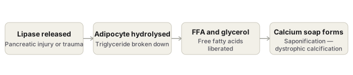
Fat necrosis. Enzymatic form: pancreatic lipase liberates free fatty acids from adipocytes, which bind calcium to form calcium soaps (saponification) — seen in acute pancreatitis. Traumatic form: occurs in fat-rich tissue such as the breast after injury, without enzymatic mediation. Gross: chalky-white foci.
Fibrinoid necrosis. Deposition of fibrin-like eosinophilic material in vessel walls, with surrounding leucocytoclasis. Seen in immune-mediated vascular injury — polyarteritis nodosa, malignant hypertension, systemic lupus erythematosus, Aschoff bodies of rheumatic fever, and pre-eclampsia. The only pattern without a distinct gross appearance.
Gummatous necrosis — firm, rubbery necrotic tissue seen in syphilis.
Gangrene
Massive necrosis with superadded putrefaction, usually following coagulative necrosis from ischaemia.
| Dry gangrene | Wet gangrene | |
|---|---|---|
| Site | Limbs | Bowel and other moist organs |
| Cause | Arterial obstruction (atherosclerosis) | Venous obstruction |
| Onset | Slow | Abrupt |
| Appearance | Dry, shrivelled, black (mummification) | Swollen, soft, moist |
| Line of demarcation | Clear | Not clear |
| Prognosis | Fair | Poor — septicaemia |
Gas gangrene is a variant of wet gangrene caused by Clostridium perfringens, entering through contaminated wounds; toxin production causes local necrosis, oedema, crepitation from gas, and systemic toxaemia. Fournier’s gangrene affects the scrotal skin.
Serum markers of necrosis
Cardiac muscle — creatine kinase (CK-MB) and troponin. Bile duct epithelium — alkaline phosphatase. Hepatocytes — transaminases (AST, ALT). Pancreas — lipase and amylase.
Necrosis compared with apoptosis
| Necrosis | Apoptosis | |
|---|---|---|
| Scope | Contiguous groups of cells | Single cells |
| Cell volume | Swells | Shrinks |
| Membrane | Ruptures | Intact until phagocytosed |
| Inflammation | Always present | Absent |
| Nucleus | Pyknosis, karyorrhexis, karyolysis | Smooth chromatin condensation |
Definition
Necrosis is the sum of morphological changes that follow cell death in living tissue, produced by the progressive degradative action of enzymes on a cell that has suffered irreversible injury. It is an unregulated, “accidental” form of cell death — the injury is too severe for any orderly signalling programme to control it, and the cell’s own membranes and ion homeostasis fail outright. Because the dying cell loses membrane integrity, its enzymes and contents leak into the surrounding tissue and provoke an acute inflammatory reaction; this inflammatory response is the feature that most reliably separates necrosis from apoptosis. Necrosis should also be distinguished from autolysis, which is self-digestion of a cell by its own lysosomal enzymes without any surrounding inflammatory reaction — the standard postmortem change, though the same process can occur in life within a cell enclosed by active inflammation.
Causes
- Hypoxia and ischaemia, the leading cause, from interruption or reduction of blood supply.
- Physical trauma — mechanical, thermal, or radiation injury.
- Chemical agents and drug toxicity.
- Microbial infection.
- Immunological injury.
General Morphology of a Necrotic Cell
Cytoplasmic Changes
The cytoplasm becomes more eosinophilic and takes on a glassy, homogeneous appearance. Once enzymes have digested the cytoplasmic organelles, the cytoplasm becomes vacuolated, producing a moth-eaten appearance.
Nuclear Changes
Breakdown of nuclear DNA and chromatin produces one of three patterns.
- Pyknosis — the nucleus shrinks and becomes densely basophilic, resembling a shrunken ink drop.
- Karyolysis — basophilic staining progressively fades as DNase activity digests the DNA, leaving a ghost nucleus that may disappear entirely within one to two days.
- Karyorrhexis — the pyknotic nucleus fragments into many smaller pieces.
Ultrastructural Findings
Discontinuities in the plasma and organelle membranes, marked mitochondrial dilatation with large amorphous densities, disruption of lysosomes, and intracytoplasmic myelin figures.
Fate of Necrotic Cells
A necrotic cell may persist for a time or may be digested and disappear. Dead cells are often replaced by myelin figures, which are either phagocytosed or degraded further into fatty acids; these fatty acids can then bind calcium salts, so a necrotic focus may itself become calcified — the basis of dystrophic calcification.
Types of Necrosis
Necrosis is classified by the gross and microscopic pattern it produces, which reflects the balance between protein denaturation and enzymatic digestion at the site. Most patterns have a distinctive gross appearance, with the exception of fibrinoid necrosis, which can only be identified microscopically. Recognising the pattern narrows the differential directly: caseous necrosis points toward tuberculosis, fibrinoid necrosis toward an immune-mediated vasculopathy, and fat necrosis with calcium soap formation toward pancreatitis or breast trauma.
Coagulative Necrosis
The most common pattern, in which the underlying tissue architecture is preserved for at least a few days after cell death. A localised area of coagulative necrosis is termed an infarct, and this pattern is characteristic of infarction in every solid organ except the brain.
- Causes: Most often ischaemia from obstruction of a vessel; less often bacterial or chemical injury.
- Mechanism: Ischaemia denatures and coagulates structural proteins and enzymes, and this denaturation blocks proteolysis of the dead cell — which is why the tissue outline survives even though the cell is no longer viable.
- Gross: The affected organ, particularly one supplied by end arteries with limited collateral circulation — heart, kidney, spleen, or a limb — appears dry, pale, yellow, and firm, often wedge-shaped, with the base resting under the capsule and the apex pointing inward; a narrow rim of tissue immediately beneath the capsule is often spared, since it draws blood from a separate capsular supply. In a renal infarct, the gross colour evolves over time — congested and red over the first two to three days, pale yellow by the fourth day, and finally depressed below the organ surface by the end of a week.
- Microscopic: Cells are converted into “tombstones” — their outlines and cell type remain recognisable, but cytoplasmic and nuclear detail is lost. Nuclei show karyolysis or may be absent altogether. Leucocytes are recruited to the site, and their lysosomal enzymes digest the necrotic cells, with the resulting debris removed by phagocytosis.
Liquefactive (Colliquative) Necrosis
Here the dead tissue rapidly softens and transforms into a liquid, viscous mass, because enzymatic digestion dominates over protein denaturation.
- Causes: Ischaemic injury to the central nervous system, and suppurative infection by bacteria or fungi, in which leucocyte enzymes accumulate and digest (“liquefy”) the tissue.
- Mechanism: Digestion by hydrolytic enzymes released from dead cells (autolysis) and from recruited leucocytes (heterolysis).
- Gross: A soft area with a liquefied centre containing necrotic debris; a cystic space may form later.
- Typical sites: In the brain, the necrotic area is soft with a liquefied centre — a pattern possibly related to the brain’s abundant lysosomal and hydrolytic enzyme content. In an abscess, bacterial infection produces an acute inflammatory reaction and liquefaction into pus, a creamy yellow collection of dead leucocytes, macrophages, and necrotic debris. The same pattern is seen in wet gangrene and in pancreatic necrosis.
Caseous Necrosis
A distinctive pattern combining features of both coagulative and liquefactive necrosis, classically associated with tuberculosis, where it develops through a type IV hypersensitivity reaction, and with systemic fungal infection such as histoplasmosis, in which the high lipid content of the organism’s cell wall contributes to the same appearance.
- Gross: Necrotic tissue is yellow-white, soft, friable, and granular, resembling dry, clumped cheese — hence the name.
- Microscopic: Unlike coagulative necrosis, tissue architecture is completely obliterated, and cellular outlines cannot be identified; the necrotic material appears as eosinophilic, coarsely granular debris of fragmented or lysed cells. It is surrounded by a granuloma — epithelioid cells, Langhans-type giant cells with nuclei arranged in a horseshoe pattern, lymphocytes, and fibroblasts. Caseous necrotic material commonly undergoes dystrophic calcification.
Fat Necrosis
Focal destruction of adipose tissue at fat-rich anatomical sites, occurring in two forms.
- Enzymatic fat necrosis develops around an acutely inflamed pancreas in acute pancreatitis. Lipase and other enzymes leak from injured acinar cells into the pancreatic substance and peritoneal cavity, splitting the triglycerides within fat cells and liberating free fatty acids, which combine with calcium to form calcium soaps — a reaction known as fat saponification.
- Traumatic fat necrosis develops in tissue with high fat content, such as a heavy, pendulous breast or the thigh, following significant trauma, without requiring enzymatic mediation.
- Gross: Chalky-white foci, which help both surgeon and pathologist identify the lesion on inspection.
- Microscopic: Necrotic fat cells appear pale, with shadowy outlines, surrounded by basophilic calcium deposits and an inflammatory reaction.
Fibrinoid Necrosis
A special pattern characterised by deposition of a pink-staining, fibrin-like proteinaceous material within the tissue matrix, obscuring the underlying cellular detail. It is the one pattern of necrosis without a distinctive gross appearance.
- Causes: Chiefly immune-mediated vascular injury, from deposition of antigen-antibody complexes in vessel walls — as in polyarteritis nodosa — as well as malignant hypertension, systemic lupus erythematosus, Aschoff bodies of rheumatic fever, the placental bed in pre-eclampsia, and hyperacute transplant rejection.
- Microscopic: A brightly eosinophilic, hyaline-like deposit within the vessel wall, best appreciated with phosphotungstic acid haematoxylin stain; nuclear debris of degenerating neutrophils (leucocytoclasis) surrounds the necrotic focus, and local haemorrhage may follow if the vessel ruptures.
Gummatous Necrosis
A pattern in which the necrotic tissue is firm and rubbery, characteristically found in syphilis.
Gangrene
Gangrene is massive necrosis of tissue with superadded putrefaction, most often following coagulative necrosis due to ischaemia. Two main types are recognised.
Dry Gangrene
- Causes: Arterial occlusion, typically from atherosclerosis.
- Sites: Usually a limb, most often the distal lower limb — leg, foot, or toe.
- Gross: The affected part is dry, shrunken, and dark brown to black, resembling the foot of a mummy; the black colour results from iron sulfide formed from liberated haemoglobin. A clear line of demarcation separates gangrenous from adjacent normal tissue, and the gangrenous segment may eventually separate spontaneously.
- Microscopic: Coagulative necrosis with smudging of the soft tissue and overlying skin; the line of demarcation shows granulation tissue with inflammatory cells.
Wet Gangrene
- Causes: Venous blockage, as with a strangulated hernia, intussusception, or volvulus. Diabetic patients may develop dry gangrene of the foot, from atherosclerosis of the tibial and popliteal arteries, as well as wet gangrene following a minor injury.
- Sites: Naturally moist tissues and organs — bowel, lung, mouth, cervix, vulva. Bedsores in an immobilised patient, at pressure points such as the sacrum, buttocks, and heel, follow the same pattern.
- Gross: The affected part is soft, swollen, putrid, and dark, without a clear line of demarcation. Toxic products absorbed from the site can produce septicaemia and death.
- Microscopic: Liquefactive necrosis, with intense acute inflammatory infiltrate and thrombosed vessels.
Gas Gangrene
A special variant of wet gangrene caused by the gas-forming, Gram-positive anaerobic bacterium Clostridium perfringens, which enters tissue through contaminated open wounds — often muscle — or as a complication of colonic surgery. Toxins produced by the organism cause local necrosis and oedema and are absorbed to produce severe systemic manifestations. The affected area is swollen, oedematous, crepitant from gas accumulation, and eventually dark and foul-smelling. Microscopically, muscle fibres show coagulative necrosis with liquefaction, abundant Gram-positive bacilli, and a peripheral zone of leucocytic infiltration, oedema, and congestion.
Fournier’s gangrene is a further named variant occurring in the scrotal skin.
Dry Gangrene Compared with Wet Gangrene
| Feature | Dry Gangrene | Wet Gangrene |
|---|---|---|
| Common site | Limbs | Bowel |
| Cause of ischaemia | Arterial obstruction | Usually venous obstruction |
| Onset | Slow | Abrupt |
| Appearance | Shrivelled, dry, black (mummification) | Swollen, soft, moist |
| Line of demarcation | Clear | Not clear |
| Spread | Slow | Rapid |
| Prognosis | Fair | Poor, owing to septicaemia |
Laboratory Detection of Necrosis
Damage to the cell membrane allows intracellular proteins to leak into the circulation, so tissue-specific necrosis can be detected in blood or serum, non-invasively and at the bedside. Cardiac muscle releases a unique isoform of creatine kinase and the contractile protein troponin; hepatic bile duct epithelium releases alkaline phosphatase; hepatocytes release transaminases. Elevated levels of these markers signal necrosis of the corresponding tissue.
Necrotic tissue is also the substrate for two subsequent processes described above — gangrene, and dystrophic calcification, in which the fatty acids and membrane phospholipids of dead cells provide the material for calcium deposition.
Diagram 1: The Patterns of Necrosis
Draw a panel of five small boxes side by side, one per pattern, each containing a simplified microscopic field.
- Coagulative: a grid of cell outlines still visible (ghost cells), each filled with uniform pink shading, faint or absent nuclei within the outlines. Label: architecture preserved.
- Liquefactive: an irregular cystic cavity lined by a thin rim of proliferating cells — label this rim “gliosis” for a brain example, or “fibroblasts” for an abscess wall — with the cavity filled with stippled debris.
- Caseous: a central structureless pink-grey amorphous zone with scattered debris, surrounded by a ring of oval cells with slipper-shaped nuclei (epithelioid cells) and a few multinucleated giant cells with horseshoe-arranged nuclei (Langhans type) at the margin, with a thin outer cuff of lymphocytes.
- Fat: rounded, empty-looking fat cell outlines with patches of granular basophilic material scattered among them, representing calcium soaps.
- Fibrinoid: a cross-section of a small blood vessel wall, stained bright pink in a smudged, homogeneous band within the wall, with small dark fragments of nuclear debris scattered around it.
Labels required
- Each panel titled with its necrosis type.
- Panel 1: note that cell outlines and architecture are retained, with no early liquefaction.
- Panel 3: label the caseous centre, the epithelioid cells, the giant cells, and the lymphocytic cuff.
- Panel 4: label the calcium soap deposits separately from the fat cell outlines.
- Panel 5: label the vessel wall and the leucocytoclasis.
Errors commonly made
- Drawing coagulative necrosis with dissolved or absent cell outlines, which is the pattern for liquefactive necrosis, not coagulative.
- Omitting the granulomatous cuff around caseous necrosis and drawing only the amorphous centre.
- Drawing ruptured fat cells without the calcium soap deposits, which are the feature that identifies fat necrosis specifically.
- Omitting leucocytoclasis around fibrinoid necrosis.
Diagram 2: Fat Necrosis — Enzymatic Pathway
Draw a short horizontal flowchart of four boxes connected by arrows.
- Pancreatic injury releases lipase (a small pancreas icon with droplet-shaped enzyme symbols being released).
- Adipocyte hydrolysed (a fat cell breaking open).
- Free fatty acids and glycerol released (two separate small molecule symbols).
- Free fatty acids bind calcium to form calcium soaps — saponification (a chalky white-grey clump forming).
Labels required
- The term saponification must appear explicitly.
- Both hydrolysis products, free fatty acids and glycerol, must be shown separately.
- The calcium should be labelled as coming from the surrounding tissue fluid, linking this diagram to dystrophic calcification.
Errors commonly made
- Skipping the free fatty acid step and moving directly from “fat cell” to “calcium soap.”
- Failing to connect the diagram to dystrophic calcification as the underlying process.
- Using the pancreatic-injury trigger box unchanged when illustrating traumatic fat necrosis of the breast — that variant is not enzyme-mediated and the trigger box should instead read “trauma to adipose tissue.”
Short Answers (1)
Multiple Choice Questions (8)
My Sticky Notes
Personal revision notes, mnemonics and reminders.
