
Multiple Myeloma
Overview
Plasma cell disorders = 6 entities (myeloma, plasmacytoma, lymphoplasmacytic lymphoma, Waldenström’s, heavy chain disease, MGUS) — monoclonal Ig-producing B-lineage neoplasms, ~16% of B-cell malignancies. Unlike other B-cell malignancies: monoclonal Ig synthesis (± Bence-Jones light chains), NO prominent lymphadenopathy.
Multiple myeloma: multifocal monoclonal plasma cell malignancy. Elderly (peak 5th-6th decade), rare <40, male predominance.
Aetiology/pathogenesis
Unknown cause. Implicated: radiation (long latency, e.g. atomic bomb survivors ~20yr later), black race, occupational (petroleum, farming, wood, leather), karyotype (t(11;14), t(4;14), del13q), MYC/RAS overexpression, p53/RB mutation.
Mechanism:
- Adhesion — myeloma cells bind marrow stroma+ECM via adhesion molecules
- Cytokine release — triggers stromal fibroblast/macrophage release of IL-6, VEGF, TGF-β, TNF-α, IL-1, lymphotoxin, MIP-1α, RANKL 3a. IL-6 = central role → proliferation+survival (antiapoptotic; also cyclin-D/p21 → M-protein production) 3b. Osteoclast activation — IL-1, lymphotoxin, VEGF, MIP-1α, RANKL, TNF = OAF → bone resorption
Morphology
Osseous: >95% start in marrow, multifocal, red-marrow bones first (skull, spine, ribs, pelvis) then long bones. Medullary cavity→cancellous erosion→cortical destruction. X-ray: punched-out lytic lesions 1-2cm. Gross: soft gelatinous reddish-grey tumour replacing marrow, osteoporosis. LM: hypercellular, myeloma cells >10% (small-differentiated to large-undifferentiated, ± bi/multinucleate). Eccentric nucleus but NO cart-wheel pattern (unlike normal plasma cell), prominent nucleoli. Cytoplasm: basophilic, perinuclear halo, vacuoles, Russell bodies. Reactive plasmacytosis (aplastic anaemia, RA, SLE, cirrhosis, cancer, TB): mature cells, NEVER >10%.
Extraosseous: Blood — atypical plasma cells (~50%), normocytic anaemia, rouleaux, ↑ESR. Myeloma kidney — Bence-Jones light chains + Tamm-Horsfall protein → tubular casts. Myeloma neuropathy — nerve root infiltration, vertebral fractures. Systemic AL amyloidosis (~10%). Hepatosplenomegaly (minority).
Clinical features
- Bone pain — MOST common (back/ribs), tumour+osteoclast activity, pathologic fractures
- ↑Infection risk — hypogammaglobulinaemia + granulocyte dysfunction
- Renal failure (~25%, pathology 50%) — hypercalcaemia, glomerular amyloid, hyperuricaemia, direct infiltration
- Anaemia (~80%) — marrow replacement (myelophthisis)
- Bleeding tendency — thrombocytopenia, platelet dysfunction, M-protein/coag interaction
- Hyperviscosity syndrome — headache, fatigue, visual changes, haemorrhage
- Neuro symptoms — hyperviscosity, cryoglobulins, amyloid
- Biochem: ↑Ca (bone destruction), ↑urate (tumour necrosis+renal failure), ↑β2-microglobulin
- POEMS syndrome (~1%): Polyneuropathy, Organomegaly, Endocrinopathy, M-protein, Skin changes
Diagnosis (classic triad)
- Marrow plasmacytosis >10%
- Radiologic lytic lesions
- Serum/urine M component
Clinical relevance
Triad = single most tested framework, each element independently testable. Myeloma kidney mechanism (light chain+Tamm-Horsfall casts) = explains why early renal failure often reversible with hydration+light-chain reduction (tubular/toxic, not glomerular initially). 10% threshold = practical rule distinguishing neoplastic from reactive plasmacytosis (common in infection/autoimmune/cirrhosis/cancer). CRAB pattern (hyperCalcaemia, Renal failure, Anaemia, Bone lesions) = clinical trigger for myeloma workup in elderly patient.
Overview
Plasma cell disorders (plasma cell dyscrasias/paraproteinaemias/monoclonal gammopathies) are neoplastic proliferations of B-lymphocyte-lineage cells producing monoclonal immunoglobulin, comprising six entities: multiple myeloma, localised plasmacytoma, lymphoplasmacytic lymphoma, Waldenström’s macroglobulinaemia, heavy chain disease, and MGUS (about 16% of all B-cell malignancies collectively). Unlike other B-cell lymphoid malignancies, they show monoclonal immunoglobulin synthesis (complete immunoglobulin, or excess free light chains — Bence-Jones proteins — or a single heavy-chain class) and characteristically lack prominent lymphadenopathy.
Multiple myeloma is a multifocal, monoclonal malignant proliferation of plasma cells producing osseous and extraosseous disease. It primarily affects the elderly (peak 5th–6th decades), rare under 40, more common in males.
Aetiology and Pathogenesis
Aetiology unknown; implicated factors: large-dose radiation exposure with a long latency (e.g. atomic bomb survivors developing myeloma ~20 years later), higher incidence in blacks, occupational exposure (petroleum products, farming, woodwork, leather work), karyotypic abnormalities (t(11;14), t(4;14), deletion 13q), and oncogene/anti-oncogene changes (MYC/RAS overexpression, p53/RB mutation).
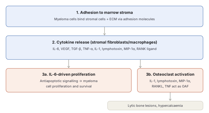
- Adhesion — cell-surface adhesion molecules bind myeloma cells to marrow stromal cells and extracellular matrix.
- Cytokine release — this binding triggers adhesion-mediated signalling, stimulating marrow fibroblasts/macrophages to release IL-6, VEGF, TGF-β, TNF-α, IL-1, lymphotoxin, MIP-1α, and RANK ligand. 3a. IL-6-driven proliferation — IL-6 plays the central role, driving myeloma cell proliferation and survival via antiapoptotic signalling (also affecting the cell cycle via cyclin D/p21, driving M-protein production). 3b. Osteoclast activation — IL-1, lymphotoxin, VEGF, MIP-1α, RANKL, and TNF act as osteoclast-activating factors (OAF), producing bone resorption. The same signalling also promotes drug resistance and tumour cell migration within the marrow.
Morphology
Osseous (Bone Marrow) Lesions
Disease begins in the marrow in >95% of cases, typically multifocal, affecting red-marrow-rich bones (skull, spine, ribs, pelvis) preferentially, later long bones. Lesions begin in the medullary cavity, erode cancellous bone, and eventually destroy cortex — radiographically, characteristic “punched-out” 1–2 cm lytic defects.
Gross: normal marrow replaced by soft, gelatinous, reddish-grey tumour tissue; focal or diffuse osteoporosis.
Light Microscopy: marrow aspirate from a bony rarefaction site is usually diagnostic (biopsy of a radiologically abnormal/tender site if aspirate gives a dry tap). Hypercellular marrow with myeloma cells >10% of cellularity — clumped, sheeted, or scattered among normal haematopoietic cells; size ranges from small differentiated (plasma-cell-like) to large undifferentiated forms; binucleate/multinucleate forms occur. Nucleus eccentric (like normal plasma cells) but typically lacking the classic cart-wheel chromatin pattern, with prominent nucleoli. Cytoplasm abundant, basophilic, with a perinuclear halo, vacuolation, and Russell bodies (hyaline globules of synthesised immunoglobulin). Reactive plasmacytosis (aplastic anaemia, rheumatoid arthritis, SLE, cirrhosis, metastatic cancer, chronic infection/TB) is distinguished by mature morphology and plasma cells never exceeding 10%.
Extraosseous Lesions
- Blood — atypical plasma cells in ~50%; normocytic normochromic anaemia; marked rouleaux formation (hyperviscosity); raised ESR.
- Myeloma kidney (myeloma nephrosis) — Bence-Jones light chains filtered and precipitated with Tamm-Horsfall protein as tubular casts in the distal convoluted tubule, sometimes surrounded by multinucleate giant cells.
- Myeloma neuropathy — nerve root infiltration → non-specific polyneuropathy; vertebral pathologic fractures → neurological compromise.
- Systemic (AL) amyloidosis — in ~10% of cases, multi-organ involvement.
- Hepatosplenomegaly — in a minority, from direct organ infiltration.
Clinical Features
- Bone pain — the most common symptom (back, ribs); from tumour proliferation and osteoclast activation; pathologic fractures cause persistent localised pain.
- Infection susceptibility — bacterial infections (pneumonia, pyelonephritis) from hypogammaglobulinaemia, plus granulocyte dysfunction/neutropenia.
- Renal failure — in ~25% (renal pathology in 50%); from hypercalcaemia, glomerular amyloid deposits, hyperuricaemia, and direct renal infiltration.
- Anaemia — in ~80%, from marrow replacement (myelophthisis) and haematopoietic inhibition.
- Bleeding tendency — thrombocytopenia, deranged platelet function, and M-protein interference with coagulation factors.
- Hyperviscosity syndrome — headache, fatigue, visual disturbance, haemorrhage.
- Neurological symptoms — from hyperviscosity, cryoglobulins, amyloid deposits.
- Biochemistry: hypercalcaemia (bone destruction); hyperuricaemia (tumour necrosis, renal failure); raised β2-microglobulin and other globulins in serum/urine.
- POEMS syndrome — ~1% of cases: Polyneuropathy, Organomegaly, Endocrinopathy, M-protein, Skin changes.
Diagnosis
Classic diagnostic triad:
- Marrow plasmacytosis >10%.
- Radiologic lytic bone lesions.
- Serum and/or urine M component (monoclonal protein).
Clinical Relevance
- The classic diagnostic triad (marrow plasmacytosis, lytic lesions, M component) is the single most exam-relevant framework in this topic — each element is independently testable and together they define the diagnosis without requiring more invasive workup.
- Myeloma kidney’s specific mechanism (Bence-Jones light chains precipitating with Tamm-Horsfall protein as tubular casts) explains why renal failure in myeloma is often initially reversible with adequate hydration and light-chain reduction — the damage is largely obstructive/toxic at the tubular level rather than an irreversible glomerular process, at least early on.
- Distinguishing neoplastic (myeloma) from reactive plasmacytosis by the 10% marrow threshold is a genuinely practical diagnostic rule, since reactive plasmacytosis occurs in a wide range of common conditions (infection, autoimmune disease, cirrhosis, malignancy) that must not be mistaken for myeloma.
- Recognising bone pain plus anaemia plus renal impairment plus hypercalcaemia as a symptom cluster (sometimes abbreviated CRAB — hyperCalcaemia, Renal failure, Anaemia, Bone lesions) is the clinical pattern that should prompt myeloma workup in an elderly patient.
Diagram 1: Molecular Pathogenesis of Bone Destruction
Draw a top box (adhesion) feeding into a cytokine-release box, which forks into two parallel outcomes (proliferation, osteoclast activation), with the osteoclast branch continuing to a final consequences box.
- Adhesion — myeloma cells bind marrow stroma/ECM.
- Cytokine release — IL-6, VEGF, TGF-β, TNF-α, IL-1, lymphotoxin, MIP-1α, RANKL, from stimulated stromal fibroblasts/macrophages. 3a. IL-6-driven proliferation — antiapoptotic signalling, tumour growth. 3b. Osteoclast activation — several of the same cytokines act as OAF.
- Consequences — lytic bone lesions, hypercalcaemia.
Labels required
- IL-6 must be singled out and labelled as playing the central role in proliferation — not just listed among the other cytokines undifferentiated.
- The osteoclast-activating-factor (OAF) role must name at least the most emphasised cytokines (IL-1, lymphotoxin, RANKL) rather than a generic “cytokines cause bone destruction” label.
Errors commonly made
- Treating myeloma-related bone destruction as a direct effect of tumour cell infiltration alone, rather than as a cytokine-mediated (OAF) stimulation of osteoclasts — the tumour cells recruit the bone-resorbing machinery rather than resorbing bone themselves.
- Listing all cytokines as functionally interchangeable, missing that IL-6 specifically drives tumour cell survival/proliferation while a different, overlapping but not identical subset specifically drives osteoclast activation.
Long Essays (2)
Multiple Choice Questions (2)
My Sticky Notes
Personal revision notes, mnemonics and reminders.
