
Megaloblastic Anaemia
Overview
Macrocytic anaemia from impaired DNA synthesis (B12 and/or folate deficiency). Marrow shows nucleus lagging behind cytoplasm maturation = megaloblast. In India, nutritional B12 deficiency (vegetarians) and folate deficiency (poor diet) are the main causes. Pernicious anaemia (IF deficiency) is the classic Western cause but is rare in India.
B12 metabolism
Source: animal foods only. Absorption: gastric R-binder → intrinsic factor (IF, from parietal cells) → absorbed in distal ileum → transcobalamin II → liver/marrow. Stores: 2-4 mg, lasts 2-4 years (so deficiency takes years to show). Functions: (1) methylcobalamin — homocysteine→methionine; (2) adenosylcobalamin — methylmalonyl-CoA→succinyl-CoA (deficiency → abnormal fatty acids in neurons → neuro symptoms).
Folate metabolism
Source: green vegetables, fruits, liver — destroyed by cooking. Absorbed duodenum/jejunum. Stores: only 10-12 mg, lasts ~4 months (so deficiency develops fast). Function: one-carbon transfer for DNA synthesis.
Biochemical basis — methyl-folate trap
2 folate-dependent reactions: (1) dUMP→dTMP (thymidylate synthetase, uses methylene-THF, makes DHF which needs DHF-reductase to recycle — target of methotrexate/pyrimethamine); (2) homocysteine→methionine (uses methylene-THF + needs B12, regenerates THF).
Folate deficiency → ↓methylene-THF directly → impaired DNA synthesis. B12 deficiency → blocks reaction 2 → folate stuck as methyl-THF (inactive, “trapped”) → can’t make methylene-THF → same DNA synthesis defect.
This is why B12 deficiency ALSO causes a folate-type block — the “methyl-folate trap.”
Causes
B12 deficiency: poor intake (strict vegetarians, breastfed infants of vegetarian mothers) OR malabsorption — gastric (pernicious anaemia, gastrectomy, congenital IF lack) or intestinal (tropical sprue, ileal resection, Crohn’s, blind loop syndrome, fish tapeworm).
Folate deficiency: poor intake (alcoholics, teens, infants, elderly, poverty) OR malabsorption (tropical sprue, coeliac disease, partial gastrectomy, jejunal resection, Crohn’s) OR excess demand (pregnancy, lactation, infancy; malignancy, ↑haematopoiesis, TB, RA) OR excess urinary loss (liver disease, CCF). Develops faster than B12 deficiency (smaller stores).
Other: drugs (methotrexate, pyrimethamine, alcohol), stem cell defects, rare enzyme deficiencies. Tropical sprue often causes combined deficiency.
Clinical features
Anaemia (insidious) · glossitis (smooth beefy-red tongue) · neurological symptoms — ONLY with B12 deficiency: subacute combined degeneration of spinal cord + peripheral neuropathy (numbness, paraesthesia, weakness, ataxia, poor coordination, ↓reflexes). Folate deficiency rarely causes neuropathy, never subacute combined degeneration. Also: mild jaundice, angular stomatitis, purpura, weight loss, anorexia.
Lab findings
Blood: macrocytosis (MCV>120fl), anisopoikilocytosis, macro-ovalocytes, basophilic stippling. Low WBC with hypersegmented neutrophils (>5 lobes) — classic clue. Low platelets in severe cases.
Marrow: hypercellular, ↓M:E ratio, megaloblasts (large nucleated cells, nucleus lags cytoplasm — OPPOSITE of iron deficiency anaemia). Giant metamyelocytes/bands. Marrow iron increased.
Biochem: ↑unconjugated bilirubin, ↑LDH (ineffective erythropoiesis) — serum iron/ferritin normal or high.
Diagnosis: serum B12 assay (<100 pg/mL = deficient) and serum/RBC folate assay. Schilling test = classical test to separate dietary vs IF-deficiency causes of B12 deficiency (mostly replaced by direct assays now).
Clinical relevance
Nucleus-lags-cytoplasm (megaloblastic) vs cytoplasm-lags-nucleus (iron deficiency) = opposite patterns, remember together. Hypersegmented neutrophils = cheap early clue on routine smear. NEVER give folate alone to a B12-deficient patient — it fixes the anaemia but neurological damage keeps progressing (dangerous, easy-to-miss pitfall).
Overview
Megaloblastic anaemias are macrocytic anaemias caused by impaired DNA synthesis, from deficiency of vitamin B12 (cobalamin) and/or folate, producing a distinctive marrow abnormality: nuclear maturation lags behind cytoplasmic maturation in erythroid precursors. Because cell division slows while cytoplasmic (haemoglobin) development proceeds normally, the nucleated red cell precursors become abnormally large — Ehrlich’s original megaloblasts (1880) — and the mature red cells released from them are macrocytic and functionally abnormal.
In India, nutritional deficiency — chiefly vitamin B12 deficiency in strict vegetarians and folate deficiency from poor dietary intake — is the dominant cause; deficiency of intrinsic factor causing pernicious anaemia, though the classic cause described in Western literature, is comparatively rare in India and more prevalent in people of European descent.
Vitamin B12 (Cobalamin) Metabolism
- Sources: exclusively animal-protein foods (liver, kidney, meat, fish, eggs, dairy) — plants contain essentially none. Strict vegetarians and exclusively breast-fed infants of vegetarian mothers are therefore at risk on diet alone.
- Absorption: dietary B12 binds gastric R-binder, then in the duodenum is transferred to intrinsic factor (IF), a glycoprotein secreted by gastric parietal cells (paralleling acid secretion). The B12-IF complex is absorbed via specific receptors in the distal ileum; inside the ileal mucosal cell, IF is degraded and B12 is transferred to transcobalamin II for portal circulation to liver, marrow, and other tissues.
- Stores: about 2–4 mg (mainly hepatic), sufficient for 2–4 years — which is why B12 deficiency takes years to become clinically manifest even after absorption completely stops.
- Functions — B12 is a cofactor for two reactions:
- As methylcobalamin, methylation of homocysteine to methionine (linked to folate metabolism, see below).
- As adenosylcobalamin, conversion of methylmalonyl-CoA to succinyl-CoA in propionate metabolism; B12 deficiency causes methylmalonyl-CoA/propionyl-CoA accumulation, generating abnormal fatty acids incorporated into neuronal lipids — a proposed basis for the neurological complications of B12 deficiency.
Folate Metabolism
- Sources: green leafy vegetables, fruits, liver, kidney — folate is heat-labile and easily destroyed by cooking/canning.
- Absorption: duodenum and upper jejunum; dietary polyglutamate folate is cleaved by folate conjugase to mono-/di-glutamates, reduced to tetrahydrofolate (THF), and circulates as methyl-THF.
- Stores: only about 10–12 mg, sufficient for roughly 4 months — far smaller than B12’s reserve, which is why folate deficiency develops much faster than B12 deficiency.
- Function: coenzyme for one-carbon (methyl/formyl) transfer reactions essential to DNA synthesis.
Biochemical Basis: the Methyl-Folate Trap
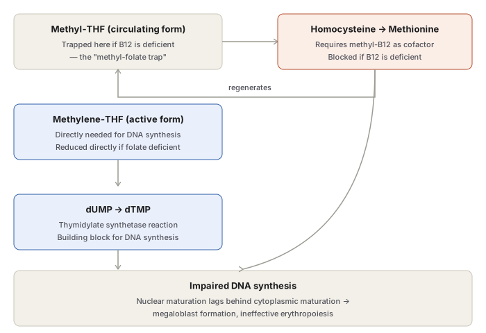
Two folate-dependent one-carbon transfer reactions build the DNA-synthesis pathway:
- Thymidylate synthetase reaction — methylene-THF donates a methyl group to convert dUMP to dTMP (a direct DNA building block); this generates dihydrofolate (DHF), which DHF-reductase must recycle back to active THF (the target of methotrexate and pyrimethamine).
- Homocysteine-methionine reaction — methylene-THF donates a methyl group to convert homocysteine to methionine, a reaction requiring vitamin B12 (methylcobalamin) as cofactor, regenerating THF in the process.
Folate deficiency directly reduces available methylene-THF, impairing DNA synthesis. Vitamin B12 deficiency blocks the homocysteine-methionine reaction, which traps folate in its inactive circulating form (methyl-THF) — unable to convert back to active methylene-THF — so a pure B12 deficiency also produces a functional folate deficiency and the identical defect in DNA synthesis. This is the methyl-folate trap hypothesis, and it explains why B12 and folate deficiency produce an identical megaloblastic picture despite being biochemically distinct starting points.
Aetiology
Vitamin B12 deficiency: inadequate intake (strict vegetarians, exclusively breast-fed infants) or malabsorption — gastric causes (pernicious anaemia, gastrectomy, congenital IF deficiency) or intestinal causes (tropical sprue, ileal resection, Crohn’s disease, blind loop syndrome, fish tapeworm infestation).
Folate deficiency: inadequate intake (alcoholics, teenagers, infancy, old age, poverty), malabsorption (tropical sprue, coeliac disease, partial gastrectomy, jejunal resection, Crohn’s disease), excess demand (pregnancy, lactation, infancy physiologically; malignancy, increased haematopoiesis, chronic exfoliative skin disease, tuberculosis, rheumatoid arthritis pathologically), or excess urinary loss (active liver disease, congestive heart failure). Folate deficiency develops far more rapidly than B12 deficiency, given its much smaller body reserve.
Other causes: drugs interfering with DNA synthesis (methotrexate, pyrimethamine, alcohol), acquired stem cell defects, rare congenital enzyme deficiencies. Tropical sprue commonly produces combined B12 and folate deficiency.
Clinical Features
- Anaemia — the cardinal feature; insidious, gradually progressive.
- Glossitis — a smooth, beefy-red tongue.
- Neurological manifestations — specific to vitamin B12 deficiency (particularly pernicious anaemia): subacute combined degeneration of the spinal cord and peripheral neuropathy, from demyelination of peripheral nerves, spinal cord, and cerebrum — numbness, paraesthesia, weakness, ataxia, poor finger coordination, diminished reflexes. Folate deficiency only occasionally produces neuropathy, and does not cause subacute combined degeneration — a clinically important distinction, since replacing folate alone in a B12-deficient patient corrects the hematologic picture while the neurological damage continues to progress, sometimes irreversibly.
- Others: mild jaundice (from ineffective erythropoiesis), angular stomatitis, purpura, melanin pigmentation, weight loss, anorexia, and features of any underlying malabsorptive disorder.
Laboratory Findings
Blood picture: macrocytosis (MCV commonly >120 fL), marked anisocytosis and poikilocytosis, macro-ovalocytes, basophilic stippling, occasional normoblasts; reticulocyte count low-to-normal untreated. Total leucocyte count may be reduced; hypersegmented neutrophils (>5 nuclear lobes) are a characteristic clue that should raise suspicion of megaloblastic anaemia on a routine film. Platelet count may be moderately reduced with bizarre forms in severe disease.
Bone marrow: hypercellular, reduced myeloid:erythroid ratio, erythroid hyperplasia with characteristic megaloblasts — large nucleated precursors with fine, open, lightly-staining (“sieve-like”) chromatin whose nuclear maturation lags behind cytoplasmic haemoglobinisation (the reverse pattern to iron deficiency anaemia, where cytoplasmic maturation lags behind). Giant metamyelocytes and band forms are seen among granulocyte precursors; megakaryocytes are usually normal in number but may show hypersegmented nuclei. Marrow iron is increased (ring sideroblasts are rare). Random chromosomal abnormalities may be seen in marrow cells.
Biochemistry: raised unconjugated bilirubin and LDH (from ineffective erythropoiesis and marrow cell breakdown); serum iron and ferritin normal or elevated.
Establishing the specific deficiency: serum vitamin B12 assay (normal 280–1000 pg/mL; <100 pg/mL is deficient) and serum/red cell folate assay, now generally performed by automated chemiluminescence/enzyme-linked fluorescence platforms rather than the traditional microbiological assay. The Schilling test (staged radiolabelled-B12 urinary excretion study) was the classical method to distinguish dietary B12 deficiency from intrinsic-factor deficiency/malabsorption, though it has been largely superseded by direct assays in current practice.
Clinical Relevance
- The nuclear-cytoplasmic asynchrony pattern is the single fact that most reliably separates megaloblastic anaemia from every other cause of macrocytosis on marrow examination, and its direction (nucleus lags cytoplasm) is the exact mirror image of iron deficiency anaemia (where cytoplasm lags nucleus) — a pairing worth holding in memory together.
- Hypersegmented neutrophils on a routine peripheral smear are a low-cost early clue to megaloblastic anaemia, often visible before marrow examination is even considered.
- Distinguishing B12 from folate deficiency matters clinically beyond the biochemistry: giving folate alone to a B12-deficient patient will correct the anaemia (masking the diagnosis on blood counts) while the neurological damage from B12 deficiency continues to progress — a genuinely dangerous pitfall if the two are not told apart before treatment starts.
Diagram 1: The Methyl-Folate Trap
Draw methyl-THF and the homocysteine-methionine reaction at the top, methylene-THF and the dUMP→dTMP reaction below it, with a regeneration arrow looping methionine synthesis back to methylene-THF, and both pathways converging on one “impaired DNA synthesis” box at the bottom.
- Methyl-THF (circulating, inactive form) — where folate accumulates and is “trapped” if B12 is deficient.
- Homocysteine → methionine — needs methylcobalamin (B12); regenerates THF, which becomes methylene-THF.
- Methylene-THF (active form) — directly needed for DNA synthesis; reduced directly if folate itself is deficient.
- dUMP → dTMP — the actual DNA-building-block reaction methylene-THF feeds.
- Both routes converge on impaired DNA synthesis → megaloblast formation.
Labels required
- The regeneration arrow (methionine synthesis → THF → methylene-THF) must be explicit — this is the step whose blockade by B12 deficiency is the entire basis of the trap, and omitting it collapses the diagram into two unconnected deficiency states rather than showing why B12 deficiency mimics folate deficiency.
- Vitamin B12 must be labelled specifically as a cofactor for the homocysteine-methionine step, not for the dUMP→dTMP step — a frequently swapped pairing.
Errors commonly made
- Believing vitamin B12 deficiency directly blocks DNA synthesis by its own separate mechanism, rather than understanding that it works through trapping folate — the “trap” is the actual tested concept, not two independent deficiency pathways that happen to look similar.
- Mixing up which folate form is active (methylene-THF) versus which is the inert circulating/storage/trapped form (methyl-THF).
- Forgetting that folate deficiency alone (with normal B12) produces the identical defect through a more direct route — simply not enough methylene-THF to begin with, no trapping required.
Short Essays (1)
Multiple Choice Questions (2)
My Sticky Notes
Personal revision notes, mnemonics and reminders.
