
Marfan Syndrome
Definition
Autosomal dominant connective tissue disorder. Affects skeleton, eyes, and cardiovascular system. Caused by fibrillin defect. Prevalence about 1 in 5000. Most cases (70-85%) run in families; rest are new mutations.
Pathogenesis
Fibrillin is made by fibroblasts. It builds microfibrils in the extracellular matrix. Microfibrils act as a scaffold for tropoelastin (part of elastic fibres). Microfibrils are richest in the aorta, ligaments, and ciliary zonules (support the eye lens) — the same tissues affected in this disease.
Gene: FBN1 (chromosome 15q21). Over 1000 different mutations found, so gene testing is hard. Diagnosis is mostly clinical. Mutant fibrillin acts as a “dominant negative” — it blocks normal microfibril assembly, not just reduces the amount.
Second mechanism: normal microfibrils also hold back TGF-β (limit how active it is). When microfibrils are lost, TGF-β signalling goes up too much. This damages vessel wall muscle and matrix. This explains features that plain structural loss cannot explain, like bone overgrowth. Proof: TGF-β receptor mutations cause a similar disease (Marfan type 2). This also matters for treatment — some angiotensin receptor blockers (which reduce TGF-β activity) are now used along with beta-blockers to protect the aorta.
Morphology
Skeletal: tall, thin build; long limbs and fingers (arachnodactyly); high-arched palate; joints too flexible; kyphoscoliosis; pectus excavatum or pigeon chest.
Eye: lens moves out of place, usually both sides (ectopia lentis). This finding is very specific for Marfan syndrome — a strong clue at bedside.
Heart and vessels (most serious system):
- Elastic fibres break down in aortic wall (cystic medionecrosis, not specific to Marfan alone) → aneurysm and dissection → aortic rupture = most common cause of death, can happen at any age.
- Weak aortic valve ring → aortic valve leaking (incompetence).
- Floppy mitral valve → prolapse, may cause heart failure.
Not every patient shows all features — some have mainly heart problems with few skeletal/eye signs. This depends on which exact FBN1 mutation is present.
Clinical relevance
Bilateral ectopia lentis in a tall, long-limbed patient is a fast bedside clue, useful because full gene testing is often impractical (too many possible mutations). TGF-β mechanism changed treatment directly — angiotensin receptor blockers are now added with beta-blockers to slow aortic widening and lower dissection risk.
Definition
Marfan syndrome is an autosomal dominant disorder of connective tissue, principally affecting the skeleton, eyes, and cardiovascular system, caused by an inherited defect in fibrillin, an extracellular glycoprotein. Prevalence is estimated at roughly 1 in 5000 worldwide; about 70–85% of cases are familial, the rest sporadic (de novo FBN1 mutation in a parental germ cell).
Pathogenesis
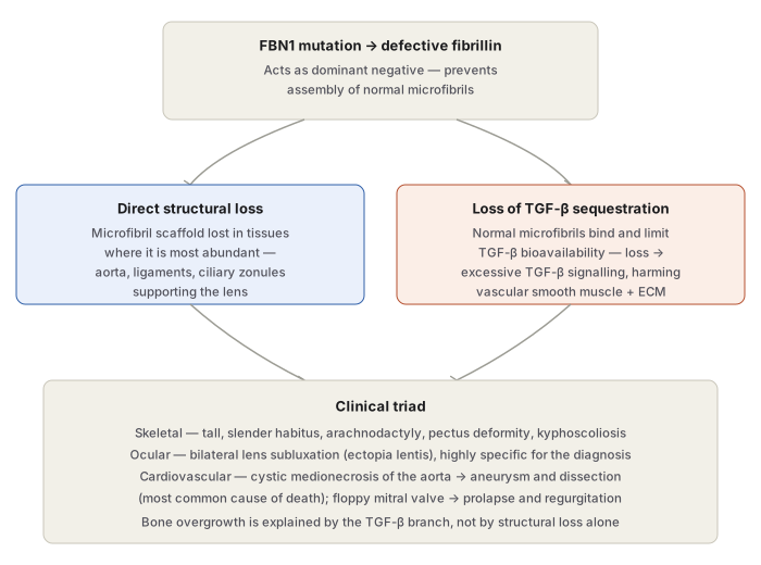
Fibrillin is secreted by fibroblasts and is the major component of microfibrils in the extracellular matrix. Microfibrils act as scaffolds for deposition of tropoelastin, an integral component of elastic fibres. Although widely distributed, they are especially abundant in the aorta, ligaments, and the ciliary zonules that support the lens — precisely the tissues affected in Marfan syndrome.
Fibrillin is encoded by FBN1 (chromosome 15q21). Mutations occur throughout this very large gene — over 1000 distinct causative mutations identified — which makes routine DNA-sequencing diagnosis impractical; diagnosis is therefore made mainly on clinical grounds. Because heterozygotes are symptomatic, the mutant fibrillin protein is thought to act as a dominant negative, preventing assembly of normal microfibrils rather than simply halving their amount.
Not every feature of Marfan syndrome follows directly from loss of structural microfibrils — bone overgrowth, for instance, does not. The explanation is a second mechanism: normal microfibrils sequester transforming growth factor-β (TGF-β), limiting its bioavailability. Loss of microfibrils therefore causes excessive TGF-β signalling, which damages vascular smooth muscle development and extracellular matrix integrity. Supporting this model, mutations in the TGF-β type II receptor produce a related disorder, Marfan syndrome type 2. This mechanistic insight has direct therapeutic relevance: certain angiotensin receptor type II blockers, which inhibit TGF-β activity, are now used clinically for cardiovascular prevention in Marfan syndrome, alongside β-adrenergic blockers to lower blood pressure and reduce the risk of aortic catastrophe.
Morphology
Skeletal
The most evident feature: a slender, elongated habitus with disproportionately long legs, arms, and fingers (arachnodactyly); a high-arched palate; and hyperextensible joints. Spinal deformity (severe kyphoscoliosis) is common. The chest may show pectus excavatum (deeply depressed sternum) or a pigeon-breast deformity.
Ocular
Bilateral dislocation or subluxation of the lens (ectopia lentis), from weakness of its suspensory ligaments, is the most characteristic ocular change — when bilateral, it is highly specific for Marfan syndrome and strongly suggests the diagnosis.
Cardiovascular
The most serious system involved:
- Fragmentation of elastic fibres in the aortic tunica media predisposes to aneurysmal dilation and aortic dissection. This microscopic change, cystic medionecrosis, is not itself specific to Marfan syndrome (similar changes occur in hypertension and with ageing), but its consequence — aortic rupture — is the most common cause of death, and can occur at any age.
- Loss of medial support dilates the aortic valve ring, producing aortic incompetence.
- The mitral valve (and other cardiac valves) may become excessively distensible and regurgitant (“floppy valve”), producing mitral valve prolapse and, less often than aortic rupture, terminal congestive cardiac failure.
Expression is markedly variable — some patients show predominantly cardiovascular disease with minimal skeletal/ocular change — a variability attributed to the specific FBN1 mutation involved, not every patient displays every feature described above.
Clinical Relevance
Recognising bilateral ectopia lentis as highly specific for Marfan syndrome gives a fast bedside route to the diagnosis in a tall, long-limbed patient, without waiting for genetic testing that is often impractical given the sheer number of possible FBN1 mutations. The TGF-β mechanism is not just an explanatory curiosity — it directly changed treatment, since angiotensin receptor blockers that reduce TGF-β signalling are now added to β-blockers specifically to slow aortic root dilation and reduce dissection risk, a rare example of a molecular mechanism translating this directly into a drug choice.
Diagram 1: Two Mechanisms From One Fibrillin Defect
Draw one top box (FBN1 mutation → defective fibrillin, dominant negative) branching into two boxes (direct structural loss; loss of TGF-β sequestration), both converging into one bottom box (clinical triad).
- Top — FBN1 mutation produces fibrillin that blocks normal microfibril assembly.
- Left branch — direct loss of the microfibril scaffold in aorta, ligaments, ciliary zonules.
- Right branch — loss of TGF-β sequestration by microfibrils → excess TGF-β signalling, harming vascular smooth muscle and ECM.
- Bottom — clinical triad (skeletal, ocular, cardiovascular), with bone overgrowth specifically attributed to the TGF-β branch.
Labels required
- The “dominant negative” mechanism must be named at the top box — this is what explains why heterozygotes (not just homozygotes) are symptomatic, a frequently tested nuance.
- The TGF-β branch must be explicitly linked to bone overgrowth — this is the detail that shows understanding goes beyond “loss of structural protein weakens tissue,” since structural loss alone cannot explain overgrowth.
- Ectopia lentis and cystic medionecrosis/aortic dissection must both appear in the outcome box, since they are the two most specific, most examined findings in the whole topic.
Errors commonly made
- Explaining every Marfan feature (including bone overgrowth) purely through loss of structural microfibrils, missing the separate TGF-β mechanism entirely.
- Treating cystic medionecrosis as specific to Marfan syndrome rather than a nonspecific aortic media change also seen in hypertension and ageing — what is specific is the clinical context and the resulting dissection risk, not the histologic lesion itself.
- Omitting that expression is highly variable between patients (some almost entirely cardiovascular, with minimal skeletal/ocular findings) — treating the triad as always fully present in every case.
My Sticky Notes
Personal revision notes, mnemonics and reminders.
