
Leprosy
Definition
Hansen’s disease — chronic, non-fatal, affects cooler body parts (skin, nerves, mucosa, eyes, testis). Bacteraemic spread → liver/spleen/marrow/nodes. Advanced → secondary amyloidosis, renal disease.
Endemic: hot/moist/poor tropics. 8 countries = 80% cases; India = 1/3 of global registered cases (TN, Bihar, Puducherry, AP, Odisha, WB, Assam).
Organism
M. leprae — resembles TB bacillus but less acid-fast (decolourised at 5% H2SO4 vs 20% for TB). Globi / cigarettes-in-pack pattern. First bacterium linked to human disease, yet still uncultivable in vitro — armadillo = animal model.
Detection: ZN · Fite-Faraco (preferred, ZN modification) · GMS · PCR · IgM anti-PGL-1 (95% LL, 60% TT).
Slit-skin smear → Bacterial Index (BI) 0-6+. BI=0 → paucibacillary; BI≥1+ → multibacillary.
Transmission
Slow, long incubation (2-20yr, avg 3yr). Direct contact (skin/nasal/oral secretions) · materno-foetal · breast milk.
Immunology
Type IV (delayed) hypersensitivity, no bacterial toxin — same principle as TB but different players:
- Antigens: PGL-1 (species-specific), LAM. Nerve invasion via trisaccharide-Schwann cell basal lamina binding.
- Host HLA/MHC → determines which antigen response develops
- T cells: BOTH CD4+ AND CD8+ (unlike TB). TT = CD4+ helper-dominant. LL = CD8+ excess but defective → fails to clear organism.
- Humoral: ↑Ig in LL but non-protective.
Lepromin test (classification, NOT diagnostic): Fernandez (24-48h, early) / Mitsuda (3-4wk, delayed granuloma) = positive in TT. Negative in LL.
Ridley-Jopling spectrum
TT (high resistance) → BT → BB (mid/dimorphic) → BL → LL (low resistance)
Across spectrum TT→LL: CMI ↓, granuloma organisation ↓, bacillary load ↑, humoral response ↑.
| LL | TT | |
|---|---|---|
| Skin | Symmetric, multiple, leonine facies | Asymmetric, few, macular |
| Nerve | Present, sensory milder | Present, distinct sensory loss |
| Histo | Foamy lepra cells + clear zone | Hard-tubercle granuloma, erodes epidermis, no clear zone |
| Bacilli | Numerous (multibacillary) | Few, in nerves (paucibacillary) |
| Immunity | Suppressed | Good |
| Lepromin | − | + |
Variants outside spectrum: indeterminate (initial nonspecific stage) · pure neural (no skin lesions) · histoid (Wade — LL variant, dermatofibroma-like, heavily bacillated).
Histopathology quick-map
LL: foamy macrophages/lepra cells, perivascular/perineural/periadnexal, clear zone, AFB+++. TT: epithelioid + Langhans’ granuloma, erodes basal epidermis, no clear zone, AFB scant (in nerves). BT: epithelioid + lymphocytes, narrow clear zone. BL: histiocytes>epithelioid, numerous bacilli. BB: epithelioid sheets, no giant cells.
Reactional leprosy
- Type I (reversal) — borderline only. Upgrading (BL→TT direction: ↑CMI, lymphocytes, oedema, necrosis, ↓BI) vs Downgrading (BT→LL direction: ↓CMI, granuloma dispersal, ↑bacilli)
- Type II (ENL) — LL patients, classically post-treatment. Tender nodules, fever, iridocyclitis, synovitis, panniculitis, neutrophils/eosinophils, vasculitis, ↑bacilli. Repeated attacks → amyloidosis risk.
Clinical relevance
5% vs 20% H2SO4 decolourisation = quick discriminator vs TB on unlabelled AFB smear. Lepromin result maps directly to spectrum position (immunity/BI/lepromin all move together). ENL vs reversal reaction = different management, different trigger (ENL = post-treatment in LL).
Definition and Distribution
Leprosy (Hansen’s disease, after Hansen’s 1874 discovery of the causative organism, though the disease itself was described in Indian texts as far back as the 6th century BC) is a chronic, non-fatal infectious disease affecting mainly the cooler parts of the body — skin, mucous membrane of the mouth, upper respiratory tract, eyes, peripheral nerves, superficial lymph nodes and testis — with skin and peripheral nerve involvement dominating clinically. Bacteraemic spread via endothelial colonisation or reticuloendothelial filtering can involve liver, spleen, bone marrow and regional nodes; advanced disease may produce secondary (immune-mediated) amyloidosis and renal disease.
The disease is endemic to hot, moist, poor tropical regions and is now almost exclusively confined to a handful of developing countries — eight countries (India, China, Nepal, Brazil, Indonesia, Myanmar, Madagascar, Nigeria) account for roughly 80% of global cases, with India alone contributing about a third of all registered cases worldwide (concentrated in Tamil Nadu, Bihar, Puducherry, Andhra Pradesh, Odisha, West Bengal and Assam).
Causative Organism
Mycobacterium leprae closely resembles M. tuberculosis but is less acid-fast — a genuinely useful discriminator, since it decolourises with a weaker 5% sulphuric acid solution compared with the 20% needed for M. tuberculosis. In tissue, the organisms appear as compact rounded masses (globi) or arranged in parallel rows resembling cigarettes-in-a-pack. M. leprae was the first bacterium ever identified as a cause of human disease, yet remains one of the very few bacterial species that has never been cultured on artificial medium; the nine-banded armadillo, which develops disease histopathologically and immunologically similar to human leprosy, serves as the experimental model.
Detection methods include Ziehl–Neelsen staining (less acid-fast than M. tuberculosis, as above); the Fite-Faraco stain, a ZN modification giving better tissue-section staining and generally preferred for leprosy; Gomori methenamine silver staining; PCR; and IgM antibodies to the PGL-1 antigen, positive in 95% of lepromatous but only 60% of tuberculoid disease. The slit-skin smear, ZN-stained and examined under oil immersion, gives a quantitative bacterial index (BI) from 0+ to 6+ — 0+ defines paucibacillary disease, while any higher score (1–10 bacilli/100 fields up to >1000/field) defines multibacillary disease, a classification with direct treatment implications.
Transmission
Leprosy is a slowly communicable disease with an unusually long incubation period — 2 to 20 years, averaging around 3 years — transmitted by direct contact with untreated patients shedding bacilli from damaged skin, nasal secretions, oral mucosa and hair follicles, and less commonly by materno-foetal (transplacental) transmission or via breast milk from an affected mother.
Immunology
As in tuberculosis, tissue damage in leprosy is driven by T-cell-mediated delayed (type IV) hypersensitivity rather than by any bacterial toxin — but the immunology differs from tuberculosis in ways that determine the entire clinical spectrum of the disease:
- Bacterial antigens. M. leprae’s cell wall carries a species-specific phenolic glycolipid (PGL-1) and lipoarabinomannan (LAM), which shape the host lymphocyte/macrophage response; a distinctive feature of the organism is its invasion of peripheral nerves via binding of a bacillary trisaccharide to the basal lamina of Schwann cells.
- Host genotype. HLA/MHC class determines which bacillary antigens engage host immune cells, explaining why host response — and hence clinical form — varies between individuals infected with the same organism.
- T-cell response, unlike tuberculosis, involves both CD4+ and CD8+ subsets, with CD4+ cells in leprosy acting cytotoxically as well as in their usual helper role. Th1 and Th2 CD4+ subpopulations elaborate different cytokines: tuberculoid leprosy is dominated by a CD4+ (helper T-cell) response, while lepromatous leprosy, despite an excess of CD8+ (suppressor) T cells, fails to clear the organism because of a functional CD8+ defect.
- Humoral response is paradoxically prominent in lepromatous disease — high IgG, IgA and IgM with antibodies against mycobacterial antigens — but confers no protection against the organism.
Lepromin test — not a diagnostic test, but a classification tool: intradermal lepromin (an M. leprae antigen extract) produces an early indurated reaction at 24–48 hours (Fernandez reaction) or a delayed granulomatous reaction at 3–4 weeks (Mitsuda reaction) in tuberculoid patients, reflecting intact cell-mediated immunity; lepromatous patients are lepromin-negative, reflecting suppressed cell-mediated immunity. This single test result is the clearest bedside proxy for where a patient sits on the immune spectrum described below.
Classification (Ridley–Jopling)
Leprosy is classified along a spectrum defined by host immune response, from high resistance to low resistance:
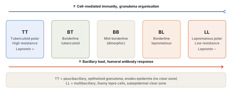
The two polar forms — tuberculoid and lepromatous — represent opposite, immunologically stable ends of this spectrum and are compared directly, since this comparison is one of the most consistently tested points in the topic:
| Feature | Lepromatous leprosy (low resistance) | Tuberculoid leprosy (high resistance) |
|---|---|---|
| Skin lesions | Symmetrical, multiple, hypopigmented/erythematous macules, papules or nodules; coalescing nodules give leonine facies | Asymmetrical, single or few hypopigmented/erythematous macules |
| Nerve involvement | Present, sensory disturbance less severe | Present, distinct sensory disturbance |
| Histopathology | Foamy macrophages (lepra cells) in dermis, separated from epidermis by a clear zone | Hard tubercle-like granuloma eroding the basal epidermal layer; no clear zone |
| Bacteriology | Numerous bacilli (globi, cigarettes-in-pack) — multibacillary | Few bacilli, seen in destroyed nerves — paucibacillary |
| Immunity | Suppressed cell-mediated immunity | Good cell-mediated immunity |
| Lepromin test | Negative | Positive |
Between the two poles lie three borderline forms — borderline tuberculoid (BT), mid-borderline/dimorphic (BB), and borderline lepromatous (BL) — completing the five-group modified Ridley–Jopling classification (TT–BT–BB–BL–LL). Cases that do not yet fit the spectrum are termed indeterminate leprosy, the non-specific initial stage of any type. Additional recognised variants outside this classification include pure neural leprosy (nerve involvement without the cardinal skin lesions) and histoid leprosy (a variant of LL, described by Wade, with dermatofibroma-like nodules densely packed with bacilli).
Histopathology by Type
- Lepromatous leprosy: dermal proliferation of foamy macrophages (“lepra cells” or Virchow cells), concentrated around vessels, nerves and adnexal structures, heavily laden with acid-fast bacilli (globi/cigarettes-in-pack); characteristically separated from the epidermis by an uninvolved subepidermal clear zone; overlying epidermis is thinned, flattened, occasionally ulcerated.
- Tuberculoid leprosy: dermal granulomas resembling hard tubercles (epithelioid cells, Langhans’ giant cells, peripheral lymphocyte mantle) with a predilection for dermal nerves, which may be destroyed and infiltrated; the granulomatous infiltrate erodes the basal epidermal layer — no clear zone; bacilli are scanty, found chiefly in destroyed nerves.
- Borderline forms: BT shows epithelioid cells with plentiful lymphocytes and a narrow clear zone, scanty nerve-confined bacilli; BL shows predominant histiocytes, few epithelioid cells, irregular lymphocytes, and numerous bacilli; BB (mid-borderline) shows sheets of epithelioid cells without giant cells, sparse perineural lymphocytes, and bacilli mostly in nerves.
- Indeterminate leprosy: non-specific lymphocytic/mononuclear infiltrate around adnexal structures and vessels; nerve involvement, when present, strongly supports the diagnosis, which is confirmed by demonstrating bacilli.
- Pure neural leprosy: the histopathology of the corresponding skin-lesion type is instead found on nerve biopsy; may be AFB-positive or -negative.
- Histoid leprosy: whorls and fascicles of foamy spindle cells in the upper dermis beneath a clear subepidermal space, heavily bacillated.
Reactional Leprosy
Two immunologically distinct reaction types occur, chiefly in borderline disease or during treatment:
- Type I (reversal reactions) — the polar forms are immunologically stable, but borderline disease can shift along the spectrum: an upgrading reaction (increased cell-mediated immunity, lymphocyte influx, oedema, central necrosis, falling bacterial index) occurs in BL patients moving toward the tuberculoid pole on treatment; a downgrading reaction (falling cellular immunity, granuloma dispersal, rising bacillary numbers) occurs in BT patients drifting toward the lepromatous pole.
- Type II — erythema nodosum leprosum (ENL) — occurs in lepromatous patients, typically after starting treatment: tender cutaneous nodules, fever, iridocyclitis, synovitis and lymphadenopathy, with neutrophilic/eosinophilic infiltration, prominent vasculitis, and inflammation extending into subcutaneous fat (panniculitis); increased bacillary load; repeated attacks predispose to secondary amyloidosis.
Clinical Relevance
- The acid-fastness gradient between the two mycobacteria that dominate this section — M. leprae decolourising at 5% sulphuric acid versus M. tuberculosis at 20% — is a small, specific fact that resolves an otherwise ambiguous “acid-fast organism in tissue” question.
- The lepromin test result is the clinical shortcut for placing a patient on the Ridley–Jopling spectrum without waiting for full histopathology: positive (Fernandez/Mitsuda) points toward the tuberculoid pole and intact cellular immunity, negative toward the lepromatous pole and suppressed cellular immunity — this single correlation (immunity ↔ lepromin ↔ bacillary load, all moving together across the spectrum) is the organising idea of the whole topic.
- Type I and Type II reactions are managed very differently from the underlying disease itself and from each other, so recognising ENL (fever, tender nodules, vasculitis, panniculitis, occurring characteristically after treatment starts) as immunologically distinct from a reversal reaction is a genuine clinical distinction, not just a classification exercise.
Diagram 1: The Ridley–Jopling Immune Spectrum
Draw a horizontal row of five boxes, left to right: TT, BT, BB, BL, LL. Above the row, draw a single arrow pointing left, labelled “increasing cell-mediated immunity and granuloma organisation.” Below the row, draw a single arrow pointing right, labelled “increasing bacillary load and humoral antibody response.” Underneath, add a short summary box contrasting TT and LL histology directly.
Labels required
- Both trend arrows must be shown running in opposite directions across the same five boxes — the entire point of the spectrum is that immunity and bacillary load move inversely, not that they are two unrelated facts about leprosy.
- TT and LL must each carry their lepromin result (+ and −, respectively), since this is the one bedside test result that maps directly onto spectrum position.
- The three borderline categories (BT, BB, BL) must be shown as genuinely intermediate, not lumped as a single “borderline” box — examiners specifically test which borderline form a described histology or reaction belongs to.
Errors commonly made
- Treating tuberculoid and lepromatous leprosy as two unrelated diseases rather than as the two stable poles of one continuous immune spectrum, which is the reason borderline reactions (upgrading/downgrading) are even possible.
- Reversing which pole has the higher bacillary load — a common slip is assuming the “high resistance” tuberculoid form has more bacteria because it sounds more severe, when in fact high resistance is exactly why bacillary load stays low there.
Short Essays (1)
Short Answers (1)
My Sticky Notes
Personal revision notes, mnemonics and reminders.
