
Langerhans Cell Histiocytosis
Overview
Histiocytosis = proliferations of dendritic cells/Langerhans cells/macrophages, benign to malignant. LCH = monoclonal Langerhans cell proliferation, more common, benign-to-intermediate end (vs rare malignant histiocytic/Langerhans/dendritic cell sarcomas — true lymphomas).
LCH = 3 related conditions in children: eosinophilic granuloma, Hand-Schüller-Christian disease, Letterer-Siwe disease. All 3: histiocytes+lymphocytes+eosinophils proliferation, differ in extent/aggressiveness.
Formerly “histiocytosis-X”
Now known:
- Cell of origin = Langerhans cell (marrow origin, normally in epidermis) — not unknown
- Three conditions = ONE disorder, different expressions. Shown by: (i) shared markers S-100, CD1a, HLA-DR; (ii) shared Birbeck granules (histiocytosis-X bodies) — rod-shaped, tennis-racket-like end, from langerin receptor-mediated endocytosis.
Three forms
1. Eosinophilic granuloma — unifocal (60%, commoner) vs multifocal (often part of Hand-Schüller-Christian). Children/young adults, male predominant. Solitary osteolytic lesion (femur, skull, vertebrae, ribs, pelvis). Micro: macrophage aggregates + eosinophils, fat/pigment-laden macrophages, Birbeck granules (EM). Benign — asymptomatic till fracture/pain; spontaneous healing or curettage/radiotherapy.
2. Hand-Schüller-Christian disease — TRIAD: multifocal bony defects + diabetes insipidus + exophthalmos. Children <5yr. Orbital lesion→exophthalmos, hypothalamic→DI. Lung/liver/spleen/node involvement common (~half). Micro: indistinguishable from eosinophilic granuloma. Benign but more disabling — fever, skin lesions, recurrent pneumonitis. Spontaneous resolution or chemo/radiation.
3. Letterer-Siwe disease — ACUTE DISSEMINATED form, infants/children <2yr. Hepatosplenomegaly, lymphadenopathy, thrombocytopenia, anaemia, leucopenia. Micro: pleomorphic macrophages with NUCLEAR ATYPIA (more atypical than other 2), vacuoles + Birbeck granules. Acute: fever, rash, weight loss, bleeding, organomegaly, cystic bone lesions (skull/pelvis/long bones). Intense chemo can control; intercurrent infection often fatal. Now regarded as unusual malignant lymphoma form.
Clinical relevance
Progression unifocal/asymptomatic (eosinophilic granuloma) → multifocal/disabling (Hand-Schüller-Christian) → disseminated/acute/fatal (Letterer-Siwe) = ONE disease spectrum, increasing extent, younger age = more aggressive — not 3 unrelated diseases sharing a name. Birbeck granules + S-100/CD1a/HLA-DR immunophenotype = the diagnostic anchor unifying all 3 clinically-different presentations. Hand-Schüller-Christian triad (bone+DI+exophthalmos) = classic tested pattern linking 3 anatomically unrelated sites to one histiocytic infiltration — learn as a unit, not 3 separate facts.
Overview
“Histiocytosis” describes a group of proliferations of dendritic cells, Langerhans cells, or macrophages, spanning both benign and malignant examples. Langerhans cell histiocytosis (LCH) refers specifically to a monoclonal proliferation of Langerhans cells — the more common, generally benign-to-intermediate end of this spectrum (as distinct from the rare, clearly malignant histiocytic sarcoma, Langerhans cell sarcoma, and dendritic cell sarcoma, which are true lymphomas).
LCH comprises three clinicopathologically related conditions occurring in children: eosinophilic granuloma, Hand-Schüller-Christian disease, and Letterer-Siwe disease. All three feature proliferation of the same three cell types — histiocytes, lymphocytes, and eosinophils — differing mainly in extent and clinical aggressiveness.
Historical Note: “Histiocytosis-X”
This group was formerly termed histiocytosis-X, but two developments clarified both the cell of origin and the unity of the three conditions:
- Cell of origin resolved — the “X” (unknown) proliferating cell is actually the Langerhans cell, of marrow origin, normally resident chiefly in the epidermis (and some other organs).
- Unity of the three conditions demonstrated — via (i) shared immunohistochemical markers (S-100 protein, CD1a, HLA-DR) across all three, and (ii) shared ultrastructural finding of Birbeck granules (histiocytosis-X bodies) — rod-shaped structures with a dilated, “tennis-racket”-like terminal end, arising from receptor-mediated endocytosis of langerin, a protein involved in Birbeck granule biosynthesis. These findings established that the three conditions are different clinical expressions of one basic disorder, not three unrelated diseases.
The Three Clinicopathologic Forms
1. Eosinophilic Granuloma
The unifocal form is more common (~60%) than the multifocal variety (which is often a component of Hand-Schüller-Christian disease). Affects children and young adults, predominantly male. Presents as a solitary osteolytic lesion — femur, skull, vertebrae, ribs, pelvis — diagnosed by biopsy of the lytic lesion.
Microscopy: closely-packed macrophage aggregates admixed with variable eosinophils; macrophages contain fat droplets or brown pigment granules (phagocytic activity), occasional multinucleate forms, and — best seen by electron microscopy — Birbeck granules.
Course: benign; the bony lesion is asymptomatic until erosion causes pain or fracture. May undergo spontaneous fibrosis/healing, or require curettage/radiotherapy.
2. Hand-Schüller-Christian Disease
Defined by the classic triad: multifocal bony defects, diabetes insipidus, and exophthalmos. Develops in children under 5 years. Multifocal lytic lesions can occur at any site; orbital involvement produces exophthalmos, hypothalamic involvement produces diabetes insipidus. Multiple spherical lung lesions are frequent; liver, spleen, and lymph node involvement occurs in about half of patients.
Microscopy: indistinguishable from unifocal eosinophilic granuloma.
Course: benign but more disabling than the unifocal form — fever, skin lesions, recurrent pneumonitis and other infections are frequent. Lesions may resolve spontaneously or require chemotherapy/radiation.
3. Letterer-Siwe Disease
The acute, disseminated form of LCH, occurring in infants and children under 2 years. Features hepatosplenomegaly, lymphadenopathy, thrombocytopenia, anaemia, and leucopenia, with generalised hyperplasia of tissue macrophages across organs.
Microscopy: involved organs show aggregates of pleomorphic macrophages with nuclear atypia — a more atypical picture than the other two forms — containing cytoplasmic vacuoles and Birbeck granules.
Course: acute presentation with fever, skin rash, weight loss, anaemia, bleeding disorders, and organomegaly; cystic bony lesions in skull, pelvis, long bones. Intense chemotherapy can control the disease, but intercurrent infections often prove fatal. This form is now regarded as an unusual form of malignant lymphoma, reflecting its more aggressive, atypical behaviour compared to the other two LCH forms.
Clinical Relevance
- The progression across the three forms — unifocal/asymptomatic (eosinophilic granuloma) → multifocal/disabling (Hand-Schüller-Christian) → disseminated/acute/potentially fatal (Letterer-Siwe) — is best understood as one disease spectrum of increasing extent and age-inverse severity (younger age at presentation correlating with more aggressive disease), rather than three unrelated conditions that happen to share a name.
- Birbeck granules and the S-100/CD1a/HLA-DR immunophenotype are the diagnostic anchor tying all three forms together — recognising that identical markers appear across clinically very different presentations is what allows a pathologist to correctly unify a solitary bone lesion in a teenager and a disseminated infantile illness under the same diagnostic umbrella.
- The Hand-Schüller-Christian triad (bony defects, diabetes insipidus, exophthalmos) is a classic, frequently tested clinical pattern precisely because it links three anatomically unrelated findings (bone, hypothalamus, orbit) to a single underlying histiocytic infiltration — recognising the triad as a unit is more useful than memorising the three findings independently.
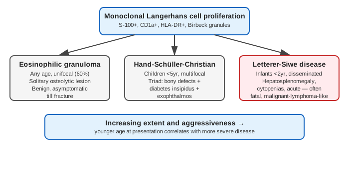
Diagram 1: The Three Forms as One Disease Spectrum
Draw a top box (the shared Langerhans cell proliferation with its markers) fanning into three branch boxes (the three named forms), with a closing bar underneath showing the severity gradient.
- Top — monoclonal Langerhans cell proliferation; S-100+, CD1a+, HLA-DR+, Birbeck granules.
- Eosinophilic granuloma — any age, unifocal (60%), solitary osteolytic lesion, benign/asymptomatic till fracture.
- Hand-Schüller-Christian disease — children <5yr, multifocal, triad (bony defects + diabetes insipidus + exophthalmos).
- Letterer-Siwe disease — infants <2yr, disseminated, hepatosplenomegaly + cytopenias, acute/often fatal, malignant-lymphoma-like.
- Bottom bar — increasing extent and aggressiveness; younger age at presentation = more severe disease.
Labels required
- The shared marker panel (S-100, CD1a, HLA-DR, Birbeck granules) must sit on the top box only, since it is what unifies all three forms diagnostically — it should not be repeated separately under each branch as if each form had its own distinct markers.
- The age ranges must be explicit for each form, since age is the variable that tracks most tightly with severity in this spectrum.
Errors commonly made
- Treating the three forms as three separate diseases that happen to share a name, rather than one disorder of varying extent — the shared immunophenotype and Birbeck granules are the evidence that unifies them.
- Assuming Letterer-Siwe disease and Hand-Schüller-Christian disease are histologically distinguishable from eosinophilic granuloma — microscopy is essentially the same across the unifocal and multifocal benign forms; it is the clinical extent, age, and (in Letterer-Siwe) nuclear atypia that separate them, not a unique histologic signature per form.
My Sticky Notes
Personal revision notes, mnemonics and reminders.
