
Iron Deficiency Anaemia
Overview
Most common nutritional deficiency worldwide. Commonest cause of microcytic hypochromic anaemia. Higher in developing countries. In India, leading cause of anaemia in women of reproductive age and young children.
Iron metabolism
Loss: ~1 mg/day (skin, bowel, GU tract); +0.5-1 mg/day in menstruation. Absorption: duodenum/jejunum, only 5-10% of dietary iron absorbed normally, up to 20-30% in pregnancy/deficiency. Hepcidin (liver) controls ferroportin — high hepcidin blocks iron export from gut cell. Vitamin C/citric acid/gastric acid help non-haem absorption; antacids/milk/phytates/tea tannins block it. Haem iron absorbed better than non-haem.
Transport: on transferrin (~33% saturated). Distribution: Hb 65%, myoglobin 3.5%, enzymes 0.5%, plasma transferrin-iron 0.5%, storage (ferritin/haemosiderin) 30%. No regulated excretion — absorption is the only control point.
Pathogenesis — 3 stages
Storage iron depletion → iron-deficient erythropoiesis (no anaemia yet) → frank anaemia (microcytic, hypochromic).
Causes: blood loss, increased requirement, poor intake, poor absorption.
Causes by group
- Women (reproductive age) — menorrhagia (most common), repeated pregnancies (~750 mg iron per pregnancy), poor diet, menarche
- Post-menopausal women — endometrial Ca, GI bleed (gastric/colon Ca, hiatus hernia)
- Adult males — almost always chronic blood loss: peptic ulcer, haemorrhoids, hookworm infestation, GI cancer, oesophageal varices, NSAID use, ulcerative colitis
- Infants/children — peak 1-2 yrs, demand > intake. Term infant iron stores last 4-6 months; preterm infants need earlier supplementation
Clinical features
Anaemia: weakness, fatigue, dyspnoea on exertion, palpitations, pallor; angina/CCF in elderly; pica; menorrhagia.
Epithelial changes (chronic): koilonychia (spoon nails), atrophic glossitis, angular stomatitis, Plummer-Vinson syndrome (dysphagia from post-cricoid web).
Lab findings
Blood film: microcytic hypochromic, anisopoikilocytosis, target cells, few normoblasts. Dimorphic picture if combined with folate/B12 deficiency.
Indices: ↓MCV, ↓MCH, ↓MCHC.
Marrow (not routine): erythroid hyperplasia, micronormoblasts (cytoplasm lags nucleus — opposite of megaloblastic anaemia), Prussian blue shows absent iron stores.
Biochemistry: ↓serum iron, ↑TIBC with <10% saturation, ↓↓ferritin, ↓red cell protoporphyrin, ↑serum transferrin receptor.
Key discriminator: iron deficiency = ↓iron + ↑TIBC + ↓ferritin. Anaemia of chronic disease = ↓iron + ↓/normal TIBC + normal/↑ferritin.
Treatment
Correct underlying cause + correct deficit. Oral iron (ferrous sulfate etc) first-line — best on empty stomach, response = reticulocytosis by day 3-4, peak day 10. Parenteral iron: oral intolerance, malabsorption, post-op, need for rapid correction (e.g. near delivery).
Overview
Iron deficiency is the single most common nutritional deficiency worldwide, and iron deficiency anaemia is by far the most frequent cause of a microcytic, hypochromic blood picture. Prevalence is markedly higher in developing countries — in India, iron deficiency remains the leading cause of anaemia across all age groups, with the highest burden in women of reproductive age (roughly a fifth of whom are iron deficient) and in young children.
Iron Metabolism
Understanding iron deficiency requires understanding the normal iron economy first — daily requirements are small, but the margin for error is narrow.
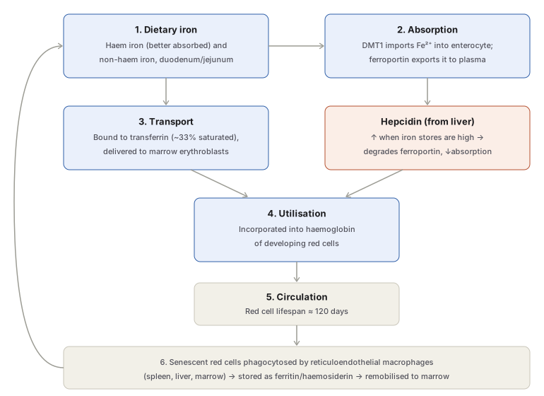
- Losses: about 1 mg/day in an adult male or non-menstruating female (from skin, bowel, genitourinary tract desquamation), with an additional 0.5–1 mg/day lost through menstruation.
- Absorption: occurs mainly in the duodenum and proximal jejunum. Of the 10–15 mg of dietary iron in a typical diet, only 5–10% is normally absorbed, rising to 20–30% in pregnancy or established iron deficiency (the “mucosal block” mechanism — absorption is upregulated when stores are low and downregulated when stores are replete). Hepcidin, released by the liver, is the principal regulatory hormone: it inversely controls ferroportin, the transporter that exports iron from the enterocyte into plasma. Ascorbic acid, citric acid, and gastric acid enhance non-haem iron absorption; antacids, milk, phytates, and tea tannates impair it. Haem iron is absorbed far more efficiently than non-haem iron.
- Transport: bound to transferrin (normally about one-third saturated); erythroid precursors carry transferrin receptors and take up iron for hemoglobin synthesis.
- Distribution: haemoglobin (65% of body iron), myoglobin (3.5%), haem/non-haem enzymes (0.5%), circulating transferrin-bound iron (0.5%), and storage iron as ferritin/haemosiderin in reticuloendothelial cells and hepatocytes (30%).
- Excretion: the body has no regulated excretory mechanism for iron — losses are fixed and independent of intake, which is why absorption is the only lever available to correct deficiency.
Pathogenesis
Iron deficiency develops in three progressive stages: storage iron depletion (iron reserves fall, erythropoiesis unaffected) → iron-deficient erythropoiesis (marrow iron supply falls, still no anaemia) → frank iron deficiency anaemia (red cells become microcytic and hypochromic). It arises from one or more of four mechanisms — increased blood loss, increased requirement, inadequate dietary intake, and decreased absorption — whose relative importance varies by age and sex.
Aetiology by Age and Sex
- Women in reproductive years — the highest-incidence group. Menorrhagia is the leading cause; also repeated pregnancies (each pregnancy siphons off roughly 750 mg of maternal iron — about 400 mg to the fetus, 150 mg to the placenta, 200 mg lost at delivery/lactation), poor dietary intake in lower socioeconomic groups, and blood loss at menarche.
- Post-menopausal women — chronic blood loss now dominates: endometrial carcinoma, or gastrointestinal bleeding (gastric/colonic carcinoma, hiatus hernia).
- Adult males — uncommon without an underlying cause; almost always chronic blood loss, chiefly gastrointestinal (peptic ulcer, haemorrhoids, hookworm infestation, carcinoma of stomach/large bowel, oesophageal varices, chronic NSAID/aspirin use, ulcerative colitis), less often urinary tract (haematuria), nose (epistaxis), or lung (haemoptysis).
- Infants and children — common, peak incidence at 1–2 years, from increased requirement outstripping poor dietary intake. Term infants have iron reserves sufficient for the first 4–6 months; premature infants have inadequate stores (maternal iron is laid down chiefly in the last trimester) and require supplementation earlier.
Clinical Features
Anaemia itself: insidious onset — weakness, fatigue, exertional dyspnoea, palpitations, pallor of skin/mucosae/sclerae; angina and cardiac failure in older patients with severe anaemia; pica (unusual dietary cravings); menorrhagia is both a cause and a symptom in affected women.
Epithelial tissue changes (long-standing deficiency): koilonychia (spoon-shaped nails), atrophic glossitis, angular stomatitis, and Plummer-Vinson syndrome — dysphagia from thin post-cricoid oesophageal webs.
Laboratory Findings
- Blood picture: hypochromic, microcytic red cells with anisocytosis and poikilocytosis; hypochromia (increased central pallor) generally precedes microcytosis. Target cells, elliptocytes, and polychromasia may be seen; normoblasts are uncommon. A dimorphic picture (mixed macrocytic and microcytic-hypochromic populations) occurs when iron deficiency coexists with folate/B12 deficiency. Reticulocyte count is normal or mildly raised. Platelet count is normal, occasionally mildly raised after recent bleeding.
- Red cell indices: MCV, MCH, and MCHC all reduced (MCV <50 fL, MCH <15 pg, MCHC <20 g/dL in significant deficiency).
- Bone marrow (not routinely required, done to exclude other hypochromic anaemias): increased cellularity from erythroid hyperplasia (reduced myeloid:erythroid ratio); micronormoblastic erythropoiesis with ragged cytoplasmic borders, cytoplasmic maturation lagging behind nuclear maturation (the reverse of megaloblastic anaemia); Prussian blue staining shows absent reticuloendothelial iron stores and absent normoblast siderotic granules.
- Biochemistry: serum iron low (often <50 µg/dL; normal 40–140); total iron-binding capacity (TIBC) high with <10% saturation (normal ~33%) — this pattern distinguishes iron deficiency from anaemia of chronic disease, where both serum iron and TIBC are reduced; serum ferritin very low (reflecting depleted tissue stores, and normal/raised in anaemia of chronic disease); red cell protoporphyrin low; serum transferrin receptor protein raised.
Treatment Principles
Two essential components: correcting the underlying cause of blood loss/deficiency, and correcting the iron deficit itself.
- Oral iron (ferrous sulfate, fumarate, gluconate, or polysaccharide-iron preparations) is first-line and highly effective; optimal absorption is on an empty stomach, though food co-administration reduces gastrointestinal side effects at the cost of some absorption. Response is monitored by reticulocytosis, appearing by 3–4 days and peaking around day 10. Poor response suggests incorrect diagnosis, non-compliance, ongoing blood loss, marrow suppression, or malabsorption.
- Parenteral iron is reserved for oral intolerance, malabsorptive GI disease, the post-operative setting, or when rapid replenishment is required (e.g. severe anaemia close to expected delivery).
Clinical Relevance
The serum iron/TIBC/ferritin pattern is the single most exam-relevant discriminator in this topic: iron deficiency shows low iron with high TIBC and low ferritin, while anaemia of chronic disease — the other common cause of a hypochromic picture — shows low iron with low or normal TIBC and normal/raised ferritin. Getting this pairing right is what actually separates the two conditions at the bedside, since both can look identical on a plain blood film.
Diagram 1: The Daily Iron Cycle and Its Regulation
Draw a closed loop of six stages, with the loop-closing arrow running back up the left margin from the last box to the first.
- Dietary iron — haem (better absorbed) and non-haem, duodenum/jejunum.
- Absorption — DMT1 in, ferroportin out, regulated by hepcidin.
- Transport — transferrin-bound, to marrow.
- Utilisation — haemoglobin synthesis in erythroblasts.
- Circulation — red cell lifespan ~120 days.
- Senescence and recycling — RE macrophage phagocytosis, storage as ferritin/haemosiderin, remobilisation back to marrow.
Labels required
- Hepcidin’s regulatory direction must be explicit: high stores → high hepcidin → ferroportin degraded → less absorption. Getting this backwards is the single most common error with this axis.
- The loop must visibly close (arrow from stage 6 back to stage 1’s transport step) — the point of the diagram is that this is a recycling system, not a one-way pathway, and internal recycling (not diet) supplies most of the iron used for erythropoiesis day to day.
Errors commonly made
- Treating dietary absorption as the dominant daily source of marrow iron, when in fact recycled iron from senescent red cell breakdown supplies far more of the marrow’s daily requirement than fresh dietary absorption does.
- Reversing hepcidin’s effect — hepcidin reduces iron absorption/release (by degrading ferroportin), it does not increase it.
- Omitting DMT1 and ferroportin by name and just writing “absorption,” losing the two specific transport proteins that are actually being tested.
Long Essays (2)
My Sticky Notes
Personal revision notes, mnemonics and reminders.
