
Invasion and Metastasis
Local invasion
Benign = encapsulated, pushes aside, no invasion. Malignant = invades/infiltrates/destroys (some well-diff. e.g. follicular thyroid Ca partially encapsulated too). Route of least resistance → eventually no boundary. Invades thin-walled veins/capillaries >> thick arteries. Resistant tissues: dense collagen, elastic tissue, cartilage.
Metastasis
Definition: discontinuous 2° tumour mass at distant site. Distinguishes malignant (nearly all can metastasise, exceptions: CNS glioma, BCC skin) from benign (never). 1/3 evident mets at presentation, +20% occult. Larger/aggressive/faster-growing → more likely (not absolute rule).
Routes
1. Lymphatic (carcinomas favour): permeation (continuous growth in channel) vs emboli (detach→ subcapsular sinus of next node). Regional node involvement (breast→axillary, thyroid→cervical, bronchogenic→hilar/paratracheal) — but not all node enlargement = mets (reactive sinus histiocytosis too). Skip mets (venous-lymphatic anastomoses/lymphatic obliteration). Retrograde mets (flow-obstruction, e.g. prostate→supraclavicular). Virchow’s node = L supraclavicular from abdominal Ca (stomach/colon/gallbladder).
2. Haematogenous (sarcomas favour, also lung/breast/thyroid/kidney/liver/prostate/ovary carcinomas): seed-soil theory (Ewing/Paget) → favourable soil: liver, lung, brain, bone, kidney, adrenal. Unfavourable: spleen (open sinusoids), heart, skeletal muscle. Drainage pattern: systemic vein sites(limb/head-neck/pelvis)→lung; portal vein organs(bowel/spleen/pancreas)→liver; pulmonary vein→systemic. Arterial spread rare (thick/elastic) but via pulmonary capillary/artery: kidney/adrenal/bone/limb/uterus→lung. Retrograde: vertebral mets from thyroid/prostate. Gross: multiple round nodules, variable size, may exceed 1° size.
3. Body cavities/natural passages (uncommon):
- Transcoelomic: gastric Ca→both ovaries (Krukenberg); ovarian Ca→whole peritoneum; pseudomyxoma peritonei (mucinous ovarian/appendiceal); bronchial/breast→pleura/pericardium.
- Epithelium-lined surfaces (rare): endometrium↔ovary(tube), bronchus→alveoli, kidney→ureter→lower UT.
- CSF: ependymoma/leptomeningeal→other CNS sites.
- Implantation: surgical instruments; lower lip Ca→upper lip (rare).
Mechanism/biology (metastatic cascade)
Breach ECM at 3 levels: tumour BM → interstitial CT → microvascular BM.
- Clonal proliferation + angiogenesis — tumour heterogeneity → metastasis-capable subclone; new vessels most invasion-prone.
- Tumour cell loosening (EMT) — loss/inactivation of E-cadherin (binds β-catenin normally) via mutation, β-catenin activation, or SNAIL/TWIST transcriptional repression → epithelial→mesenchymal shape shift, ↑vimentin (RK detail).
- Tumour cell-ECM attachment — bind laminin+fibronectin via receptors; ↓integrins.
- ECM degradation — ↑MMPs (collagenase, MMP-9 gelatinase→cleaves type IV collagen, releases VEGF), ↑cathepsin D, ↓MMP inhibitors.
- Intravasation/locomotion — AMF (autocrine motility factor) + matrix cleavage chemotactic products + HGF/scatter factor (↑at invasive edge of glioblastoma).
- Thrombus formation — platelet coat nourishes+shields from immune attack; <0.1% circulating tumour cells survive (liquid-biopsy studies confirm massive daily shedding — RK).
- Extravasation — adhesion to distant endothelium, reverse of invasion sequence.
- Survival/growth of deposit — PDGF/FGF/TGF-β/VEGF; seed-soil compatibility; tumour dormancy possible (melanoma, breast, prostate) before outgrowth; may further metastasise.
Deterministic (early intrinsic mutation set) vs probabilistic (chance×cell number×time) models of “why metastasise” — not mutually exclusive, unresolved (RK).
Prognostic indicators
Clinical: size, grade, nodal status, vascular invasion. Molecular: C-met oncogene, CD44, oestrogen receptor, EGFR, angiogenesis factors, MAGNA expression.
Clinical relevance
Virchow’s node + predictable drainage patterns → locate primary on exam. <0.1% survival + dormancy → explains liquid biopsy rationale + late recurrence years after “cure.” E-cadherin/SNAIL/TWIST loss (IHC) = invasive-potential marker. MMP-9 malignant-vs-benign distinction = rationale (limited success) for protease-inhibitor anti-metastatic therapy.
Local Invasion
Benign tumours form encapsulated or circumscribed masses that expand and push aside surrounding tissue without invading, infiltrating, or metastasising.
Malignant tumours also enlarge by expansion, and some well-differentiated cancers may be partially encapsulated (e.g. follicular carcinoma of thyroid), but are characteristically distinguished by invasion, infiltration, and destruction of surrounding tissue, besides distant spread. Tumours generally follow the route of least resistance, though eventually recognise no anatomic boundary — extending through tissue spaces, permeating lymphatics, blood vessels, and perineural spaces, and occasionally penetrating bone via nutrient foramina. Thin-walled capillaries and veins are invaded far more readily than thick-walled, elastic-rich arteries; dense compact collagen, elastic tissue, and cartilage resist invasion.
Metastasis
Metastasis (meta = transformation, stasis = residence) is spread by invasion producing discontinuous secondary tumour mass(es) at the site of lodgement. Along with anaplasia and invasiveness, it is one of the defining features separating malignant from benign tumours — benign tumours never metastasise, while nearly all malignant tumours can, with rare exceptions (CNS gliomas, basal cell carcinoma of skin). About a third of malignant tumours have evident metastases at presentation, with a further ~20% harbouring occult metastasis. Larger, more aggressive, faster-growing tumours are generally more likely to metastasise, though exceptions exist — tumour size and type alone cannot reliably predict individual tumour behaviour.
Routes of Metastasis
1. Lymphatic spread — the dominant route for carcinomas (sarcomas favour the haematogenous route, though some also spread via lymphatics). Occurs by lymphatic permeation (continuous tumour growth within lymphatic channels) or lymphatic emboli (detached tumour cells carried to the next draining node, lodging first in the subcapsular sinus). Regional nodes draining the tumour are typically involved (breast → axillary nodes; thyroid → lateral cervical nodes; bronchogenic carcinoma → hilar/paratracheal nodes) — though not every enlarged regional node reflects metastasis, since necrotic tumour products/antigens can also incite reactive sinus histiocytosis. Skip metastasis (bypassing the nearest node, due to venous-lymphatic anastomoses or lymphatic obliteration) and retrograde metastasis (against lymph flow after lymphatic obstruction — e.g. prostate cancer to supraclavicular nodes) both occur. Virchow’s node — metastasis specifically to the left supraclavicular node from abdominal organ cancers (stomach, colon, gallbladder) — is a classic clinical sign. Regional nodes play a dual role: initially a barrier/filter that may destroy tumour cells, later providing fertile soil for tumour growth.
2. Haematogenous spread — the common route for sarcomas, but also frequent for carcinomas of lung, breast, thyroid, kidney, liver, prostate, and ovary. Common metastatic sites — liver, lungs, brain, bones, kidney, adrenals — provide “good soil” for “good seeds” (the seed-soil theory, Ewing and Paget). Spleen, heart, and skeletal muscle are generally resistant (spleen’s open sinusoidal architecture doesn’t retain tumour cells long enough to seed). Drainage anatomy predicts spread pattern: systemic-vein-drained sites (limbs, head/neck, pelvis) → lungs; portal-vein-drained organs (bowel, spleen, pancreas) → liver; pulmonary-vein blood carrying lung cancer cells → systemic circulation → distant secondaries. Arterial spread is less common (thick, elastic, resistant walls) but occurs via thin-walled pulmonary capillaries/arterial branches — kidney, adrenal, bone, limb, and uterine cancers reach the lungs this way. Retrograde venous spread also occurs (e.g. vertebral metastases from thyroid/prostate cancer). Grossly, blood-borne metastases appear as multiple, rounded nodules of varying size scattered through the organ, sometimes exceeding the primary tumour in size; metastatic deposits may occasionally present before any evident primary is found.
3. Spread along body cavities and natural passages (uncommon):
- Transcoelomic spread — tumour fragments break off through the serosal wall into coelomic fluid and implant elsewhere in the cavity (peritoneal cavity most often, occasionally pleural/pericardial). Classic examples: gastric carcinoma seeding both ovaries (Krukenberg tumour); ovarian carcinoma spreading throughout the peritoneal cavity without infiltrating underlying organs; pseudomyxoma peritonei (gelatinous peritoneal coating from mucin-secreting ovarian/appendiceal carcinoma); bronchial/breast carcinoma seeding pleura/pericardium.
- Spread along epithelium-lined surfaces — unusual, since intact epithelium and mucus resist penetration; exceptions include endometrium↔ovary via fallopian tube, bronchus into alveoli, kidney into lower urinary tract via ureter.
- Spread via cerebrospinal fluid — ependymal/leptomeningeal tumours releasing cells into CSF, seeding other CNS sites.
- Implantation — rare, via surgical instruments or prolonged direct contact (e.g. lower-lip cancer implanting onto the apposing upper lip).
Mechanism and Biology of Invasion and Metastasis
Local invasion and lymphovascular spread both require the tumour to breach extracellular matrix (ECM) barriers at three levels — the tumour’s own basement membrane, the interstitial connective tissue, and the microvascular basement membrane — through the following sequential steps:
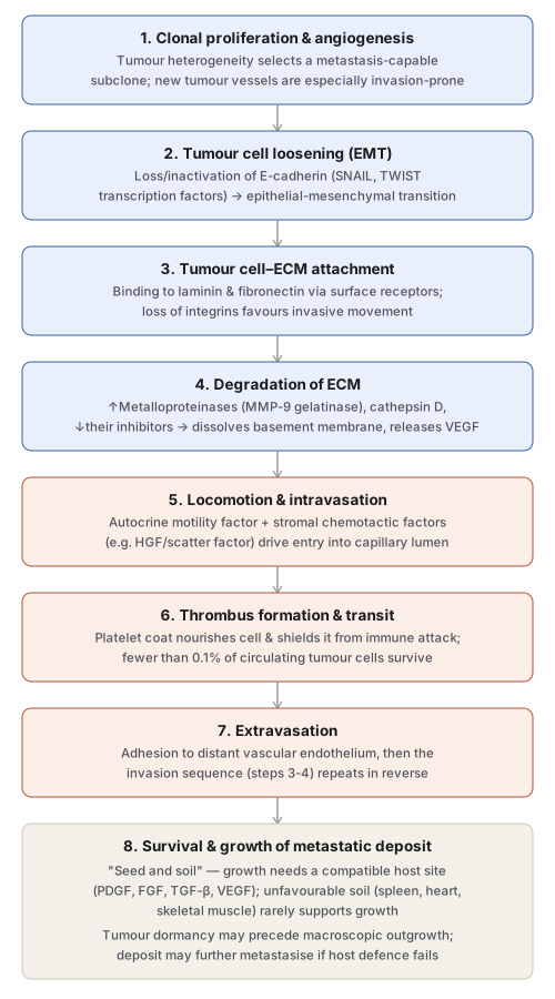
- Aggressive clonal proliferation and angiogenesis — tumour heterogeneity means only a subpopulation (subclone) has the right biologic properties to complete metastasis; newly formed tumour vessels are especially vulnerable to invasion since they are in direct contact with cancer cells.
- Tumour cell loosening (epithelial-mesenchymal transition, EMT) — normal cells stay adherent via cell adhesion molecules, chiefly E-cadherin, whose cytoplasmic tail binds β-catenin. In epithelial cancers, E-cadherin is lost or inactivated (mutational inactivation, β-catenin gene activation, or inappropriate expression of the transcriptional repressors SNAIL and TWIST), loosening cell-cell adhesion and driving a phenotypic shift from polygonal epithelial cells toward a spindly, migratory mesenchymal shape with upregulated mesenchymal markers (e.g. vimentin).
- Tumour cell–ECM interaction — loosened cells attach to ECM proteins laminin and fibronectin via surface receptors; concurrent loss of integrins (transmembrane receptors that normally maintain polarised, differentiated resting state) further favours invasion.
- Degradation of ECM — tumour cells overexpress proteases and matrix-degrading enzymes — matrix metalloproteinases (e.g. collagenases, MMP-9 gelatinase which cleaves type IV collagen) and cathepsin D — while metalloproteinase inhibitor levels fall, tilting the balance toward degradation. This dissolves the tumour’s own basement membrane, then the interstitial matrix, then the vessel wall basement membrane. MMPs also release ECM-sequestered growth factors such as VEGF, adding chemotactic, angiogenic, and growth-promoting effects.
- Entry into capillary lumen (intravasation) — driven by autocrine motility factor (AMF), a tumour-cell-derived cytokine stimulating receptor-mediated motility, and by chemotactic/growth-promoting cleavage products of degraded matrix. Stromal-cell-derived cytokines such as hepatocyte growth factor/scatter factor (HGF/SCF) also potentiate migration — HGF/SCF levels are notably elevated at the advancing edge of highly invasive tumours such as glioblastoma.
- Thrombus formation — tumour cells protruding into the capillary lumen become coated by circulating blood constituents (chiefly platelets), forming a thrombus that nourishes them and shields them from immune attack. This protection matters enormously: although huge numbers of tumour cells enter the circulation daily (recent “liquid biopsy” studies confirm this), fewer than 0.1% survive to establish a metastasis — most are destroyed by host immune cells.
- Extravasation — circulating tumour cells mechanically lodge in and adhere to distant vascular endothelium, then extravasate into extravascular tissue, repeating the same ECM-breaching sequence as local invasion, now in reverse.
- Survival and growth of the metastatic deposit — extravasated cells grow under the influence of growth factors from host tissue, the tumour itself, and matrix cleavage products (PDGF, FGF, TGF-β, VEGF), continuing only if host immune defence fails to eliminate them. Growth is governed by seed-soil compatibility — spleen, heart, and skeletal muscle rarely support metastatic growth despite receiving blood flow, while liver, lung, brain, bone, kidney, and adrenal provide favourable “soil.” Even after successful extravasation, metastatic cells may enter a prolonged non-progressing state — tumour dormancy (well described in melanoma, breast, and prostate carcinoma) — representing a possible last line of host defence before macroscopic outgrowth. Metastatic deposits may themselves further metastasise, to the same organ or elsewhere.
Why some tumours metastasise and others don’t remains incompletely answered — competing “deterministic” (metastatic potential is an early, intrinsic tumour property) and “probabilistic” (a matter of chance × tumour cell number × time) models are not mutually exclusive and remain under study.
Prognostic Indicators
Metastasis is a genetically programmed, stepwise phenomenon involving only a tumour subpopulation, correlating with:
- Clinical prognostic markers: tumour size, histologic grade, nodal involvement, vascular invasion.
- Molecular prognostic markers: oncogene expression (C-met), CD44, oestrogen receptor status, epidermal growth factor receptor, angiogenesis factors/degree of neovascularisation, expression of metastasis-associated gene/nucleic acid (MAGNA).
Clinical Relevance
- Virchow’s node and the seed-soil predictable spread patterns (breast→axillary, thyroid→cervical, bronchogenic→hilar) are direct clinical shortcuts to locating a primary tumour or staging spread on examination.
- Recognising that fewer than 0.1% of circulating tumour cells actually establish metastasis, and that tumour dormancy can persist for years, explains both the biological basis of “liquid biopsy” surveillance and the clinical phenomenon of late metastatic recurrence long after apparently curative primary treatment.
- The EMT/E-cadherin-SNAIL-TWIST axis is the molecular explanation for why poorly differentiated, E-cadherin-low tumours behave more aggressively — directly relevant to interpreting immunohistochemistry (E-cadherin loss) as a marker of invasive potential.
- MMP-9 overexpression distinguishing malignant from benign counterparts in breast, colon, and stomach tumours is the rationale behind exploring protease-inhibitor-based anti-metastatic therapy, even though clinical success with this approach has been limited.
Diagram 1: The Metastatic Cascade
Draw a single downward column of eight sequential stages, grouped by shading into three phases: local invasion/ECM breach (steps 1-4, blue), vascular transit (steps 5-7, coral), and colonisation (step 8, neutral).
- Clonal proliferation and angiogenesis — subclone selection from tumour heterogeneity.
- Tumour cell loosening (EMT) — E-cadherin loss via SNAIL/TWIST.
- Tumour cell–ECM attachment — laminin/fibronectin binding, integrin loss.
- ECM degradation — MMPs (MMP-9), cathepsin D, VEGF release.
- Locomotion and intravasation — AMF, HGF/scatter factor.
- Thrombus formation and transit — platelet coat, immune evasion, <0.1% survival.
- Extravasation — reverse of the invasion sequence at the distant site.
- Survival and growth of the metastatic deposit — seed-soil compatibility, possible dormancy.
Labels required
- Step 2 must name E-cadherin as the specific adhesion molecule lost, and SNAIL/TWIST as the transcription factors responsible — this is the molecular detail examiners target to separate rote learners from those who understand EMT.
- Step 6 must carry the “<0.1% survive” figure — it is the number that explains why metastasis is such an inefficient process despite massive daily shedding of tumour cells into circulation.
- Step 8 must explicitly name the unfavourable sites (spleen, heart, skeletal muscle) alongside the favourable ones, since “seed-soil theory” is frequently tested by asking why a specific organ is spared.
Errors commonly made
- Treating steps 3 and 4 (ECM attachment and ECM degradation) as a single step — they involve different molecules (integrins/laminin/fibronectin vs MMPs/cathepsin D) and are frequently tested as separate sequential events.
- Assuming a favourable metastatic site is simply “wherever blood flow is highest” rather than seed-soil compatibility — the spleen receives substantial blood flow yet is a classically unfavourable site, precisely because of its open sinusoidal architecture rather than perfusion.
- Forgetting that extravasation (step 7) is not a distinct new mechanism but a mirror-image repeat of the invasion machinery (steps 3-4) at the distant site.
Short Essays (2)
My Sticky Notes
Personal revision notes, mnemonics and reminders.
