
Hyperaemia and Congestion
Definition
Both = ↑blood volume in dilated vessels of organ/tissue. Hyperaemia = ↑arterial inflow (active). Congestion = ↓venous outflow (passive). Acute or chronic.
Active hyperaemia
Arteriolar/capillary dilatation (sympathetic/vasoactive substances) → pink/red, warm. Examples: inflammation, blushing, menopausal flush, exercise, fever, goitre, AV malformations.
Passive hyperaemia (venous congestion)
Impaired venous drainage → bluish/cyanotic. Usually chronic = CVC.
- Local — portal obstruction (cirrhosis), external compression (bandage/tumour/pregnancy), thrombosis
- Systemic — heart failure or pulmonary disease. Side of heart failure determines site: LEFT HF → CVC LUNG (pulmonary venous). RIGHT HF → CVC systemic organs (liver/spleen/kidney). Congestion accumulates UPSTREAM of failing chamber.
CVC morphology by organ
Lung (L heart failure, e.g. mitral stenosis): gross = heavy, firm, brown induration. Micro = widened septa (oedema+congestion+fibrosis), capillary rupture → intra-alveolar haemorrhage → “heart failure cells” (haemosiderin macrophages).
Liver (R heart failure / IVC-hepatic vein occlusion): gross = enlarged, tender, tense capsule, nutmeg mottling (red centrilobular + yellow peripheral). Micro = worst at centrilobular zone 3 (farthest from blood supply) — distended central vein/sinusoids → hepatocyte degeneration → centrilobular haemorrhagic necrosis; long-standing → centrilobular fibrosis + regeneration = cardiac cirrhosis. Periportal zone (less hypoxic) = fatty change only.
Spleen (R heart failure / portal HTN): gross = congestive splenomegaly (150g→250g early, →500-1000g chronic), congested/tense/cyanotic, grey-tan. Micro = red pulp sinusoidal dilatation + haemorrhage (capillarisation of sinusoids), RE hyperplasia, capsular/trabecular fibrosis, Gamna-Gandy bodies (siderofibrotic nodules = organised old haemorrhage + haemosiderin + calcium). Cause of hypersplenism.
Kidney: mild — slight enlargement, congested medulla, tubular cloudy swelling/fatty change, mesangial proliferation.
Haemorrhage (related terms)
Escape of blood from vessel. Terms by extent: haematoma (tissue extravasation+swelling) · ecchymoses (large skin/mucosal) · purpura (≤1cm) · petechiae (pinhead) · diapedesis (microscopic RBC escape, follows marked congestion).
Causes: trauma · spontaneous (aneurysm rupture, septicaemia, bleeding diathesis, leukaemia, pernicious anaemia, scurvy) · inflammatory vessel erosion (peptic ulcer, TB cavity, syphilitic aortitis, PAN) · neoplastic invasion · atherosclerosis · raised pressure (HTN, varices).
Effects depend on: amount + speed + site. ~20% loss well tolerated; sudden 33% may be fatal; 50% over 24h may not be.
Clinical relevance
L HF→lung, R HF→liver/spleen/kidney = direct testable mapping (matches RHD/JVD/hepatosplenomegaly vignettes). Nutmeg liver→cardiac cirrhosis = mechanism of eventual real hepatic dysfunction from chronic RHF. Gamna-Gandy bodies = marker of LONGSTANDING congestive splenomegaly/portal HTN. Diapedesis from congestion alone (not a clotting disorder) explains petechiae with normal coagulation.
Definition
Hyperaemia and congestion both describe a localised increase in the volume of blood within the dilated vessels of an organ or tissue, but they differ fundamentally in mechanism: hyperaemia (active hyperaemia) results from increased arterial/arteriolar inflow, whereas congestion (passive hyperaemia) results from impaired venous outflow. Both may be acute (rapid onset) or chronic (prolonged, gradual).
Active Hyperaemia
Arteriolar and capillary dilatation, mediated by sympathetic neurogenic mechanisms or vasoactive substances, increases blood inflow; the affected tissue appears pink or red (erythema) and is warm to the touch. Examples: inflammation (e.g. congested alveolar wall vessels in pneumonia), blushing (emotional flushing of facial skin), menopausal flush, muscular exercise, high-grade fever, goitre, and arteriovenous malformations.
Passive Hyperaemia (Venous Congestion)
Impaired venous drainage causes venous and capillary dilatation; the affected tissue appears bluish (cyanotic) from accumulated deoxygenated venous blood. More often chronic than acute — chronic venous congestion (CVC) — and classified by extent:
- Local venous congestion — obstruction confined to one organ/region: portal venous obstruction in liver cirrhosis, external compression (tight bandage, plaster cast, tumour, pregnancy, hernia), or intraluminal thrombosis.
- Systemic (general) venous congestion — from heart failure or pulmonary disease impairing pulmonary blood flow (fibrosis, emphysema). The site of congestion follows directly from which side of the heart fails: left heart failure (e.g. aortic stenosis, myocardial infarction weakening the LV) causes pulmonary venous congestion (CVC lung); right heart failure (e.g. pulmonary stenosis, pulmonary hypertension) causes systemic venous congestion (CVC of liver, spleen, kidney and other organs). Congestion always accumulates upstream of the failing chamber.
Morphology of Chronic Venous Congestion by Organ
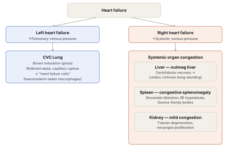
CVC Lung (left heart failure, e.g. rheumatic mitral stenosis — raised pulmonary venous pressure): Grossly, lungs are heavy, firm, with a dark rusty-brown cut surface — brown induration. Microscopically: widened alveolar septa (interstitial oedema, dilated congested capillaries, mild fibrosis); rupture of congested capillaries causes minute intra-alveolar haemorrhage, with haemosiderin from breakdown erythrocytes taken up by alveolar macrophages — “heart failure cells” — whose pigment plus the fibrosis together produce the brown induration.
CVC Liver (right heart failure, or IVC/hepatic vein occlusion): Grossly, liver enlarged, tender, tense capsule; cut surface shows the characteristic nutmeg mottling — red (congested centrilobular) alternating with yellow (fatty peripheral) zones. Microscopically, changes are worst in the centrilobular zone (zone 3) — farthest from the periportal blood supply and thus most hypoxic: distended central veins/sinusoids, centrilobular hepatocyte degeneration progressing to centrilobular haemorrhagic necrosis; long-standing cases develop centrilobular fibrosis with hepatocyte regeneration — cardiac cirrhosis. The less-hypoxic peripheral (periportal) zone instead shows fatty change.
CVC Spleen (right heart failure, or portal hypertension from cirrhosis): Grossly, moderate splenomegaly early (up to 250 g vs normal 150 g), progressing to marked enlargement (500–1000 g) in long-standing cases — congestive splenomegaly, a leading cause of hypersplenism. Deeply congested, tense, cyanotic; grey-tan cut surface. Microscopically: enlarged, sinusoid-dilated red pulp with recent/old haemorrhage (sinusoids may undergo “capillarisation”); reticuloendothelial (splenic macrophage) hyperplasia; fibrous thickening of capsule/trabeculae; and Gamna–Gandy bodies (siderofibrotic nodules) — organised foci of old haemorrhage encrusted with haemosiderin and calcium.
CVC Kidney: mild changes — slight enlargement, congested medulla; tubular cloudy swelling/fatty change, mesangial proliferation in glomeruli.
Haemorrhage (Related Concept)
Haemorrhage is escape of blood from a vessel — external, or internal into serous cavities (haemothorax, haemoperitoneum, haemopericardium) or a hollow viscus. Terminology by extent/site: haematoma (extravasation into tissue with swelling), ecchymoses (large skin/mucosal extravasation), purpura (haemorrhages up to 1 cm), petechiae (pinhead-sized), and diapedesis (microscopic RBC escape through vessel walls, typically following marked congestion — the direct conceptual link back to this topic).
Causes: trauma; spontaneous rupture (aneurysm, septicaemia, bleeding diathesis, leukaemia, pernicious anaemia, scurvy); inflammatory vessel wall erosion (peptic/typhoid ulcer, tuberculous cavity, syphilitic aortitis, polyarteritis nodosa); neoplastic vascular invasion; vascular disease (atherosclerosis); and raised intravascular pressure (hypertensive cerebral/retinal haemorrhage, variceal bleeding). Clinical effect depends on amount, speed, and site of blood loss — up to 20% loss is generally well tolerated by compensatory mechanisms, a sudden 33% loss may be fatal, while an equivalent 50% loss over 24 hours may not be.
Clinical Relevance
- Predicting which organs will show CVC purely from knowing which side of the heart has failed (left → lung; right → liver/spleen/kidney) is a direct, testable application of the “congestion accumulates upstream of the failing chamber” principle, and is exactly what Decipher’s right-heart-failure clinical vignettes (distended jugular veins, hepatosplenomegaly) are testing.
- Nutmeg liver progressing to cardiac cirrhosis is the pathologic explanation for why long-standing, poorly controlled right heart failure can eventually produce genuine hepatic synthetic dysfunction, not just transient hepatic congestion.
- Gamna–Gandy bodies are a specific, recognisable marker of longstanding congestive splenomegaly (chronic organised haemorrhage), useful for dating the duration of portal hypertension or chronic right heart failure on a spleen specimen.
- Recognising diapedesis as a consequence of marked congestion (rather than a primary bleeding disorder) helps explain why petechial haemorrhage can appear in severely congested tissue even when clotting function is entirely normal.
Diagram 1: Chronic Venous Congestion by Side of Heart Failure
Draw a top “heart failure” box branching into two columns, left heart failure and right heart failure, each leading down to its characteristic organ findings.
- Left heart failure → CVC lung: brown induration grossly; widened septa and heart failure cells microscopically.
- Right heart failure → systemic organ congestion, itself containing three grouped boxes: liver (nutmeg liver → cardiac cirrhosis), spleen (congestive splenomegaly, Gamna-Gandy bodies), kidney (mild changes only).
Labels required
- The branch point must be labelled with which side of the heart fails, since this single fact is what determines every downstream finding — the diagram exists to test that mapping, not the organ findings in isolation.
- “Heart failure cells” must be labelled specifically as haemosiderin-laden alveolar macrophages, not left as an unexplained name.
- Nutmeg liver’s mottling must be labelled with both colours and their cause (red = centrilobular congestion, yellow = peripheral fatty change), since the two-tone pattern is the actual diagnostic feature on gross inspection.
Errors commonly made
- Reversing the mapping — assigning liver/spleen congestion to left heart failure or lung congestion to right heart failure, rather than remembering that congestion is always upstream of the failing chamber.
- Describing nutmeg liver as uniformly congested rather than showing the alternating centrilobular (congested/necrotic) and peripheral (fatty) pattern that gives it its name and its gross mottled appearance.
Multiple Choice Questions (1)
My Sticky Notes
Personal revision notes, mnemonics and reminders.
