
Hereditary Spherocytosis
Overview
Autosomal dominant haemolytic anaemia. Red cell membrane defect → rigid spherical cells (microspherocytes) → trapped and destroyed in spleen.
Pathogenesis
Defect in membrane-anchoring proteins:
- Spectrin deficiency — almost all cases; severity matches deficiency degree. Recessive α-spectrin = severe. Dominant β-spectrin = mild.
- Ankyrin defect — ~half of cases. Homozygous recessive = severe. Heterozygous dominant (more common) = mild.
Unstable lipid bilayer → progressive membrane loss in circulation → same Hb volume in smaller surface → spherical, rigid microspherocyte (not flexible biconcave disc) → can’t pass splenic cords → more membrane lost each pass → phagocytosed by splenic macrophages.
Clinical features
Any age, equal sex ratio, often +ve family history.
- Anaemia — mild to moderate
- Splenomegaly — constant
- Jaundice — ↑unconjugated bilirubin (congenital haemolytic jaundice)
- Pigment gallstones — frequent
Splenectomy = only reliable treatment (doesn’t fix membrane defect, but removes site of destruction).
Lab findings
Reticulocytosis 5-20%. Blood film: microspherocytes (no central pallor). MCV normal/↓slightly, MCHC ↑. Osmotic fragility test: ↑ (lyse easily in hypotonic saline). Autohaemolysis test: ↑ (10-15% vs <4% normal), corrected by glucose. Direct Coombs’ test: NEGATIVE — key test to separate from acquired (AIHA) spherocytosis, where Coombs’ is POSITIVE.
Related disorders
Hereditary elliptocytosis — AD, spectrin or protein 4.1 defect, milder, oval cells. Hereditary stomatocytosis — AD, stomatin defect, overhydrated cells with mouth-like slit, mild anaemia+splenomegaly.
Clinical relevance
Negative Coombs’ test is the key discriminator: spherocytes + splenomegaly + jaundice could be hereditary spherocytosis (Coombs’ negative, treat with splenectomy) or autoimmune haemolytic anaemia (Coombs’ positive, treat with immunosuppression) — same picture, opposite management, one test tells them apart.
Overview
Hereditary spherocytosis is a common autosomal dominant haemolytic anaemia caused by an inherited defect in the red cell membrane, producing rigid, spherical red cells (microspherocytes) that are prematurely trapped and destroyed in the spleen.
Pathogenesis
The molecular defect involves proteins that anchor the lipid bilayer to the underlying cytoskeleton:
- Spectrin deficiency — present in almost all cases, and the degree of deficiency correlates with anaemia severity. Recessive α-spectrin mutations cause more severe disease; dominant β-spectrin mutations cause milder disease.
- Ankyrin abnormality — present in about half of cases (ankyrin binds spectrin to band 3 protein). Homozygous recessive disease is severe; the more common heterozygous dominant pattern is milder.
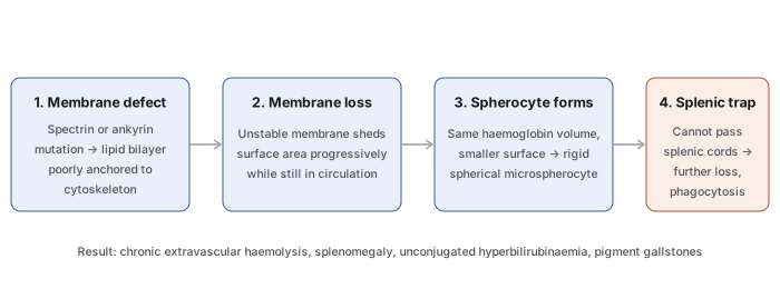
The defective anchoring destabilises the lipid bilayer. Red cells released into circulation with normal volume progressively lose membrane surface area, and since they must still contain the same haemoglobin volume within a smaller surface, they assume a spherical, poorly deformable shape — the microspherocyte — rather than the normal flexible biconcave disc. These rigid cells cannot pass easily through the splenic cords; each passage strips further membrane, producing an increasingly hyperspheroidal subpopulation that is ultimately phagocytosed by splenic macrophages.
Clinical Features
Onset can occur at any age from infancy to old age, with equal sex incidence and often a positive family history.
- Anaemia — usually mild to moderate.
- Splenomegaly — a constant feature.
- Jaundice — from raised unconjugated bilirubin (“congenital haemolytic jaundice”).
- Pigment gallstones — frequent, from chronically increased bile pigment production.
Splenectomy is the only reliable treatment, since it removes the site of red cell destruction — the intrinsic membrane defect itself is not corrected, but the anaemia and jaundice resolve because the spherocytes are no longer being trapped and destroyed.
Laboratory Findings
- Mild-to-moderate anaemia with reticulocytosis (5–20%).
- Blood film: characteristic microspherocytes — small, densely staining red cells lacking the normal central pallor.
- MCV normal or slightly reduced; MCHC increased (from the relative dehydration/loss of membrane relative to haemoglobin content).
- Osmotic fragility test: increased — spherocytes lyse more readily than normal biconcave cells in hypotonic saline, since they have no reserve surface area to accommodate osmotic swelling.
- Autohaemolysis test: increased spontaneous lysis after incubation (10–15% vs <4% in normal cells), correctable by adding glucose.
- Direct Coombs’ test: negative — this is the key test distinguishing hereditary spherocytosis from acquired (autoimmune) spherocytosis, in which the Coombs’ test is positive. Spherocytes can also appear after red cell transfusion.
Related Membrane Disorders
- Hereditary elliptocytosis (ovalocytosis) — also autosomal dominant, involving spectrin or the linker protein 4.1 (which connects spectrin to actin); clinically milder than hereditary spherocytosis, with oval/elliptical rather than spherical red cells. Elliptocytosis can also be acquired, in iron deficiency and myeloproliferative disorders.
- Hereditary stomatocytosis — autosomal dominant defect in the membrane protein stomatin, causing overhydrated red cells (increased Na⁺/K⁺ permeability) with a central mouth-like slit; mild anaemia and splenomegaly.
Clinical Relevance
The negative direct Coombs’ test is the single most decision-relevant laboratory finding in this topic: a spherocytic blood film with splenomegaly and jaundice could equally represent hereditary spherocytosis or autoimmune haemolytic anaemia, and the Coombs’ test — positive only in the acquired, antibody-mediated form — is what actually separates a condition treated by splenectomy from one treated by immunosuppression.
Diagram 1: Pathogenesis of Spherocyte Formation
Draw a single horizontal sequence of four stages, ending in a short results line beneath.
- Membrane defect — spectrin or ankyrin mutation, poor anchoring of lipid bilayer to cytoskeleton.
- Membrane loss — unstable membrane sheds surface area progressively in circulation.
- Spherocyte forms — same haemoglobin volume, smaller surface → rigid microspherocyte.
- Splenic trap — cannot pass splenic cords, further membrane loss, phagocytosis.
Results line: chronic extravascular haemolysis, splenomegaly, unconjugated hyperbilirubinaemia, pigment gallstones.
Labels required
- Both membrane proteins (spectrin, ankyrin) must be named at step 1 — the diagram should not just say “membrane defect” generically.
- Step 3 must make explicit why the cell becomes spherical — same volume, less surface area — since “loses membrane → becomes round” without the volume/surface-area reasoning is the part students most often can’t reconstruct on demand.
Errors commonly made
- Describing the spherocyte as an intrinsically abnormal shape from the mutation itself, rather than a secondary consequence of progressive membrane loss acting on a cell that started with a normal shape.
- Omitting that the spleen is not just where destruction happens but also actively contributes to further injury (each splenic passage strips more membrane) — it is both the site of final destruction and a participant in worsening the defect.
Multiple Choice Questions (2)
My Sticky Notes
Personal revision notes, mnemonics and reminders.
