
Haemophilia
Overview
Two X-linked recessive bleeding disorders, clinically look the same:
- Haemophilia A — factor VIII deficiency. ~80% of cases.
- Haemophilia B (Christmas disease) — factor IX deficiency. ~20% of cases, incidence 1 in 100,000 male births.
Name “haemophilia” given by Schönlein in 1839. Factor IX deficiency recognised as a separate disease only in 1952 (named after the first patient, “Christmas”).
Inheritance
X-linked recessive. Males affected, females usually carriers.
- Carrier mother × unaffected father → each son 50% chance affected; each daughter 50% chance carrier.
- Haemophilic father × unaffected mother → NO son affected (sons get his Y, not X); ALL daughters become carriers (obligate carriers).
- True female haemophilia is rare — needs homozygous state, usually from consanguinity (e.g. daughter of haemophilic father + carrier mother).
- ~30% of cases are new spontaneous mutations, no family history needed.
Factor VIII–vWF complex (background)
Factor VIII made in liver cells. Acts as cofactor for activated factor IX, which activates factor X (intrinsic pathway). Factor VIII circulates bound to von Willebrand factor (vWF, made by endothelium, ~99% of complex mass). vWF binding stabilises factor VIII — so vWF deficiency (von Willebrand disease) also lowers factor VIII secondarily. Normal haemostasis needs only ~25% factor VIII activity; most symptomatic patients have <5%.
Haemophilia A
Gene: F8. Many different mutation types → wide range of severity. ~10% of patients: normal factor VIII level but poor function (functional mutation).
Clinical features: bleeding hours-to-days after injury, severity matches factor VIII level. Classic: recurrent painful haemarthrosis, muscle haematoma, sometimes haematuria. Rare but most feared: spontaneous intracranial or oropharyngeal bleed.
Lab findings: whole blood clotting time ↑ (only severe cases) | PT normal | APTT ↑ (typical finding) | specific factor VIII assay ↓ confirms diagnosis. Carrier females: ~half-normal factor VIII activity. If PTT does not correct on mixing with normal plasma → suspect factor VIII inhibitor (antibody against replacement factor VIII) — happens in ~15% of severe cases.
Treatment: factor VIII concentrate or cryoprecipitate. Brought life expectancy close to normal, though transfusion-transmitted HIV was a historic setback (now prevented by screening). New option for inhibitor cases: bispecific antibody linking factor IX to factor X, bypasses need for factor VIII.
Haemophilia B (Christmas disease)
Gene: F9. Same clinical picture and inheritance as haemophilia A, but needs its own lab test (factor IX assay) — treatment differs, so mixing up A and B matters.
Treatment: fresh frozen plasma or factor IX concentrate/recombinant factor IX. Risks: hepatitis, chronic liver disease, infection (like any plasma product) plus a specific risk — replacement can trigger thrombosis.
Clinical relevance
30% new-mutation rate means “no family history” does NOT rule out haemophilia — go by clinical picture and factor assay, not pedigree alone. A and B look identical clinically but need different treatment — specific factor assay is essential, not optional. Factor VIII inhibitor (PTT not correcting on mixing study) is a major treatment-changing finding — standard factor VIII replacement stops working, need bypass therapy instead.
Overview
Haemophilia refers to two clinically indistinguishable, X-linked recessive inherited coagulation disorders — haemophilia A (deficiency of factor VIII) and haemophilia B (deficiency of factor IX, also called Christmas disease, after the surname of the first patient in whom the distinct deficiency was recognised in 1952, before which all haemophilia was assumed to be factor VIII deficiency). Haemophilia A is far more common, accounting for roughly 80% of cases; haemophilia B for the remaining 20% (estimated incidence 1 in 100,000 male births). Both are named after Schönlein, who gave this ancient “bleeder’s disease” its present name in 1839.
Inheritance
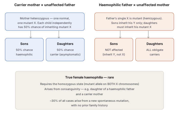
Both forms are X-linked recessive, manifesting clinically in males, with females usually the (asymptomatic) carriers. A carrier mother transmits the mutant X chromosome to each child independently at a 50:50 chance — so each son has a 50% chance of being affected, and each daughter a 50% chance of being a carrier. An affected father cannot transmit the disorder to his sons (who inherit only his Y chromosome) but transmits his mutant X to all his daughters, who therefore become obligate carriers. Approximately 30% of haemophilia A cases arise from a new, spontaneous mutation with no prior family history. True female haemophilia is rare, requiring the homozygous state — for example, a daughter born to a haemophilic father and a carrier mother, typically within a consanguineous family.
Frequency of haemophilia varies across populations, historically highest in Britain and Northern Europe.
Molecular Background — the Factor VIII–vWF Complex
Factor VIII is synthesised in hepatic parenchymal cells and acts as an essential cofactor for activated factor IX, which in turn activates factor X in the intrinsic coagulation pathway. Circulating factor VIII travels non-covalently bound to von Willebrand factor (vWF), a much larger protein (comprising ~99% of the mass of the factor VIII–vWF complex) synthesised chiefly by endothelial cells. This binding stabilises factor VIII — a deficiency of vWF (von Willebrand disease) therefore causes a secondary reduction in factor VIII, distinct from primary haemophilia A. Normal haemostasis requires only about 25% of normal factor VIII activity; most symptomatic haemophilia A patients have levels below 5%.
Haemophilia A — Factor VIII Deficiency
Caused by mutation in the F8 gene, producing deficient or reduced factor VIII activity; the underlying mutations vary widely (deletions, inversions, splice-junction mutations), explaining the range of clinical severity seen. In about 10% of patients, factor VIII protein levels are normal but coagulant activity is low due to a functional mutation.
Clinical features: bleeding for hours to days after injury, correlating well with plasma factor VIII activity. Bleeding can affect any organ, but characteristically occurs as recurrent, painful haemarthroses and muscle haematomas, and sometimes haematuria. Spontaneous intracranial and oropharyngeal bleeding are rare but the most feared complications.
Laboratory findings:
- Whole blood coagulation time — prolonged, but only in severe cases.
- Prothrombin time (PT) — usually normal.
- Activated partial thromboplastin time (APTT/PTTK) — typically prolonged.
- Specific factor VIII assay — reduced activity confirms diagnosis. Carrier females show roughly half-normal factor VIII activity; manifest disease is associated with activity below 25%.
- A PTT that fails to correct on mixing with normal plasma raises suspicion of a factor VIII inhibitor (neutralising antibody), which complicates replacement therapy in roughly 15% of patients with severe disease — factor VIII, administered repeatedly as treatment, is recognised by the immune system as foreign.
Treatment: factor VIII replacement (factor VIII concentrates or cryoprecipitate) for bleeding episodes. This treatment brought severe haemophiliacs’ life expectancy close to normal, though historically, transfusion-transmitted HIV in multi-transfused patients set this progress back — modern screening has since restored the safety of blood products. Where factor VIII inhibitors complicate treatment, a bispecific antibody bridging factor IX to factor X (bypassing the need for factor VIII entirely) is now available and appears more effective and easier to administer, though costly.
Haemophilia B — Factor IX Deficiency (Christmas Disease)
Caused by mutation in the F9 gene, producing deficiency of factor IX (Christmas factor, plasma thromboplastin component). Clinically and in inheritance pattern, indistinguishable from haemophilia A — but accurate laboratory distinction is essential, since the two require different replacement products. Screening tests mirror haemophilia A (prolonged APTT), with a specific factor IX assay confirming the diagnosis.
Treatment: infusion of fresh frozen plasma or factor IX concentrate/recombinant factor IX. Besides the general risks of any plasma-product therapy (hepatitis, chronic liver disease, transfusion-transmitted infection), factor IX replacement carries a specific additional risk of activating the coagulation system and precipitating thrombosis.
Clinical Relevance
The 30% new-mutation rate means a family history cannot be relied upon to raise suspicion — a boy presenting with unexplained haemarthrosis and no known family history of bleeding can still have haemophilia, and the diagnosis must rest on the clinical picture and specific factor assays rather than pedigree alone. Distinguishing haemophilia A from B by specific factor assay (not just by the shared prolonged APTT) is not an academic exercise — the two conditions require entirely different replacement products, and treating one as the other leaves the patient unprotected. The emergence of a factor VIII inhibitor, suspected whenever a patient’s PTT fails to correct on a mixing study, is a critical treatment-changing finding, since it renders standard factor VIII replacement ineffective and requires bypass strategies instead.
Diagram 1: X-Linked Recessive Inheritance Pattern
Draw two parallel columns (carrier mother’s cross, haemophilic father’s cross), each branching into a sons box and a daughters box, plus one shared bottom box for true female haemophilia.
- Left column — carrier mother × unaffected father → sons 50% affected, daughters 50% carrier.
- Right column — haemophilic father × unaffected mother → sons unaffected (0%), daughters 100% obligate carriers.
- Bottom — true female haemophilia requires homozygous state, from consanguinity (haemophilic father + carrier mother).
Labels required
- The reason sons of an affected father are never affected must be stated explicitly (they inherit his Y chromosome, not his X) — this is the fact that most directly tests whether the mechanism, not just the pattern, is understood.
- The “obligate carrier” status of all daughters of an affected father must be labelled as 100%, distinct from the 50% figure for daughters of a carrier mother — conflating these two different probabilities is a common error.
- The 30% new-mutation rate is worth a side note near the carrier-mother column, since it explains why a negative family history never excludes the diagnosis.
Errors commonly made
- Assuming an affected father can pass the disorder to a son — a direct contradiction of X-linked inheritance that examiners specifically probe for.
- Treating “carrier” and “affected” as interchangeable for daughters — a carrier daughter is clinically asymptomatic (heterozygous), not haemophilic.
- Forgetting that true female haemophilia exists at all, or describing it as arising from ordinary (non-consanguineous) inheritance rather than requiring the rarer homozygous combination.
Multiple Choice Questions (2)
My Sticky Notes
Personal revision notes, mnemonics and reminders.
