
Gaucher's Disease
Definition
Autosomal recessive lysosomal storage disease. Enzyme defect: acid β-glucosidase (old name: glucocerebrosidase). This enzyme normally removes glucose from ceramide. Defect → glucocerebroside builds up in lysosomes of phagocytic cells (spleen, liver, marrow, nodes), sometimes in neurons too. In phagocytes, the stored material comes from old WBC/RBC membranes. In neurons, stored material is gangliosides (different substance).
Three types (by CNS involvement)
- Type I (adult/classic) — commonest, ~80% of cases. Only viscera affected (spleen, liver, marrow, nodes). NO brain involvement.
- Type II (infantile) — severe, progressive brain (CNS) involvement from infancy.
- Type III (juvenile) — in between. Has visceral disease like Type I, plus slower brain disease like Type II.
Clinical features
Depends on type. Common: splenomegaly, hepatomegaly, lymphadenopathy. Hypersplenism → pancytopenia or thrombocytopenia. Bone pain and pathologic fractures from marrow involvement.
Morphology
Key finding: Gaucher cell — large, distended macrophage. Cytoplasm looks like “crumpled tissue paper.” Usually one nucleus, sometimes 2-3. Found in spleen, liver, marrow, lymph nodes; also around brain vessels (Virchow-Robin space) if CNS involved. Stains: PAS positive, oil red O positive, Prussian blue positive (shows glycolipid + iron mix). Rich in acid phosphatase. Often shows erythrophagocytosis.
Clinical relevance
Gaucher cell’s triple stain pattern (PAS + oil red O + Prussian blue) plus strong acid phosphatase helps tell it apart from similar-looking macrophages in other storage diseases, like the foamy cells of Niemann-Pick disease. Gaucher’s disease is one standard cause to remember on the differential list for splenomegaly, along with Niemann-Pick disease and mucopolysaccharidoses.
Definition
Gaucher’s disease is an autosomal recessive lysosomal storage disorder caused by mutation in acid β-glucosidase (formerly called glucocerebrosidase), the enzyme that normally cleaves glucose from ceramide. The defect causes lysosomal accumulation of glucocerebroside (ceramide-glucose) chiefly within phagocytic cells throughout the body, and sometimes within neurons. In phagocytic cells, the accumulating glucocerebroside derives mainly from the membrane glycolipids of aged, degraded leucocytes and erythrocytes; the deposits within neurons, where they occur, consist instead of gangliosides.
Clinical Subtypes
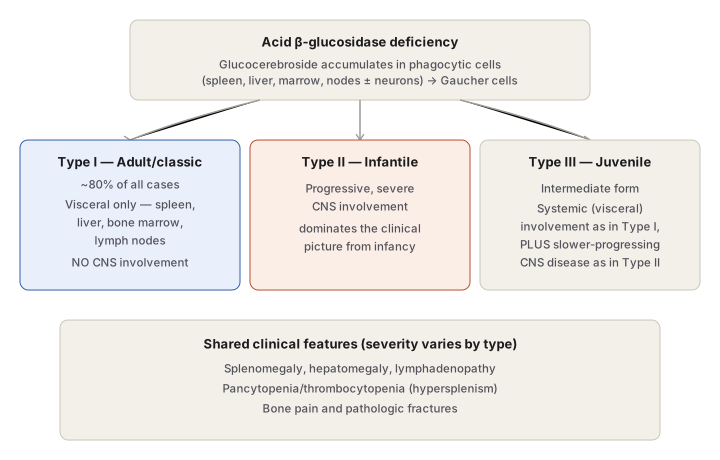
Three clinical subtypes are recognised, distinguished by the degree of neuronopathic (CNS) involvement:
- Type I (classic/adult form) — the most common, accounting for roughly 80% of all cases. Glucocerebroside storage is confined to phagocytic cells of the spleen, liver, bone marrow, and lymph nodes; no CNS involvement.
- Type II (infantile form) — marked by progressive, severe involvement of the central nervous system, dominating the clinical picture from infancy.
- Type III (juvenile form) — an intermediate phenotype, combining the systemic (visceral) involvement of type I with a slower-progressing form of the CNS disease seen in type II.
Clinical Features
Features depend on the subtype, but in general reflect the organs storing glucocerebroside — splenomegaly, hepatomegaly, lymphadenopathy, and involvement of bone marrow (and, in types II/III, the brain). Hypersplenism secondary to splenic enlargement can produce pancytopenia or thrombocytopenia. Skeletal involvement causes bone pain and pathologic fractures.
Morphology
Light Microscopy
The diagnostic finding is the Gaucher cell — a characteristically distended, enlarged macrophage, found in the spleen, liver, bone marrow, and lymph nodes, and, when neurons are involved, in the perivascular Virchow-Robin space. The cytoplasm is abundant, granular, and fibrillar, described as resembling crumpled tissue paper; the cell usually has a single nucleus, though two or three nuclei are occasionally seen. Gaucher cells stain positively with PAS, oil red O, and the Prussian blue reaction, reflecting the mixed nature of the stored material (glycolipid admixed with hemosiderin), and frequently show erythrophagocytosis. They are rich in acid phosphatase.
Clinical Relevance
Recognising the Gaucher cell’s specific staining pattern — PAS-positive, oil-red-O-positive, and Prussian-blue-positive together, with dense acid phosphatase activity — is what distinguishes it on a marrow or spleen biopsy from other storage-disease macrophages that can look superficially similar (such as the foamy, non-wrinkled macrophages of Niemann-Pick disease). Splenomegaly is also a standard differential-diagnosis entry point: Gaucher’s disease belongs to the storage-disease category of causes of splenomegaly, alongside Niemann-Pick disease and the mucopolysaccharidoses, a grouping worth recalling whenever hepatosplenomegaly in a child or young adult needs a systematic cause list.
Diagram 1: Three Clinical Subtypes Compared
Draw one top box (the shared enzyme defect and accumulation mechanism) fanning into three branch boxes (Type I, II, III), converging below into one shared-features box.
- Top — acid β-glucosidase deficiency → glucocerebroside accumulates in phagocytic cells, forming Gaucher cells.
- Type I — ~80% of cases, visceral only, no CNS involvement.
- Type II — infantile, severe progressive CNS involvement.
- Type III — juvenile, visceral (as Type I) plus slower CNS disease (as Type II).
- Bottom — shared clinical features common to all three (organomegaly, cytopenias, bone pain/fractures).
Labels required
- CNS involvement status must be explicit for all three types — present/absent/intermediate — since this single axis is what the classification is actually organised around, not an incidental detail.
- Type I’s ~80% prevalence figure must appear, since it is the detail that makes Type I the “default” clinical picture to think of first.
Errors commonly made
- Treating the three types as differing in severity of the same single disease process rather than differing specifically in whether and how fast the CNS is involved — the visceral disease itself is present in all three types.
- Forgetting that Type III combines features of both Type I and Type II rather than being a simply “medium severity” version of one or the other — it is genuinely both a visceral disease and a (slower) neurodegenerative disease at once.
Multiple Choice Questions (1)
My Sticky Notes
Personal revision notes, mnemonics and reminders.
