
Free Radical Injury
Definition
A free radical carries a single unpaired electron in its outer orbit, making it unstable and reactive; reaction with a neighbouring molecule converts that molecule into a further radical, so the reaction is self-propagating. Injury results when production outpaces elimination — oxidative stress.
Types
Reactive oxygen species — superoxide (O₂⁻), hydrogen peroxide (H₂O₂), hydroxyl radical (OH⁻). Reactive nitrogen species — nitric oxide (NO) and peroxynitrite (ONOO⁻), formed when NO combines with superoxide. Drug- and chemical-derived radicals — for example, carbon tetrachloride metabolised to CCl₃.
Generation in all cells — the sequence
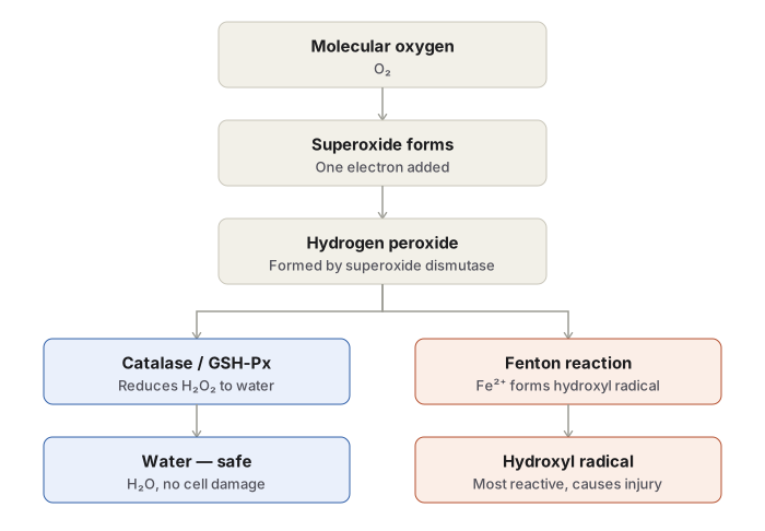
O₂ receives one electron → superoxide (O₂⁻), formed during mitochondrial electron transport or via xanthine oxidase and cytochrome P450 → superoxide dismutase converts it to hydrogen peroxide (H₂O₂) → in the presence of ferrous iron, the Fenton reaction converts hydrogen peroxide to the hydroxyl radical (OH⁻), the most reactive of the group. Transition metals, particularly iron and copper, catalyse this last step by readily donating and accepting electrons.
Generation in phagocytic leucocytes
Neutrophils and macrophages generate reactive oxygen species within the phagosome to kill ingested microbes, through a process called the respiratory burst. NADPH oxidase in the phagosomal membrane produces superoxide, which is converted to hydrogen peroxide; myeloperoxidase then converts hydrogen peroxide to hypochlorite (HOCl), the cytotoxic compound used against ingested organisms.
Mechanisms of damage
Lipid peroxidation of membrane polyunsaturated fatty acids, propagating along the membrane. Protein oxidation, with cross-linking and abnormal folding. DNA strand breaks, contributing to both cell injury and carcinogenesis. Interference with cytoskeletal elements and mitochondrial oxidative phosphorylation, worsening ATP depletion.
Conditions with increased free radical generation
Inflammation and microbial killing · chemical and drug injury, including carcinogens · radiation injury · ischaemia-reperfusion injury · cellular ageing · hyperoxia (oxygen therapy) · destruction of tumour cells · atherosclerosis.
Antioxidant defence
| Enzymatic | Non-enzymatic |
|---|---|
| Superoxide dismutase — O₂⁻ → H₂O₂ | Vitamins E, A, ascorbic acid; sulfhydryl compounds (cysteine, glutathione) |
| Catalase — H₂O₂ → H₂O + O₂ | Iron- and copper-binding proteins — transferrin, ferritin, albumin, ceruloplasmin |
| Glutathione peroxidase — neutralises H₂O₂, hydroxyl radical |
Ischaemia-reperfusion injury
Restoring blood flow to an already injured but still viable cell can worsen rather than reverse the injury. Three linked mechanisms:
- Calcium overload — reperfused blood delivers calcium to a cell with depleted ATP, triggering membrane lipid peroxidation.
- A surge of free radical generation as oxygen suddenly returns to tissue laden with ADP and pyruvate.
- Recruitment of neutrophils, whose own respiratory burst adds further free radicals.
Classic sites are the myocardium and the brain, where reperfusion after critical delay can itself contribute to irreversible cell death.
Definition
A free radical is an unstable chemical species carrying a single unpaired electron in its outer orbit. Once formed, a free radical tends to react with nearby molecules and convert them into further free radicals, so the process is self-propagating (autocatalytic). Free radicals produced within cells are ordinarily unstable and are destroyed almost as soon as they form; injury results only when their rate of production outpaces the rate at which the body eliminates them, a state described as oxidative stress. Free radical-mediated injury is implicated in a wide range of processes — chemical and radiation injury, hypoxia, cellular ageing, tissue damage caused by inflammatory cells, and ischaemia-reperfusion injury — and can activate both necrosis and apoptosis.
Types of Free Radicals
- Reactive oxygen species, the oxygen-derived free radicals, comprising the superoxide anion (O₂⁻), hydrogen peroxide (H₂O₂), and the hydroxyl radical (OH⁻).
- Reactive nitrogen species, derived from nitric oxide (NO), which is generated physiologically by endothelial cells, macrophages, neurons, and other cell types. Nitric oxide itself behaves as a free radical and can also combine with superoxide to form the highly reactive peroxynitrite anion (ONOO⁻), along with related species such as nitrogen dioxide and nitrate.
- Free radicals generated from drugs and chemicals, produced by enzymatic metabolism of exogenous compounds. These are not reactive oxygen species themselves but act similarly — the classic example is metabolism of carbon tetrachloride (CCl₄) to the free radical CCl₃, which produces severe hepatotoxicity.
Generation of Reactive Oxygen Species
In All Cells
Reactive oxygen species are produced in small amounts as a normal by-product of mitochondrial respiration, during the reduction-oxidation reactions that generate cellular energy. Complete reduction of molecular oxygen (O₂) to water (H₂O) proceeds through a four-electron transfer occurring in sequential one-electron steps, catalysed by cytochrome oxidase in the inner mitochondrial membrane, and each partial step yields a distinct intermediate.
| Species | Formed by |
|---|---|
| Superoxide (O₂⁻) | Partial (one-electron) reduction of O₂, occurring spontaneously during mitochondrial electron transport, or enzymatically via xanthine oxidase and cytochrome P450 |
| Hydrogen peroxide (H₂O₂) | Conversion of superoxide, occurring spontaneously and through the action of superoxide dismutase (SOD) |
| Hydroxyl radical (OH⁻) | Reaction of hydrogen peroxide with ferrous iron (Fe²⁺), termed the Fenton reaction; also produced by radiolysis of water |
| Peroxynitrite (ONOO⁻) | Reaction of superoxide with nitric oxide |
Hydroxyl radical is the most reactive species of the group. Transition metals such as iron and copper, by readily donating or accepting electrons, catalyse free radical formation and are essential participants in the Fenton reaction (H₂O₂ + Fe²⁺ → Fe³⁺ + OH⁻ + OH•).
In Phagocytic Leucocytes
Neutrophils and macrophages generate reactive oxygen species deliberately, within the phagosome and phagolysosome, as a weapon against ingested microbes — a process termed the respiratory burst, mechanistically similar to mitochondrial respiration. The enzyme NADPH oxidase (phagocyte oxidase), present in the phagosomal and phagolysosomal membrane, synthesises superoxide, which is converted to hydrogen peroxide. In the presence of the leucocyte enzyme myeloperoxidase, hydrogen peroxide is then converted to hypochlorite (HOCl), a highly reactive and cytotoxic compound used to kill ingested organisms.
Conditions Associated with Increased Free Radical Generation
- Inflammation and microbial killing by phagocytes.
- Chemical and drug injury, including chemical carcinogens.
- Radiation injury, such as ultraviolet light and X-rays, through radiolysis of water.
- Ischaemia-reperfusion injury, on restoration of blood flow to previously ischaemic tissue.
- Reduction-oxidation reactions catalysed by transition metals such as iron and copper.
- Cellular ageing.
- Hyperoxia, as in prolonged high-concentration oxygen therapy.
- Destruction of tumour cells and atherosclerosis are further settings in which free radical activity contributes to tissue change.
Mechanisms of Cell Damage by Free Radicals
| Mechanism | Effect |
|---|---|
| Lipid peroxidation | Polyunsaturated fatty acids within cell membranes are attacked repeatedly, generating destructive lipid hydroperoxides; the reaction propagates along the membrane, spreading damage and destroying organelles |
| Protein oxidation | Cross-linking and oxidative modification of proteins damages enzyme activity and produces abnormal protein folding, with direct fragmentation of polypeptides |
| DNA damage | Single-strand breaks occur in nuclear and mitochondrial DNA, contributing to cell injury and to malignant transformation |
| Cytoskeletal interference | Reactive oxygen species interact with cytoskeletal elements and interfere with mitochondrial oxidative phosphorylation, compounding ATP depletion |
Neutralisation of Free Radicals: Antioxidants
The body limits free radical damage through both enzymatic and non-enzymatic mechanisms present in serum, tissue fluid, and within cells themselves.
| Enzymatic antioxidants | Non-enzymatic antioxidants |
|---|---|
| Superoxide dismutase (SOD) — converts superoxide to hydrogen peroxide | Exogenous: vitamin E, vitamin A, ascorbic acid, sulfhydryl-containing compounds such as cysteine and glutathione |
| Catalase — converts hydrogen peroxide to water and oxygen, active in peroxisomes | Endogenous: iron- and copper-binding storage and transport proteins, including transferrin, ferritin, albumin, and ceruloplasmin, which limit the availability of these reactive metals for radical-generating reactions |
| Glutathione peroxidase — neutralises hydrogen peroxide, hydroxyl radical, and certain drug-derived radicals |
Ischaemia-Reperfusion Injury
Restoring blood flow after a period of ischaemia can, paradoxically, worsen an already injured but still-viable cell rather than rescuing it, and this is the clearest clinical illustration of free radical-mediated injury. Three linked mechanisms are involved.
- Calcium overload. Reperfused blood carries calcium to a cell whose ATP stores remain depleted, adding to the calcium burden and triggering lipid peroxidation of the membrane.
- A surge in free radical generation. The sudden return of oxygen to tissue that has accumulated ADP and pyruvate during ischaemia drives a burst in the production of superoxide, hydrogen peroxide, hydroxyl radical, and peroxynitrite.
- An inflammatory reaction. Neutrophils recruited to the reperfused tissue mount a respiratory burst of their own, consuming oxygen rapidly and releasing further free radicals.
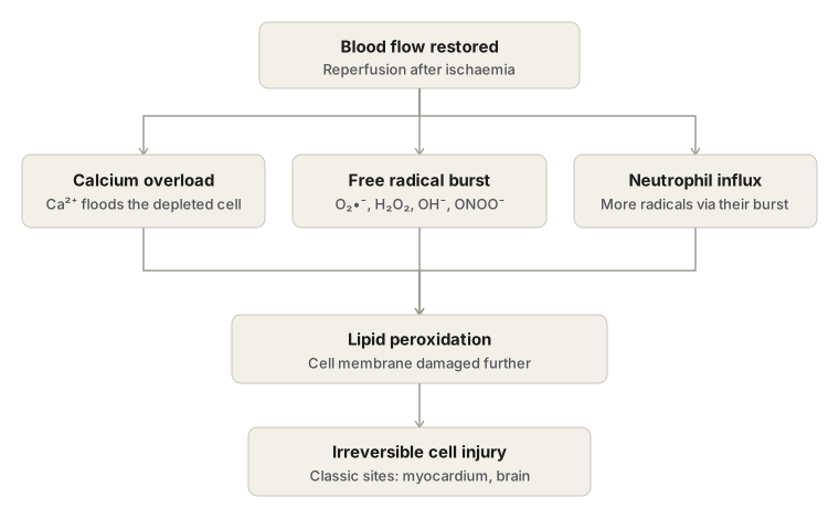
Classic examples of ischaemia-reperfusion injury occur in myocardial and cerebral ischaemia, where restoration of blood flow after a critical delay can itself contribute to irreversible cell death.
Clinical Relevance
- Free radical injury is a shared final mechanism linking several apparently unrelated conditions — radiation injury, drug- and toxin-induced organ damage, atherosclerosis, ageing, and reperfusion injury following thrombolysis or revascularisation.
- Because the same reactive oxygen species that damage host tissue are deliberately generated by phagocytes to kill microbes, excessive or prolonged neutrophil activation in chronic inflammation becomes a mechanism of collateral tissue injury.
- Recognising ischaemia-reperfusion injury explains a clinical observation that can otherwise seem contradictory: restoring blood flow does not always rescue tissue, and may transiently worsen the injury it is intended to treat.
Diagram 1: Stepwise Reduction of Oxygen to Water
Draw a single horizontal reaction chain of molecule boxes connected by arrows, each arrow labelled with the enzyme or reaction responsible.
Box 1: O₂ (molecular oxygen) → arrow labelled: mitochondrial electron transport, or xanthine oxidase / cytochrome P450 Box 2: superoxide, O₂⁻ → arrow labelled: superoxide dismutase (SOD) Box 3: hydrogen peroxide, H₂O₂ → two branching arrows: one labelled catalase (peroxisome) or glutathione peroxidase, leading to a side box “H₂O” (safe outcome); one labelled Fenton reaction (Fe²⁺), continuing forward Box 4: hydroxyl radical, OH⁻ — outline this box distinctly and caption it as the most reactive species → final arrow to Box 5: H₂O, the final product
Add a small side box showing Fe³⁺ reduced to Fe²⁺ by superoxide, to indicate the source of the ferrous iron used in the Fenton reaction.
Labels required
- Each enzyme name positioned directly on its arrow — superoxide dismutase, catalase or glutathione peroxidase, and the Fenton reaction with Fe²⁺ shown as a co-reactant.
- Both the detoxification branch (to water) and the injurious branch (to hydroxyl radical) shown from the same hydrogen peroxide box, since the intermediate has two possible fates.
Errors commonly made
- Omitting the detoxification branch and drawing only the pathway to hydroxyl radical.
- Leaving the Fenton reaction unnamed.
- Failing to show where the ferrous iron for the Fenton reaction originates.
Diagram 2: Reactive Oxygen Species Generation in the Phagocytic Vacuole
Draw a cross-section of a neutrophil or macrophage with an ingested microbe enclosed in a phagosome.
- Show NADPH oxidase embedded in the phagosomal membrane, converting O₂ to superoxide (O₂⁻) inside the vacuole.
- Show superoxide converted to hydrogen peroxide (H₂O₂).
- Show myeloperoxidase, released from cytoplasmic granules into the phagolysosome, converting hydrogen peroxide (with chloride ion, Cl⁻) into hypochlorite (HOCl).
- Show the hypochlorite acting on the ingested microbe within the vacuole.
Labels required
- Name NADPH oxidase and myeloperoxidase explicitly.
- Label the process as the respiratory burst.
- Show chloride ion as a co-reactant for the myeloperoxidase step.
Errors commonly made
- Omitting myeloperoxidase and stopping the sequence at hydrogen peroxide, without producing hypochlorite.
- Drawing the reaction as occurring in the cytoplasm rather than within the phagosome/phagolysosome.
Diagram 3: Ischaemia-Reperfusion Injury
Draw a two-column flowchart: left column labelled “during ischaemia,” right column labelled “on reperfusion,” joined by a downward arrow into a single convergence box.
Left column: blood flow interrupted → falling ATP, accumulation of ADP and pyruvate → cell injured but still viable.
Right column: blood flow restored → three parallel boxes: calcium overload (reperfused blood delivers Ca²⁺ to the ATP-depleted cell); a burst of free radical generation (O₂⁻, H₂O₂, OH⁻, ONOO⁻); neutrophil recruitment with its own respiratory burst adding further free radicals.
Convergence box: lipid peroxidation of the membrane leading to irreversible cell injury, with a note beneath: classic sites are the myocardium and the brain.
Labels required
- All three components of the reperfusion mechanism shown as separate labelled boxes.
- A caption stating that reperfusion can worsen an already injured but viable cell.
Errors commonly made
- Collapsing the three mechanisms into a single box labelled “free radicals cause damage.”
- Omitting the neutrophil/inflammatory contribution.
- Failing to specify that the cell must be viable but injured going into reperfusion — if the cell had already died during prolonged ischaemia, this mechanism does not apply.
Multiple Choice Questions (1)
My Sticky Notes
Personal revision notes, mnemonics and reminders.
