
Fetal Hydrops
Definition
Fluid collects in at least two serous cavities plus under the skin, in the fetus. Severe form: hydrops fetalis (usually fatal). Milder forms: isolated pleural/peritoneal fluid, or neck fluid (cystic hygroma). No cause found in up to 20% of cases.
Two groups: immune (Rh/ABO) and non-immune. Now non-immune causes are more common, because Rh prophylaxis works well.
Immune hydrops
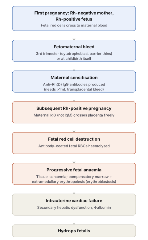
Rh incompatibility: Only the D antigen matters clinically. Rh-negative mother + Rh-positive fetus → fetal red cells cross to mother (3rd trimester or at delivery) → mother makes anti-Rh IgG (sensitisation) → next Rh-positive pregnancy → IgG crosses placenta → destroys fetal red cells → fetal anaemia → tissue ischaemia → heart failure → fluid pooling (hydrops).
Points to remember:
- ABO incompatibility together with Rh actually protects against Rh sensitisation (fetal cells cleared fast by anti-A/B IgM).
- Needs >1mL fetal blood to sensitise mother.
- Only IgG crosses placenta, not IgM. First exposure makes IgM → first pregnancy usually safe. Second exposure makes IgG → later pregnancy at risk.
- Prevention: give Rh immune globulin (RhIg) to Rh-negative mother at 28 weeks and within 72 hours after delivery.
ABO incompatibility: Now the commonest cause of immune haemolytic disease, since Rh prophylaxis reduced Rh cases. But milder than Rh disease. Seen in group A/B baby born to group O mother. Unlike Rh disease, first baby can also be affected (some O mothers have IgG anti-A/B even without earlier exposure). No prevention available.
Non-immune hydrops (now the bigger group)
| Cause type | Examples |
|---|---|
| Cardiac | Structural defects, arrhythmia, high-output failure |
| Chromosomal | Turner syndrome, trisomy 21, trisomy 18 |
| Thoracic | Diaphragmatic hernia |
| Fetal anaemia | Homozygous α-thalassaemia (Hb Bart’s), Parvovirus B19 |
| Twins | Twin-to-twin transfusion |
| Infection | CMV, syphilis, toxoplasmosis |
- Chromosomal causes usually work through a heart defect. In Turner syndrome, abnormal neck lymph drainage also adds cystic hygroma.
- Homozygous α-thalassaemia (all 4 α-genes lost, Hb Bart’s disease) is the top cause of fetal hydrops in regions where it is common.
- Parvovirus B19 infects red cell precursors in marrow, causes red cell aplasia (large nuclear inclusions seen in precursors).
All causes lead to the same final path: fetal anaemia → tissue ischaemia → heart failure → oedema. Liver also gets stressed doing extra blood-making work, so albumin drops too, adding to oedema.
Morphology
Severe: generalised fluid (hydrops fetalis). Milder: isolated fluid collections. In anaemic hydrops: pale fetus and placenta, big liver and spleen (from extra blood-making work, i.e. extramedullary haematopoiesis — also seen in kidney, lung, nodes, even heart). Many immature red cells enter blood (normoblasts, erythroblasts) — called erythroblastosis fetalis. Exception: in parvovirus B19 cases, marrow is suppressed, not overactive.
In Rh/ABO disease specifically: high bilirubin from red cell breakdown. If very high (>20 mg/dL term baby), bilirubin deposits in brain (basal ganglia, brain stem) — turns them yellow — called kernicterus. Can cause permanent brain damage.
Diagnosis and management
Rh hydrops severity tracks with rising maternal Rh antibody titre, so it can be predicted. Direct Coombs test on fetal cord blood confirms antibody-coated red cells. Fetal Rh status can now be checked from fetal DNA in maternal blood, no needle required. Severe cases: fetal blood transfusion through umbilical cord, early delivery. After birth: phototherapy converts bilirubin to a form easy to excrete. ABO disease is harder to predict before birth; check parents’ blood groups and check baby’s Hb/bilirubin after birth. If hydrops causes death, do a full postmortem to find the cause, especially to rule out a chromosomal problem that could repeat in next pregnancy.
Definition
Fetal hydrops is the accumulation of oedema fluid in at least two serous cavities together with subcutaneous oedema during intrauterine growth. Severity ranges widely — from progressive, generalised fetal oedema (hydrops fetalis, usually lethal) to more limited, often survivable collections such as isolated pleural or peritoneal effusion, or fluid at the back of the neck (cystic hygroma). In up to 20% of cases no cause is found.
Hydrops is divided by mechanism into immune and non-immune forms. Immune (Rh-alloimmune) hydrops was historically the leading cause; with effective Rh prophylaxis now widely practised, non-immune causes have become the dominant group.
Immune Hydrops
Results from antibody-mediated haemolytic anaemia in the fetus/newborn, caused by blood group incompatibility between mother and fetus — most often Rh and ABO antigens.
Rh Incompatibility
Of the numerous Rh antigens, only the D antigen is clinically important as a major cause of Rh disease. Fetal red cells carrying paternally-inherited, maternally-foreign Rh antigen can reach the maternal circulation during the third trimester (when the cytotrophoblast barrier thins) or at childbirth (fetomaternal bleed). The mother becomes sensitised, producing anti-Rh IgG. In a subsequent Rh-positive pregnancy, this maternal IgG (unlike IgM, which cannot cross the placenta) freely traverses the placenta and destroys fetal red cells. Progressive fetal anaemia produces tissue ischaemia, intrauterine cardiac failure, and peripheral pooling of fluid — the same final pathway responsible for oedema in most other causes of fetal anaemia.
Factors that shape the immune response:
- Concurrent ABO incompatibility protects against Rh sensitisation — fetal red cells are rapidly coated by maternal isohaemagglutinins (preformed anti-A/anti-B IgM) and cleared from the maternal circulation before they can sensitise her to the Rh antigen.
- Dose-dependence — clinically significant sensitisation requires a transplacental bleed exceeding roughly 1 mL of Rh-positive red cells.
- Antibody isotype — only IgG crosses the placenta. The first exposure to Rh antigen generates IgM, so Rh disease is rare in a first pregnancy; a subsequent pregnancy provokes a brisk IgG response.
Prevention: Rh immune globulin (RhIg) given to Rh-negative mothers at 28 weeks and within 72 hours of delivering an Rh-positive baby masks the antigenic sites on any leaked fetal red cells, preventing long-term maternal sensitisation.
ABO Incompatibility
Because Rh prophylaxis has succeeded, ABO incompatibility is now the most common cause of immune haemolytic disease of the newborn, though it produces disease far less often and far more mildly than Rh incompatibility — expression of A/B antigens on many non-red-cell tissues acts as a “sponge” absorbing much of the transferred antibody. Occurs almost exclusively in a group A or B infant born to a group O mother; usual anti-A/anti-B isohaemagglutinins in group O individuals are IgM (placenta-impermeable), but some group O women unpredictably carry IgG anti-A/anti-B even without prior sensitisation — so, unlike Rh disease, the first-born child can be affected. There is no equivalent preventive intervention.
Non-Immune Hydrops
Now the dominant category overall. Major causes:
| Category | Examples |
|---|---|
| Cardiovascular | Structural malformations, tachyarrhythmia, high-output failure |
| Chromosomal | Turner syndrome, trisomy 21, trisomy 18 |
| Thoracic | Diaphragmatic hernia |
| Fetal anaemia (non-immune) | Homozygous α-thalassaemia, parvovirus B19 infection |
| Twin gestation | Twin-to-twin transfusion |
| Infection (non-parvovirus) | Cytomegalovirus, syphilis, toxoplasmosis |
- Cardiovascular causes produce hydrops through intrauterine cardiac failure directly, the same downstream mechanism as in immune hydrops.
- Chromosomal anomalies — 45,X (Turner syndrome) and trisomies 21/18 — usually cause hydrops via an underlying structural cardiac defect; in Turner syndrome specifically, an additional mechanism is abnormal lymphatic drainage from the neck, producing the characteristic cystic hygroma.
- Homozygous α-thalassaemia (all four α-globin genes deleted, producing Hb Bart’s disease, in which γ-globin chains form non-functional tetramers unable to carry oxygen) causes severe fetal anaemia and is, in parts of the world where it is prevalent, the single most common cause of fetal hydrops.
- Parvovirus B19 infects and replicates within erythroid precursors (normoblasts), driving their apoptosis and producing red cell aplasia; characteristic large, homogeneous intranuclear viral inclusions are seen in bone marrow erythroid precursors.
Regardless of cause — immune or non-immune — the final common pathway to hydrops is tissue ischaemia from fetal anaemia, producing myocardial dysfunction and circulatory failure; secondary hepatic failure, from the liver’s diversion of resources into compensatory extramedullary haematopoiesis, adds hypoalbuminaemia and reduced plasma oncotic pressure to the oedema-forming mechanism.
Morphology
Findings vary with severity and cause. Hydrops fetalis is the most severe, generalised form; lesser degrees produce isolated pleural, peritoneal, or postnuchal fluid collections. In hydrops from fetal anaemia, both fetus and placenta are pale; liver and spleen are enlarged from cardiac congestion and from compensatory extramedullary haematopoiesis, which is also demonstrable in the kidney, lung, lymph nodes, and even heart. This heightened erythropoietic drive floods the peripheral circulation with immature erythroid precursors, including normoblasts and even more immature erythroblasts — the classic picture of erythroblastosis fetalis (parvovirus-associated cases are a notable exception, since here the virus is destroying erythroid precursors rather than driving their proliferation). Dysmorphic features on examination suggest an underlying chromosomal cause; postmortem may reveal a structural cardiac anomaly.
In Rh/ABO haemolytic disease specifically, red cell breakdown adds a further complication — a markedly raised circulating bilirubin. When unconjugated hyperbilirubinaemia is severe (typically >20 mg/dL in a full-term infant, often less in a premature one), bilirubin deposits in the brain, particularly the basal ganglia and brain stem, imparting a characteristic yellow discolouration — kernicterus — with potential for permanent neurological damage.
Diagnosis and Management
Early recognition is essential, since even severe cases can sometimes be salvaged. Rh-mediated immune hydrops can be anticipated with reasonable confidence, as its severity tracks rising maternal Rh antibody titres. Amniocentesis, chorionic villus sampling, and fetal blood sampling allow antenatal risk assessment: a positive direct Coombs (antiglobulin) test on fetal cord blood confirms antibody-coated fetal red cells, and fetal Rh status can now be determined non-invasively by sequencing cell-free fetal DNA in maternal blood. Severe intrauterine haemolysis may be treated with fetal intravascular transfusion via the umbilical cord and early delivery; postnatal phototherapy converts bilirubin into readily excreted forms. ABO haemolytic disease is harder to predict antenatally but is anticipated from parental blood group incompatibility and confirmed by neonatal haemoglobin/bilirubin measurement. Whenever fetal hydrops proves fatal, thorough postmortem examination is essential to identify the cause and exclude a potentially recurring one, such as a chromosomal abnormality.
Diagram 1: Mechanism of Immune Hydrops from Rh Sensitisation
Draw a single downward column of seven sequential stages, spanning two pregnancies, connected by arrows as one continuous sequence.
- First pregnancy: Rh-negative mother, Rh-positive fetus.
- Fetomaternal bleed (3rd trimester or delivery).
- Maternal sensitisation — anti-Rh(D) IgG produced.
- Subsequent Rh-positive pregnancy — IgG crosses placenta.
- Fetal red cell destruction.
- Progressive fetal anaemia (with compensatory erythroblastosis).
- Intrauterine cardiac failure → hydrops fetalis.
Labels required
- The antibody isotype (IgG, not IgM) must be explicitly named at the placental-crossing step — this is the single fact that explains why a first pregnancy is nearly always safe and a second Rh-positive pregnancy is the one at risk.
- The dose threshold for sensitisation (>1 mL fetal blood) and the protective effect of concurrent ABO incompatibility are worth a side annotation at the sensitisation step, since both explain why not every Rh-incompatible pregnancy produces a sensitised mother.
Errors commonly made
- Describing Rh disease as affecting the first pregnancy — it is the second and subsequent Rh-positive pregnancies that are at risk, because the first exposure only generates IgM (placenta-impermeable).
- Omitting that concurrent ABO incompatibility protects against Rh sensitisation, which looks counterintuitive (two incompatibilities co-occurring reducing risk rather than adding to it) and is exactly the kind of nuance exams test.
- Conflating the mechanism of immune hydrops with non-immune hydrops — the cascade above is specific to antibody-mediated haemolysis and does not apply to the cardiac, chromosomal, or infective causes of non-immune hydrops, which reach the same endpoint (fetal anaemia/cardiac failure → oedema) by different upstream routes.
My Sticky Notes
Personal revision notes, mnemonics and reminders.
