
Fatty Change
Definition
Steatosis = intracellular accumulation of neutral fat (triglyceride) in parenchymal cells — a genuine increase in cytosolic lipid, not infiltration from outside (old terms “fatty degeneration/infiltration” abandoned). Most often in liver; also heart, skeletal muscle, kidney (lipoid nephrosis).
Fatty liver — etiology
Excess fat delivery: obesity · diabetes mellitus · congenital hyperlipidaemia.
Liver cell damage: alcohol (most common) · starvation · protein-calorie malnutrition · chronic illness (TB) · acute fatty liver of pregnancy · hypoxia (anaemia, cardiac failure) · hepatotoxins (CCl₄, chloroform, ether, aflatoxins) · drugs (methotrexate, steroids, halothane, tetracycline) · Reye syndrome.
Pathogenesis — normal pathway and 6 defect points
FFA enters hepatocyte (diet as chylomicrons/FFA, or adipose tissue as FFA, small amount synthesised locally) → esterified to triglyceride (most) or converted to cholesterol/phospholipid/ketones (less) → triglyceride packaged into lipoprotein (needs lipid acceptor protein) → released as plasma lipoprotein (LDL, VLDL).
Fatty liver = defect at one or more of:
- ↑FFA entry into liver
- ↑FFA synthesis by liver
- ↓conversion of FFA to ketone bodies → ↑esterification to triglyceride
- ↑α-glycerophosphate → ↑esterification to triglyceride
- ↓lipid acceptor protein synthesis → ↓lipoprotein formation
- Block in lipoprotein excretion
Alcohol acts through multiple of these simultaneously (multifactorial) — unlike most other causes, which act through just one.
Reversible if fibrosis hasn’t developed yet — basis for “intermittent drinking less harmful,” and abstinence reversing an enlarged fatty liver.
Morphology
Gross: enlarged, tense glistening capsule, rounded margins, pale yellow, greasy cut surface.
Microscopic: non-staining cytoplasmic vacuoles (fat dissolves during processing) — microvesicular (small, perinuclear) → macrovesicular (large, nucleus pushed to periphery). Rupture → fatty cysts. Lipogranulomas (lymphocytes, macrophages, giant cells) at sites of extravasated fat. Fat stains on frozen section: Sudan III/IV/black, Oil Red O, osmic acid.
Clinical relevance
Silent and reversible early — but with continued injury (esp. alcohol) can progress through fibrosis to irreversible cirrhosis. Presence should prompt a search for the driving cause (metabolic, toxic, infective, nutritional). Acute fatty liver of pregnancy is an obstetric emergency, distinct in urgency from the chronic causes despite the same morphology.
Definition
Fatty change, also called steatosis or fatty metamorphosis, is the intracellular accumulation of neutral fat (triglyceride) within parenchymal cells. The deposit sits in the cytosol and represents a genuine, absolute increase in intracellular lipid, not simply an appearance of fat that was already there — which is why the older terms fatty degeneration and fatty infiltration have been abandoned, since the change is neither a form of degeneration nor an infiltration from outside the cell. Fatty change is most familiar in the liver, which occupies the central role in fat metabolism, but it also occurs in the heart, skeletal muscle, and kidney, where it is termed lipoid nephrosis or minimal change disease.
Fatty Liver
The liver is the organ most often affected, since nearly every step of fat transport and metabolism passes through it.
Etiology
Fatty change in the liver arises from one of two broad categories of cause.
Conditions with excess fat delivery, in which the liver’s capacity to metabolise fat is overwhelmed by supply:
- Obesity.
- Diabetes mellitus.
- Congenital hyperlipidaemia.
Conditions with liver cell damage, in which the injured hepatocyte can no longer process fat normally:
- Alcoholic liver disease — the single most common cause.
- Starvation.
- Protein-calorie malnutrition.
- Chronic illness, such as tuberculosis.
- Acute fatty liver of late pregnancy.
- Hypoxia, as in anaemia or cardiac failure.
- Hepatotoxins — carbon tetrachloride, chloroform, ether, aflatoxins, and related poisons.
- Drug-induced liver injury — methotrexate, corticosteroids, halothane, tetracycline, among others.
- Reye syndrome.
Pathogenesis
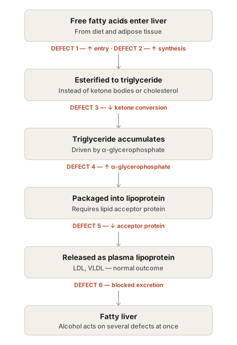
Understanding fatty liver requires tracing the normal pathway of hepatic fat metabolism, since the mechanism by which any given cause produces fatty change is a defect at one or more points along that pathway. Free fatty acids enter the hepatocyte from two sources — the diet, as chylomicrons and free fatty acids, and adipose tissue, also as free fatty acids — with a small additional contribution synthesised locally from acetate. Most of this fatty acid is esterified to triglyceride; a smaller portion is converted to cholesterol, phospholipids, and ketone bodies. Triglyceride is then packaged into lipoproteins, a process requiring a lipid acceptor protein, and released from the hepatocyte into the circulation as plasma lipoproteins (LDL, VLDL).
Fatty liver develops when a defect occurs at one or more of six points in this sequence:
- Increased entry of free fatty acids into the liver.
- Increased synthesis of fatty acids by the liver itself.
- Decreased conversion of fatty acids to ketone bodies, which increases esterification of fatty acids to triglyceride instead.
- Increased α-glycerophosphate, which likewise increases esterification of fatty acids to triglyceride.
- Decreased synthesis of the lipid acceptor protein, reducing formation of lipoprotein from triglyceride.
- A block in the excretion of lipoprotein from the liver into the plasma.
Most causes of fatty liver operate through one of these six mechanisms in isolation, but liver injury from chronic alcoholism is multifactorial, acting simultaneously through increased lipolysis, increased free fatty acid synthesis, decreased triglyceride utilisation, decreased fatty acid oxidation to ketone bodies, and a block in lipoprotein excretion.
Fatty liver, even in a severe form, is potentially reversible: if the liver is given time to regenerate and progressive fibrosis has not yet developed, the accumulated fat can clear. This is the basis for the clinical observation that intermittent drinking is less harmful than continuous drinking, and that a chronic alcoholic who becomes abstinent can see an enlarged fatty liver return toward normal, provided fibrosis has not already set in.
Morphology
Gross. The liver is enlarged, with a tense, glistening capsule and rounded margins. The cut surface bulges slightly and is pale yellow to yellow, greasy to the touch.
Microscopic. The defining feature is numerous lipid vacuoles within hepatocyte cytoplasm, seen as non-staining vacuoles on routine haematoxylin and eosin sections because the fat dissolves out during processing.
- The vacuoles begin small and clustered around the nucleus — the microvesicular pattern.
- As the process advances, the vacuoles enlarge and coalesce, pushing the nucleus to the periphery of the cell — the macrovesicular pattern.
- Hepatocytes distended by large lipid vacuoles may occasionally rupture, and the released lipid can coalesce to form fatty cysts.
- Lipogranulomas may form as a reaction to extravasated fat, consisting of lymphocytes, macrophages, and occasional multinucleated giant cells.
- Fat can be demonstrated directly in fresh, unfixed tissue by frozen section using fat stains such as Sudan III, Sudan IV, Sudan black, or Oil Red O; osmic acid can serve as both fixative and stain for this purpose.
Clinical Relevance
- Fatty liver is usually clinically silent and reversible, but it is the first stage of a spectrum that, with continued injury (chronic alcoholism being the classic driver), can progress through fibrosis to irreversible cirrhosis — recognising fatty change early is therefore an opportunity to intervene before that transition occurs.
- Because fatty liver reflects a defect somewhere in a shared six-step metabolic pathway, its presence in a patient should prompt a search for the specific driving cause — metabolic (obesity, diabetes), toxic (alcohol, drugs), infective, or nutritional — rather than being treated as a diagnosis in itself.
- Acute fatty liver of pregnancy is an obstetric emergency distinct from the more chronic causes of steatosis, underscoring that the same morphological finding can represent very different clinical urgency depending on its cause.
Diagram 1: Pathogenesis of Fatty Liver (six defect points)
Draw the normal pathway of hepatic fat metabolism as a flowchart, with each of the six possible defect points marked directly on the arrow or step where it occurs.
Main pathway, left to right: “Free fatty acids enter hepatocyte” (from two small feeder arrows above it, one labelled “diet — chylomicrons/FFA,” one labelled “adipose tissue — FFA”) → “Esterified to triglyceride” → “Packaged into lipoprotein (needs lipid acceptor protein)” → “Released as plasma lipoprotein (LDL, VLDL).”
Numbered defect annotations, placed as small numbered flags directly on the relevant arrow or box:
- On the entry arrow: ”↑ FFA entry into liver.”
- Inside the first box: ”↑ FFA synthesis by liver.”
- On the arrow to triglyceride: ”↓ conversion to ketone bodies → ↑ esterification.”
- On the same arrow: ”↑ α-glycerophosphate → ↑ esterification.”
- On the arrow to lipoprotein packaging: ”↓ lipid acceptor protein synthesis.”
- On the final arrow: “Block in lipoprotein excretion.”
Labels required
- All six numbered defects must appear individually positioned at the correct step of the pathway, not listed separately off to the side disconnected from the mechanism.
- The two sources of free fatty acid entry (diet, adipose tissue) must both be shown feeding into the pathway.
- A closing note that any one or more of these six defects, alone or in combination, produces fatty liver — and that alcohol is the example that acts through several simultaneously.
Errors commonly made
- Listing the six defects as a separate numbered list beside the diagram rather than anchoring each one to the specific pathway step it disrupts — this is a mechanism diagram, and the point is showing where each defect intervenes.
- Omitting the lipid acceptor protein as the specific requirement for step 3 (lipoprotein packaging), which is a commonly asked specific detail.
- Forgetting to note that alcohol operates through multiple defects at once, unlike most other causes.
Diagram 2: Microscopic Progression — Microvesicular to Macrovesicular Fatty Change
Draw three hepatocytes in sequence showing increasing fat accumulation.
- Normal hepatocyte: central nucleus, no vacuoles.
- Microvesicular change: several small, discrete clear vacuoles clustered around a still-central nucleus.
- Macrovesicular change: a single large clear vacuole filling most of the cytoplasm, nucleus flattened and pushed to the periphery of the cell.
Labels required
- Each stage titled: normal, microvesicular, macrovesicular.
- The nucleus position must be explicitly labelled at each stage — central in stages 1 and 2, peripheral in stage 3 — since this shift is the specific feature distinguishing the two patterns.
- A note that the vacuoles are non-staining on routine H&E because the fat dissolves during tissue processing, and that fat stains (Sudan, Oil Red O) on frozen section are needed to demonstrate it directly.
Errors commonly made
- Drawing macrovesicular change with the nucleus still central — the peripheral displacement of the nucleus by the single large vacuole is the defining feature examiners check for.
- Failing to note that the vacuoles are artifactually clear (fat dissolved out) rather than genuinely empty spaces, which is a frequently tested technical point.
Multiple Choice Questions (1)
My Sticky Notes
Personal revision notes, mnemonics and reminders.
